Journal List > Anat Cell Biol > v.55(4) > 1516080681

Nayak and Vasudeva: Terminal bifurcation of the external jugular vein: a rare variation

Abstract

Variations of external jugular vein are common. Here, we present a rare terminal bifurcation of the left external jugular vein. The left external jugular vein was formed by the union of entire retromandibular vein and posterior auricular vein. One inch above the clavicle, it bifurcated into medial and lateral divisions. The medial division terminated into the internal jugular vein and the lateral division terminated into the subclavian vein. Medial division received a common vein formed by the union of anterior jugular vein and an anonymous vein lying under the sternocleidomastoid muscle. The lateral division received a common vein formed by the union of suprascapular and transverse cervical veins. The knowledge about this variation could be useful to head and neck surgeons, radiologists and plastic surgeons.

Introduction

External jugular vein is one among the most variable veins of the neck. Though it doesn’t have a specific way of formation, in most of the individuals it is formed by the union of posterior division of retromandibular vein and the posterior auricular vein. It runs down vertically in the superficial facia of the neck and pierces the investing layer of deep cervical fascia just above the middle of the clavicle and terminates into the subclavian vein. Its tributaries include the posterior external jugular, anterior jugular, suprascapular and transverse cervical veins. External jugular vein is used to assess the right atrial pressure as this vein does not possess any valves. The vein may be entirely absent unilaterally or bilaterally [1], duplicated [2], or fenestrated [3]. Apart from this, many variations in its origin, course and termination have been reported. Reported variations about its formation include formation by union of both divisions of retromandibular vein with facial vein [4], by the union of superficial temporal vein with the posterior auricular vein [5], and by the union of submandibular, facial, anterior jugular and both divisions of retromandibular vein [6]. Variations that have been reported about the termination of the external jugular vein include termination of the vein of one side into the subclavian vein of opposite side [7], or by formation of a jugulocephalic vein after receiving the cephalic vein [8]. We report a rare, terminal bifurcation of the external jugular vein here. This report could be useful to plastic surgeons and head and neck surgeons.

Case Report

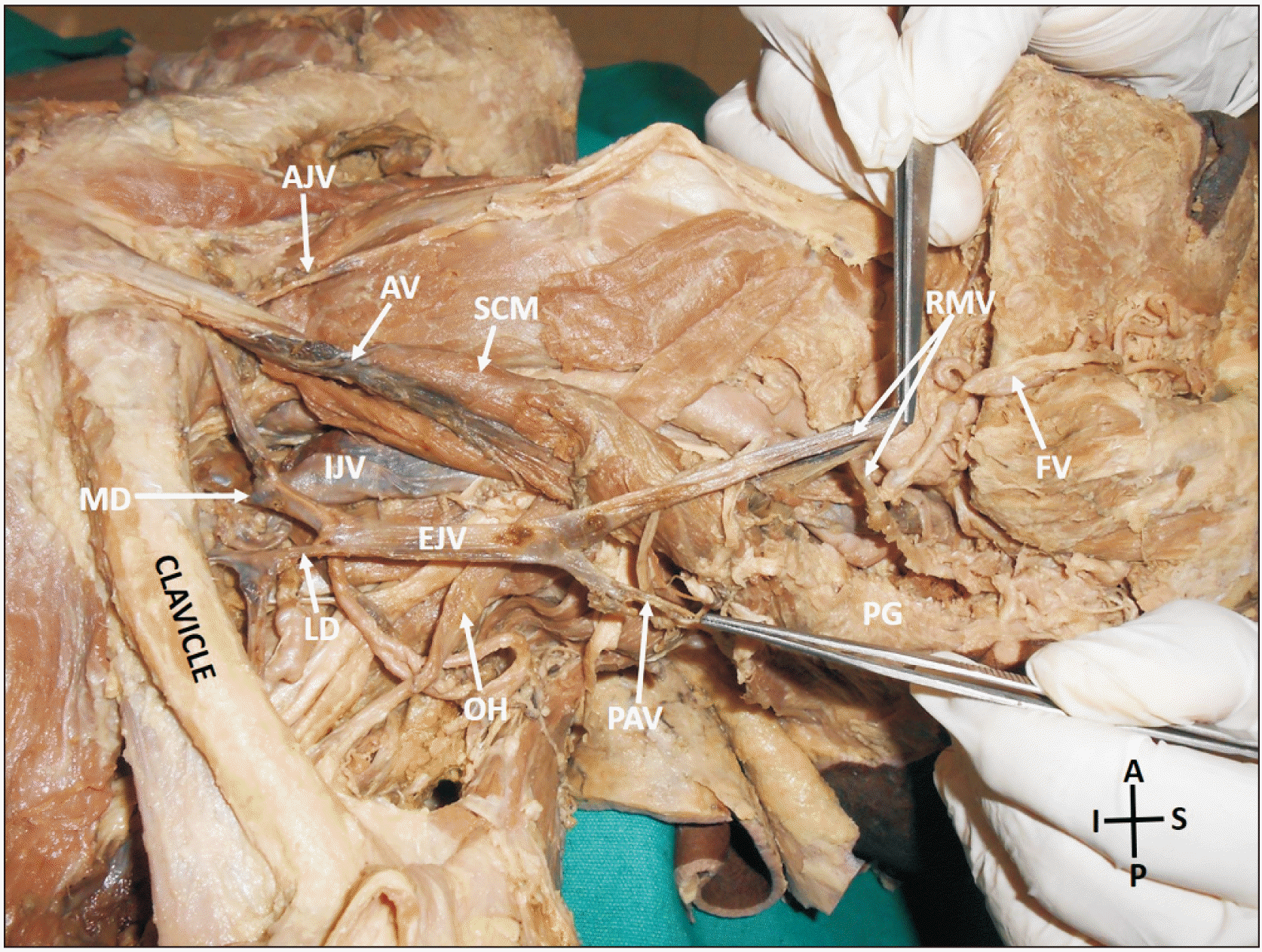
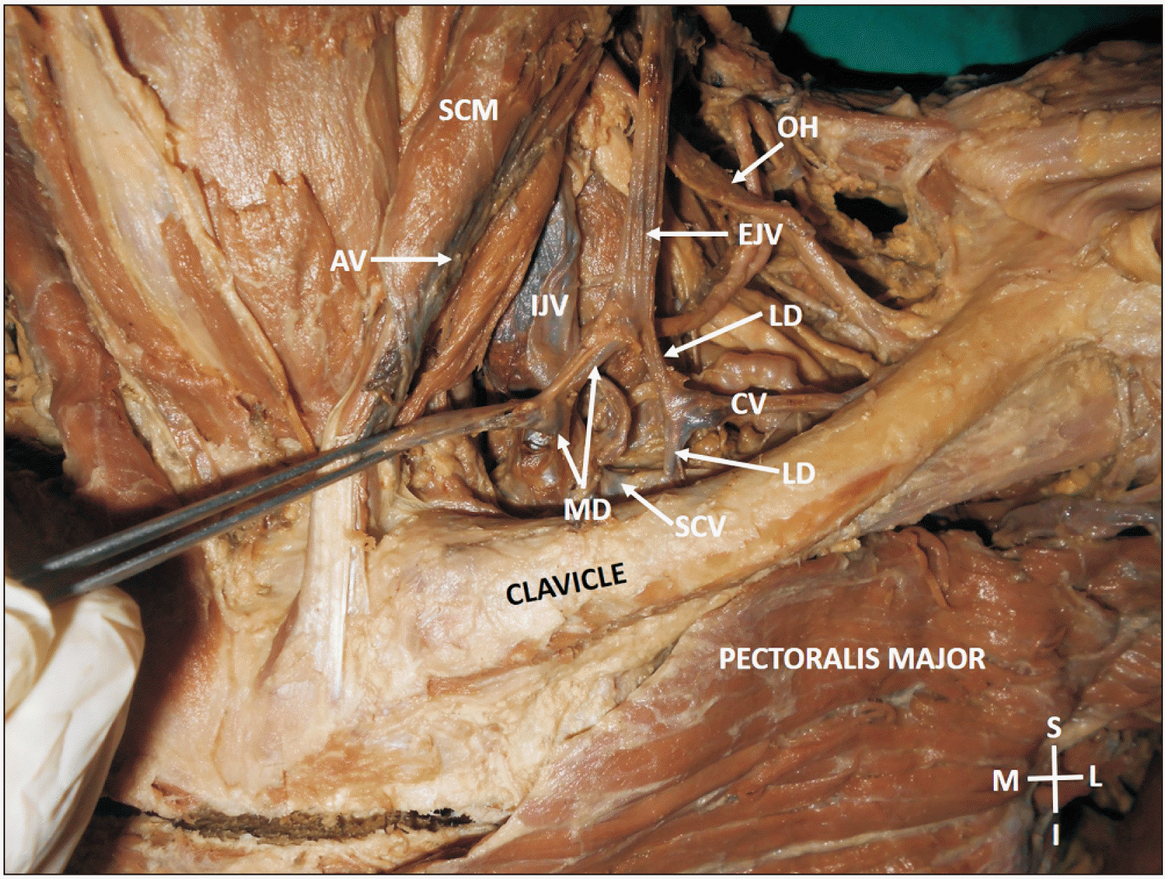
During or dissection classes for medical undergraduates, we found an unilateral variation in the formation and termination of the external jugular vein in an adult male cadaver aged 75 years. The external jugular vein of left side was only 3 inches long. It was formed in the posterior triangle of the neck, by the union of entire retromandibular vein and the posterior auricular vein. The retromandibular vein received the facial vein just below the angle of the mandible and continued down to meet the posterior auricular vein at middle of the posterior border of the sternocleidomastoid muscle. One inch above the clavicle, it bifurcated into a medial and a lateral division. The medial division received a common vein formed by the union of anterior jugular vein and an anonymous vein lying deep to the sternocleidomastoid muscle. The lateral division received a common vein formed by the union of suprascapular and transverse cervical veins. The medial division terminated by opening into the internal jugular vein and the lateral division terminated into the subclavian vein. The variations are shown in the Figs. 1, 2.

Discussion

Variations in the termination of the external jugular vein are less common compared to the formation. Knowledge of these variations is of paramount importance in various disciplines of medicine. Several cases of duplication and fenestration of the external jugular vein have been reported. In one of the latest reports [9], the external jugular vein duplicated but reunited before the termination. In another recent study, the external jugular vein united with common facial vein in the lower part of the neck to form a jugulo-facial venous circle [10]. Although both external and internal jugular veins can be palpated, external jugular vein is a reliable vein for central venous pressure assessment as it is superficial and easily visible [11]. External jugular vein is also used in diagnostic and treatment procedures through cannulation [6]. External jugular vein is one of the veins used as a recipient vessel for venous outflow in free flap head and neck reconstruction [12]. In neck dissections, preservation of the external jugular vein reduces postoperative edema of the face and neck and it could be utilized for anastomosis in reconstructive microsurgery in cases requiring resection of the internal jugular vein [13]. The superficial veins of the head and neck develop from the superficial plexus of capillaries found in the neck region. During the process of development, dilation of individual capillaries or the confluence of adjacent ones happens. Regression of some veins and diversion of the flow from one to other too can happen. However, these obvious factors are not yet completely understood [14].
Knowledge about the current variation could be useful for the surgeons free flap neck reconstruction surgeries. The external jugular vein is one of the preferable routes compared to cubital veins in emergencies due to more rapid circulation time to heart and faster cardiac responses. However, a variation such as the current one might complicate the procedure due to the terminal bifurcation of the vein. Catheterisation through internal jugular vein in a case like the current one could be difficult due to the termination of the medial division of external jugular vein into the internal jugular vein at the root of the neck.
In conclusion, this case describes an extremely rare type of termination of the external jugular vein. Terminal bifurcation of the external jugular veins is one of the rarest variations. The knowledge of this variation is of importance in radiological and surgical procedures.

Notes

Author Contributions

Conceptualization: SBN. Data acquisition: SBN. Data analysis or interpretation: SBN. Drafting of the manuscript: SBN. Critical revision of the manuscript: SBN, SKV. Approval of the final version of the manuscript: all authors.

Conflicts of Interest

No potential conflict of interest relevant to this article was reported.

References

1. Cvetko E. 2015; A case of left-sided absence and right-sided fenestration of the external jugular vein and a review of the literature. Surg Radiol Anat. 37:883–6. DOI: 10.1007/s00276-014-1398-z. PMID: 25432662.
2. Comert E, Comert A. 2009; External jugular vein duplication. J Craniofac Surg. 20:2173–4. DOI: 10.1097/SCS.0b013e3181bf0248. PMID: 19884835.
3. Ponnambalam SB, Karuppiah DS. 2020; Unilateral external jugular vein fenestration with variant anatomy of the retromandibular and facial vein. Anat Cell Biol. 53:117–20. DOI: 10.5115/acb.19.172. PMID: 32274258. PMCID: PMC7118259.
4. Singh R. 2020; Abnormal formation of external jugular vein and its repercussions. J Craniofac Surg. 31:e354–5. DOI: 10.1097/SCS.0000000000006293. PMID: 32149966.
5. Patil J, Kumar N, Swamy RS, D'Souza MR, Guru A, Nayak SB. 2014; Absence of retromandibular vein associated with atypical formation of external jugular vein in the parotid region. Anat Cell Biol. 47:135–7. DOI: 10.5115/acb.2014.47.2.135. PMID: 24987551. PMCID: PMC4076421.
6. Chauhan NK, Rani A, Chopra J, Rani A, ivastava AK Sr, Kumar V. 2011; Anomalous formation of external jugular vein and its clinical implication. Natl J Maxillofac Surg. 2:51–3. DOI: 10.4103/0975-5950.85854. PMID: 22442610. PMCID: PMC3304223.
7. Vadgaonkar R, Rai R, Ranade AV, Pai MM, Prabhu LV, Ashwin K, Jiji PJ. 2008; An anomalous left external jugular vein draining into right subclavian vein. Bratisl Lek Listy. 109:461–2. DOI: 10.4067/s0717-95022008000400018. PMID: 19166133.
8. Shetty P, Nayak SB, Thangarajan R, D'Souza MR. 2016; A rare case of persistent jugulocephalic vein and its clinical implication. Anat Cell Biol. 49:210–2. DOI: 10.5115/acb.2016.49.3.210. PMID: 27722015. PMCID: PMC5052231.
9. Ono K, Yoshioka N, Hage D, Ibaragi S, Tubbs RS, Iwanaga J. 2021; Duplication of the external jugular vein: a language barrier of database search in classic anatomical studies. Surg Radiol Anat. 43:1721–8. DOI: 10.1007/s00276-021-02717-6. PMID: 33620594.
10. Satheesha NB. 2017; Jugulo-facial venous circle, accessory slip of trapezius and absence of typical facial vein- clinically important anatomical variations. J Anat Soc India. 66(Suppl 2):S11–2. DOI: 10.1016/j.jasi.2017.10.008.
11. Nagata T, Masumoto K, Watanabe Y, Katou F. 2012; End-to-side anastomosis to the external jugular vein: preservation of external jugular vein blood flow. Br J Oral Maxillofac Surg. 50:e31–2. DOI: 10.1016/j.bjoms.2011.07.024. PMID: 21871698.
12. Ibrahim AE, Adelman DM, Parham C, Hong Z, Villa M, Chahine FM, Ghieh FM. 2019; The external jugular vein used as recipient vessel in head and neck free flap reconstruction: outcomes compared to the internal jugular vein. J Craniofac Surg. 30:178–83. DOI: 10.1097/SCS.0000000000004873. PMID: 30394970.
13. Sugiyama S, Iwai T, Tohnai I. 2017; Empty fenestration of the external jugular vein: a rare variant. J Anat Soc India. 66(Suppl 2):S1–3. DOI: 10.1016/j.jasi.2017.10.005.
14. Yadav S, Ghosh SK, Anand C. 2000; Variations of superficial veins of head and neck. J Anat Soc India. 49:61–2.

Fig. 1
Dissection of the left side of the neck showing the variation of the EJV. EJV, external jugular vein; RMV, retromandibular vein; PAV, posterior auricular vein; MD, medial division of the terminal part of the retromandibular vein; LD, lateral division of the terminal part of the retromandibular vein; FV, facial vein; PG, parotid gland; SCM, sternocleidomastoid; AJV, anterior jugular vein; AV, anonymous vein deep to sternocleidomastoid; IJV, internal jugular vein; OH, inferior belly of omohyoid; A, anterior; P, posterior; S, superior; I, inferior.
acb-55-4-501-f1.tif
Fig. 2
Dissection of the left side of the neck showing the termination of the EJV. EJV, external jugular vein; MD, medial division of the terminal part of the retromandibular vein; LD, lateral division of the terminal part of the retromandibular vein; CV, common vein formed by the union of suprascapular and transverse cervical veins; SCM, sternocleidomastoid; AV, anonymous vein deep to sternocleidomastoid; IJV, internal jugular vein; OH, inferior belly of omohyoid; SCV, supraclavicular vein; M, medial; S, superior; L, lateral; I, inferior.
acb-55-4-501-f2.tif
TOOLS
Similar articles