Journal List > Anat Cell Biol > v.55(4) > 1516080677

Kim, Hayashi, Jin, Murakami, and Rodríguez-Vázquez: Umbilical cord vessels other than the umbilical arteries and vein: a histological study of midterm human fetuses

Abstract

At birth, the umbilical cord contains various types of thin vessels that are near and outside the umbilicus and separate from the umbilical arteries and vein. These vessels are regarded as the remnant “vitelline vessels” and are often called “umbilical vessels”, although this terminology could lead to confusion with the true umbilical arteries and vein. No study has yet comprehensively examined these vessels using histological sections. Our examination of these vessels in 25 midterm fetuses (gestational age: 10–16 weeks) led to five major findings: (i) all specimens had umbilical branches of the inferior epigastric artery; (ii) 5 specimens had vitelline vein remnants; (iii) 4 specimens had a thin artery originating from the left hepatic artery that ran along the umbilical vein; (iv) 2 specimens had a so-called “para-umbilical vein” that was along the umbilical vein and reached the umbilicus; and (v) all specimens had lymphatic vessels originating from the umbilicus that ran caudally along the umbilical artery. The pelvic vein tributaries were well developed along the intra-abdominal umbilical artery, but did not reach the umbilicus. The lymphatic vessel was distinguished from the veins by an intraluminar cluster of lymphocytes attaching to the endothelium. The arterial branch in the umbilical cord did not accompany veins and lymphatic vessels, in contrast to the mother artery in the rectus abdominis. All these thin vessels seemed to be obliterated when the fibrous umbilical ring grew during late-term. The para-umbilical collateral vein in adults might develop outside the fibrous umbilical ring after birth.

Introduction

At birth, the proximal part of the umbilical cord contains various types of thin surface vessels near and outside the umbilicus, in addition to the umbilical arteries and vein. These surface vessels are referred to as “persistent vitelline vessels” [1-3], although there is no evidence that they are connected to the root of the midgut mesentery and thereby form a junction between the vitelline and hepatic portal veins. On the other hand, a vein present in and along the umbilical cord is sometimes termed the “umbilical vein” [4]. However, this name can lead to confusion with the true umbilical vein, which arises from the ductus venosus. Moreover, adults with portal vein hypertension often have additional veins near or along the umbilical vein that are termed “para-umbilical veins”, although this is rare in healthy adults [5, 6]. We are unaware of any study that examined whether the para-umbilical veins run along the true umbilical vein in fetuses or whether they develop after birth in the abdominal wall.
Consequently, the aim of this study was to analyze all types of vessels, other than the true umbilical arteries and vein, at, near, and especially outside the umbilicus of midterm human fetuses. Additional research to be published elsewhere examined the persistent vitelline arteries and veins of midterm fetuses.

Materials and Methods

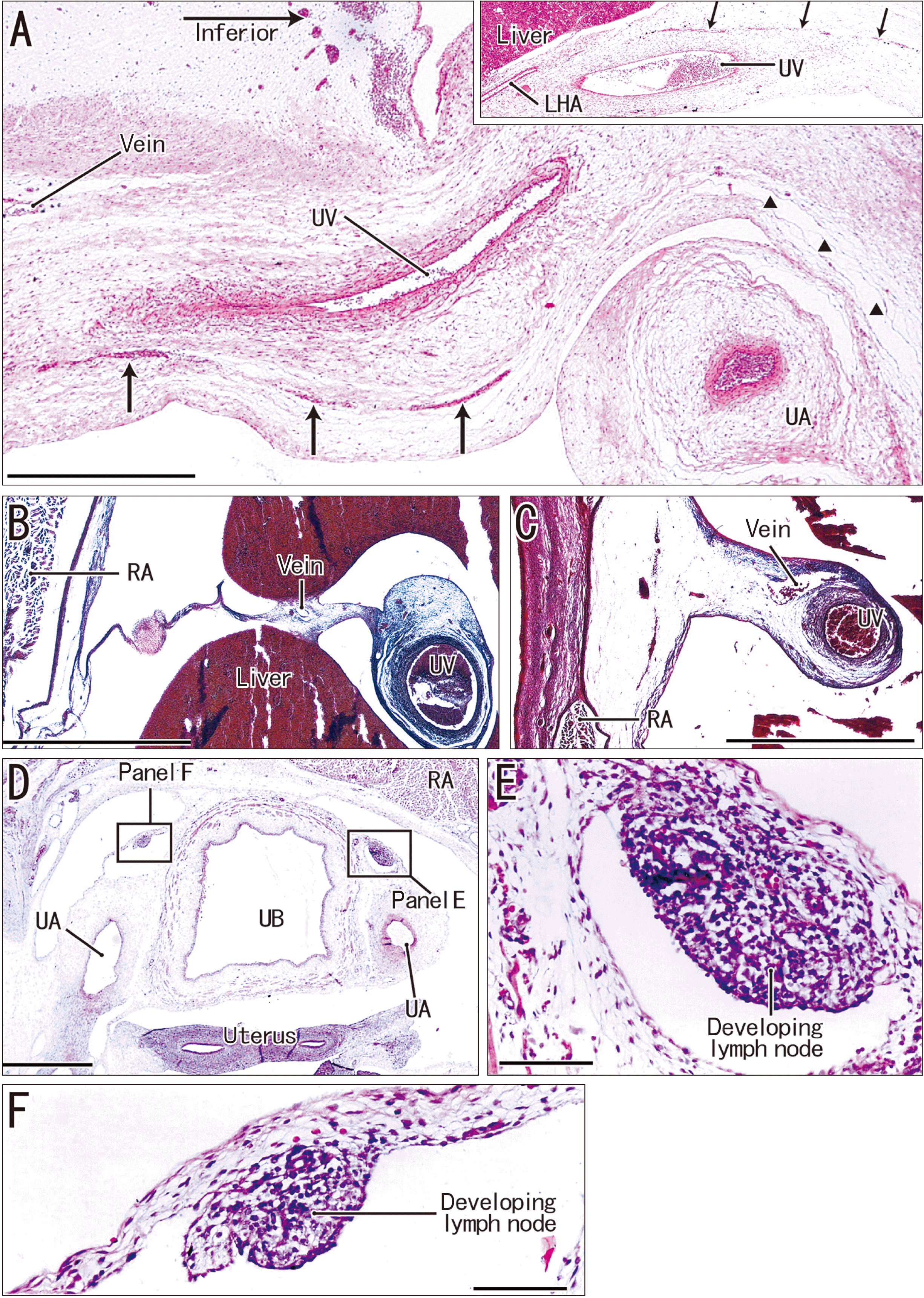
The study was performed in accordance with the provisions of the 2013 revision of the Declaration of Helsinki. Paraffin-embedded histological sections of the abdomen and pelvis of 25 midterm fetuses (gestational age approximately 10–16 weeks, crown-rump length 52–125 mm) were examined. All sections were part of the large collection at the Department of Anatomy of the Universidad Complutense (Madrid), and the embryos were from miscarriages or ectopic pregnancies at the Department of Obstetrics of the University. No information was available on the genetic background of the embryos or the abortion. The sections were stained with hematoxylin and eosin (H&E), Azan, or Goldner-Masson trichrome. Goldner-Masson trichrome stain [7] shows collagenous fibers in light green, instead of aniline-blue (as in Masson trichome). The sectional planes were horizontal (15 fetuses) or sagittal (10 fetuses) and section thickness ranged from 5 to 10 μm. Each specimen had one umbilical vein and two umbilical arteries, and none had absent or supernumerary vessels. This study was approved by the Ethics Committee of Complutense University (B08/374). All histological photographs were taken using a Nikon Eclipse 80i (Nikon, Tokyo, Japan).

Results

Umbilical branch of the inferior epigastric artery

An arterial branch (the umbilical branch) was consistently present, and it arose from the rectus abdominis. In 5 of 25 fetuses, the arterial branch entered the umbilical cord at 2 to 3 mm from the future umbilicus (Figs. 1A, C, E, 2F, G, 3C). This arterial branch in the cord did not tightly attach to the umbilical artery (Fig. 1F). At the gestational stage of the examined specimens, the superior epigastric artery did not reach the umbilicus because this region was restricted in the superior side of the uppermost tendinous intersection of the rectus abdominis muscle (Fig. 4C, D). Therefore, the umbilical branch originated from the inferior epigastric artery, and the inferior epigastric artery accompanied veins and lymphatic vessels in the rectus abdominis (Fig. 4E). However, the umbilical branch did not accompany veins in the umbilical cord (Fig. 1B, D) nor at the umbilicus (Fig. 2F, G).

Vitelline vein remnants

The vitelline vein remnant in 5 of 25 fetuses originated from the umbilical cord, ran through the peritoneal cavity between the liver and jejunum, and attached to the pancreatic head or duodenum (Figs. 2B, E, 3A–D). In these five specimens, the venous remnant did not take a long course in the cord, and disappeared near the umbilicus. The venous lumen was obliterated to various degrees (Fig. 3G, H), and the venous remnant did not connect to any other vessel such as a para-umbilical vein (see below) and a cutaneous vein.

Thin artery along the umbilical vein

The left hepatic artery always issued one or two thin arteries that ran along the umbilical vein. However, the thin artery reached the umbilicus in only 4 of 25 fetuses (Fig. 5A), but did not enter the umbilical cord and did not communicate with a branch of the inferior epigastric artery.

Venous tributary of pelvic veins along the umbilical artery and/or allantois

The pelvic vein tributaries were well-developed along the proximal part of the umbilical artery (Fig. 1G), but they usually drained the urinary bladder and its continuation, the proximal part of the allantois. The pelvic vein tributary reached a peripheral site at or near the umbilicus in only a few fetuses (Fig. 2D).

Para-umbilical vein running along the umbilical vein

The so-called “para-umbilical vein” ran along the umbilical vein and drained into thin veins at the porta hepatis. This vein was consistently present along the proximal part of the umbilical vein (Fig. 5B, C), but reached the site at or near the umbilicus in only 2 of 25 fetuses (Fig. 5A). Conversely, veins in most sections although they usually appeared as spotty subcutaneous hemorrhages until fixation (Figs. 4D, 5A).

Pelvic lymphatic vessels coming along the umbilical artery

The lymphatic plexus was consistently present along the urinary bladder and allantois, near the umbilicus. Moreover, bilaterally thick lymphatic vessels ran along the lateral or anterior aspect of the umbilical artery in most fetuses (Fig. 5D); they were distinguishable from veins by a cluster of primitive lymphocytes (initial lymph nodes) in the lumen (Fig. 5E, F). Lymphatic vessels along the umbilical artery extended to the umbilicus in most fetuses (Figs. 2A, C, 3D, E, 5A).

Discussion

Our results suggest that various types of thin vessels were present at the fetal umbilicus. The umbilical branch of the inferior epigastric artery was consistently present, and our results may be the first to show that this artery ran into the umbilical cord. In the rectus abdominis, the inferior epigastric artery accompanied the thick vein and lymphatic vessels. However, the arterial branch near the umbilical cord did not accompany veins or lymphatic vessels. We identified the vitelline vein remnant based on its intra-abdominal course. The so-called “para-umbilical vein” rarely reached the umbilicus and made no anastomosis with the pelvic vein tributaries. However, venous drainage of the umbilicus was mostly via the abdominal cutaneous veins. Although the placenta contains no lymphatic tissues [8], it is likely that thick lymphatic vessels along the umbilical artery drained the umbilical cord at and near the umbilicus. These thick lymphatic vessels are presented in classical textbooks of anatomy, as described by Monie [9]. Because of their thickness, even in adults, an internal hernia is likely under the superior vesical artery (a continuation of the umbilical artery) after lymphadenectomy [10].
Clarke [11] and Malas et al. [12] report that the umbilical arteries carried multiple vasa vasorum (7–9 on average) in the arterial wall, and that they maintained the thick wall. However, we found no evidence that the intramural vessel connected to the umbilical branch of the inferior epigastric artery and vein. However, Malas et al. [12] perform a histological examination of the umbilical artery between the umbilicus and superior vesical artery, but we examined sites at and outside of the umbilicus (Figs. 1, 2). Likewise, Clarke [11] performs dye injection into the umbilical artery and demonstrates thin arterial twigs of the thick wall of the “intra-abdominal umbilical artery”. Therefore, the vasa vasorum seemed to be rare in the umbilical cord. Moreover, their descriptions indicated the supplying artery did not originate from the inferior epigastric artery, but instead originated from the umbilical artery itself. The vasa vasorum of the umbilical arteries may be derived to varying degrees from a thin recess of the arterial lumen, due to the specific structure termed the “subendothelial cushion” [9].
Our group previously describes a fibrous umbilical ring in late-term fetuses [13]. However, this fibrous ring or the rim of the fetal umbilicus rarely contained vessels. Instead, the tight fibrous tissue seemed to obliterate the thin vessels that we identified in the mid-term fetuses of the present study. Indeed, for infants and children, recent descriptions of surgical techniques seem to ignore the supernumerary or variant vessels at the umbilicus [14, 15]. Likewise, the so-called “para-umbilical vein” reported in healthy adults [5, 6] might not correspond to the fetal structure, but instead to a secondary vein that develops postnatally in the subperitoneal loose tissue behind the anterior abdominal wall. Moreover, the para-umbilical vein in healthy adults does not always run through the falciform ligament. The para-umbilical collateral vein, which is associated with portal vein hypertension in adults, might develop outside the fibrous umbilical ring.

Notes

Author Contributions

Conceptualization: JHK, GM. Data acquisition: JHK. Data analysis or interpretation: SH, ZWJ, GM, JFR. Drafting of the manuscript: JHK, GM. Critical revision of the manuscript: ZWJ, JFR. Approval of the final version of the manuscript: all authors.

Conflicts of Interest

No potential conflict of interest relevant to this article was reported.

References

1. Wright JR Jr. 2019; Prevalence, morphology, embryogenesis, and diagnostic utility of umbilical cord vitelline vascular remnants. Pediatr Dev Pathol. 22:279–87. DOI: 10.1177/1093526618811734. PMID: 30541420.
2. De Guzman JK, Yu W, Horn C, Brundler MA, Wright JR Jr. 2021; Characterization of vitelline vessel remnant circulation in the umbilical cord. Placenta. 111:97–104. DOI: 10.1016/j.placenta.2021.06.009. PMID: 34225217.
3. Lemke C, Biedermann U. 2021; A persistent vitelline artery in an adult. Case report and review of literature. Transl Res Anat. 22:100080. DOI: 10.1016/j.tria.2020.100080.
4. Jaiman S, Nalluri HB. 2013; Abnormal continuation of umbilical vein into extra-hepatic portal vein: report of three cases. Congenit Anom (Kyoto). 53:170–5. DOI: 10.1111/cga.12020. PMID: 24712478.
5. Martin BF, Tudor RG. 1980; The umbilical and paraumbilical veins of man. J Anat. 130(Pt 2):305–22. PMID: 7400038. PMCID: PMC1233134.
6. Ibukuro K, Fukuda H, Tobe K, Akita K, Takeguchi T. 2016; The vascular anatomy of the ligaments of the liver: gross anatomy, imaging and clinical applications. Br J Radiol. 89:20150925. DOI: 10.1259/bjr.20150925. PMID: 27163944. PMCID: PMC5124872.
7. Huang Y, Chen H, Zhang H, Lu Y, Qin C. 2022; FAM20C plays a critical role in the development of mouse vertebra. Spine J. 22:337–48. DOI: 10.1016/j.spinee.2021.07.022. PMID: 34343663.
8. Becker J, Tchagou Tchangou GE, Schmidt S, Zelent C, Kahl F, Wilting J. 2020; Absence of lymphatic vessels in term placenta. BMC Pregnancy Childbirth. 20:380. DOI: 10.1186/s12884-020-03073-w. PMID: 32600346. PMCID: PMC7325062. PMID: f6de6edbb1e54159822142296568bed7.
9. Monie IW. 1945; Some observations on the subendothelial cushions of the umbilical arteries. J Anat. 79(Pt 4):137–144. PMID: 17104979. PMCID: PMC1272580.
10. Ai W, Liang Z, Li F, Yu H. 2020; Internal hernia beneath superior vesical artery after pelvic lymphadenectomy for cervical cancer: a case report and literature review. BMC Surg. 20:312. DOI: 10.1186/s12893-020-00985-4. PMID: 33267803. PMCID: PMC7709390. PMID: 31f1d3f439434b19991d03ca85e52f88.
11. Clarke JA. 1965; An X-ray microscopic study of the vasa vasorum of the human umbilical arteries. Z Zellforsch Mikrosk Anat. 66:293–9. DOI: 10.1007/BF00344341. PMID: 14311517.
12. Malas MA, Sulak O, Gökçimen A, Sari A. 2003; Morphology of umbilical vessels in human fetuses: a quantitative light microscope study. Eur J Morphol. 41:167–74. PMID: 16229159.
13. Xu D, Jin ZW, Kim JH, Rodríguez-Vázquez JF, Murakami G, Hayashi S. 2020; Umbilicus and the rectus sheath: a study using human fetuses. Surg Radiol Anat. 42:461–71. DOI: 10.1007/s00276-019-02398-2. PMID: 31897654.
14. Tröbs RB, Vahdad MR, Cernaianu G. 2016; Transumbilical cord access (TUCA) for laparoscopy in infants and children: simple, safe and fast. Surg Today. 46:235–40. DOI: 10.1007/s00595-015-1191-6. PMID: 26031233. PMCID: PMC4722059.
15. Sisti A, Huayllani MT, Boczar D, Restrepo D, Cinotto G, Lu X, Cuomo R, Grimaldi L, Nisi G, Forte AJ. 2021; Umbilical reconstruction techniques: a literature review. Aesthetic Plast Surg. 45:1078–96. DOI: 10.1007/s00266-020-01989-4. PMID: 33098045.

Fig. 1
A thin artery running along the umbilical artery (UA) in the umbilical cord in sagittal sections of a fetus with a crown-rump length of 59 mm. Goldner-Masson trichrome staining. (A) Shows the most medial plane and (G) shows the most lateral plane. Panels (B), (D), and (F) are higher magnification views of the rectangles in (A), (C), and (E), respectively. The thin artery originates from the inferior epigastric artery in the upper rim of the umbilicus (A), and its 3 mm course is traceable along the upper margin of the wall of the UA (C, E). This artery is clearly outside the UA (F). (G) Shows veins that accompanied the UA, which does not reach the umbilicus. (A, C, E, G) Scale bars=1 mm; (B, D, F) scale bars=0.1 mm. RA, rectus abdominis; UB, urinary bladder; OV, ovary.
acb-55-4-467-f1.tif
Fig. 2
Umbilical branch of the inferior epigastric artery and the vitelline vein remnant in horizontal sections of five fetuses with crown-rump length (CRL)s of 52 to 97 mm. (A–C, E, F) Azan staining. (D, G) H&E staining. (A, B) Fetus with CRL of 52 mm. (C, E) Fetus with CRL of 55 mm. (D) Fetus with CRL of 72 mm. (F) Fetus with CRL of 83 mm. (G) Fetus with CRL of 97 mm. Each panel contains the umbilical branch (artery) of the inferior epigastric artery. The remnant of the vitelline vein (VV) is present in two fetuses (A–C, E). (A–E) High magnification (scale bar in A=1 mm). (F, G) Low magnification (scale bar in F=1 mm). lymph, lymphatic vessels; AL, allantois; RA, rectus abdominis; UA, umbilical artery.
acb-55-4-467-f2.tif
Fig. 3
VV remnant and umbilical branch of the inferior epigastric artery in sagittal sections of a fetus with a crown-rump length of 46 mm. H&E staining. (A) Shows a plane in the right-lateral side of (F). Panels (E) and (G, H) are higher magnification views of the rectangles in (D) and (F), respectively. The insert in the left-bottom of (C) shows the umbilical branch (artery) of the inferior epigastric artery (square in C). The remnant of the VV originates from the anterior surface of the PA (D), runs between the liver and transverse colon (TC in D) or between the liver and JE (D, F), and reaches the superior side of the umbilical artery (A–D). (E) Shows a candidate lymphatic vessel (lymph) along the umbilical artery at the umbilicus. Scale bars in A to D, F=1 mm; in C-insert, E, G, and H=0.1 mm. VV, vitelline vein; AL, allantois; RA, rectus abdominis; UA, umbilical artery; UV, umbilical vein; PA, pancreas; JE, jejunum.
acb-55-4-467-f3.tif
Fig. 4
Superior and inferior epigastric arteries in sagittal sections of a fetus with a crown-rump length of 61 mm. H&E staining. The arrow in each panel indicates the most superior intersection of the rectus abdominis. (C) Magnified view of the rectangle in (B). (D) Magnified view of the rectangle in (A). (E) Magnified view of the rectangle in (D). The superior epigastric artery (sup.epig.a) approaches the intersection in (D), and the inferior epigastric artery (inf.epig.a) approaches the intersection in (C). Thus, the umbilicus is in the region of the inferior artery. (E) Shows the inferior epigastric artery and the accompanying veins and lymphatic vessels. (A, B) Low magnification (scale bar in A=1 mm). (C, D) High magnification (scale bar in D=1 mm). (E) Highest magnification (scale bar=0.1 mm).
acb-55-4-467-f4.tif
Fig. 5
The paraumbilical vein, a thin artery along the umbilical vein (UV), and thick lymphatic vessels along the umbilical artery (UA) of four fetuses with crown-rump length (CRL) of 46 mm to 92 mm. Panels other than panel B, H&E staining; (B) Azan staining. (A) Shows a sagittal section of a fetus with a CRL of 46 mm CRL (same specimen a s in Fig. 3). (B) Shows a horizontal section of a fetus with a CRL of 92 mm. (C) Shows a horizontal section of a fetus with a CRL of 90 mm. (D–F) Show horizontal sections of a fetus with a CRL of 75 mm. (A) Shows a thin artery along the UV and a candidate lymphatic vessel (triangles) along the UA; the insert shows the thin artery (arrows) that originates from the left hepatic artery (LHA). The vein along the UV in panels A to C is the so-called “para-umbilical vein”. (D) Shows bilateral thick lymphatic vessels along the urinary bladder (UB). Panels E and F are higher magnification views of the rectangles in panel D, and show that the lymphatic vessels contain a cluster of primitive lymphocytes. Scale bars in A to D=1 mm; in E and F=0.1 mm. RA, rectus abdominis.
acb-55-4-467-f5.tif
TOOLS
Similar articles