Abstract
The pectoralis minor (PMn) muscle originates from the third, fourth, and fifth ribs near the costochondral junctionusually and gets inserted on the medial margin and upper surface of the coracoid process of the scapula. To look at the morphological insertion patterns and sites of attachment of the PMn muscle in the donated cadavers. Over all 19 limbs were included in the study (9 right and 10 left). Out of 19 limbs, 10 belonged to female and 9 belonged to male cadavers.The cadavers were meticulously dissected to determine the morphological insertion types and location of the attachment of the muscle. Unusual pattern of insertion was observed in 6 limbs (31.6%) out of total 19 limbs included in the study. The variations we observed does not fall completely in the classification by Le Double, hence variations we observed can be considered as new and rare variant which to our knowledge is not reported in literature. We propose this new variant to be type 4 of Le Double classification. The potential of ectopic PMn tendon should be taken into consideration and tested out, especially in patients with shoulder discomfort and stiffening who have ruled out the more frequent diseases. For proper surgical planning, a preoperative magnetic resonance imaging or USG examination of the shoulder joint is required considering the prevalence of variation in the insertion pattern of PMn muscle. Preoperative identification of any abnormal PMn insertion can help to reduce the risk of iatrogenic tendon injury and post-operative problems.
The pectoralis minor (PMn) is a thin triangular muscle that lies beneath the pectoralis major (PMj) in pectoral region. It arises from the superior margins and outer surfaces of the 3rd, 4th, and 5th ribs, close to their costal cartilages, as well as from the fascia overlying the external intercostal muscles. Its fibres run upwards laterally beneath the cover of the PMj, ending in a flat tendon that is inserted to the superior surface and medial border of the coracoid process of scapula. The tendon in part or as a whole may sometime seem to crossover the coracoid process into the coracoacromial ligament (CAL) [1]. PMn helps in stabilizing the shoulder joint, depresses, rotates downwards and tilts the scapula anteriorly [2]. If inserted other than coracoid process, the PMn is considered as “hidden culprit of rotator cuff disorder” [3]. To repair the rotator cuff after injury, the PMn tendon is used and for facial and breast contouring its muscle belly is commonly used [4].
As such, there are only a few published studies on the variations in PMn muscle especially in relation to its insertion. The abnormal insertion of the PMn muscle tendon has been demonstrated to have a significant role in producing shoulder pain and range-of-motion (ROM) restrictions [5]. Evidence suggests that insertion of PMn muscle to shoulder joint capsule results in shoulder stiffness, subacromial and antero-medial sub-coracoid impingement and adhesive capsulitis [6-8]. The radiologists and surgeons treating patients with these complaints needs to be aware of variations of PMn in order to avoid misdiagnosis and iatrogenic damage. If the coracohumeral ligament (CHL) is non-existent, as it is in some PMn variations, due to the elimination of this supportive ligament component, excessive scar tissue might develop in the rotator interval, placing the patient at risk for a superior labral tear from anterior to posterior (SLAP) lesion [6]. For coracoid process osteotomy, the PMn insertion is used as a landmark and its tendon has been employed in the restoration of open or arthroscopically aided subscapularis tears, reconstruction of the acromioclavicular joint, and even in the management of irreversible supraspinatus injuries [9, 10].
The initial categorization was proposed by Le Double [11]. Only four occurrences of bilateral abnormal insertions of the PMn have been described in the literature, according to the authors’ knowledge, three of which were detected during cadaveric dissection and one of which was done radiologically. With this study we report rare aberrant insertions of PMn muscle bilaterally in the 6 limbs out of total 19 limbs included in the study. The variations we observed do not fall completely in the classification by Le Double, hence variations we observed can be considered as new and rare variant which to our knowledge is not reported in literature. We propose this new variant to be type 4 of Le Double classification.
This study was conducted on the cadavers that were donated to the All India Institute of Medical Sciences (AIIMS) Bibinagar through voluntary body donation program. All donors or their relatives provided informed written consent prior to their death so that their bodies can be utilised for medical education and research. The specimens with intact PMn muscle and shoulder region with appropriate tissue quality were included in the study. Specimens with broken shoulder joint or with some degenerative changes of upper limb or specimens with removed PMn muscle or with some surgical procedures done in the region were excluded from the study.
The study was done on a total of 22 limbs (11 right and 11 left) which were dissected from January 2019 to March 2022 for routine anatomy practical teaching. Three limbs (2 right and 1 left) where the shoulder region was distorted and PMn details were not clear, were excluded from the study. Hence, over all 19 limbs included in the study, of which 9 were of right side and 10 were of left side. Out of 19 limbs, 10 belonged to female and 9 belonged to male cadavers.
The steps for dissection were as follows. After reflection of skin and superficial fascia of pectoral region, PMj muscle was identified and pectoral fascia was removed. Clavicular and manubriosternal part of PMj was reflected, anterior attachment of deltoid muscle was cut and reflected. PMn muscle with clavipectoral fascia was identified. The fascia was removed and PMn muscle was traced way up to the coracoid process, the CAL, the glenohumeral joint capsule, the clavicle, and the humeral tubercles that surround the shoulder carefully. Length and width of the tendon and the aponeurotic part were measured in centimetre (Table 1). Presence or absence of CAL and CHL was noticed. Furthermore, the origin of the PMn was noticed carefully for any variation in all the cadavers.
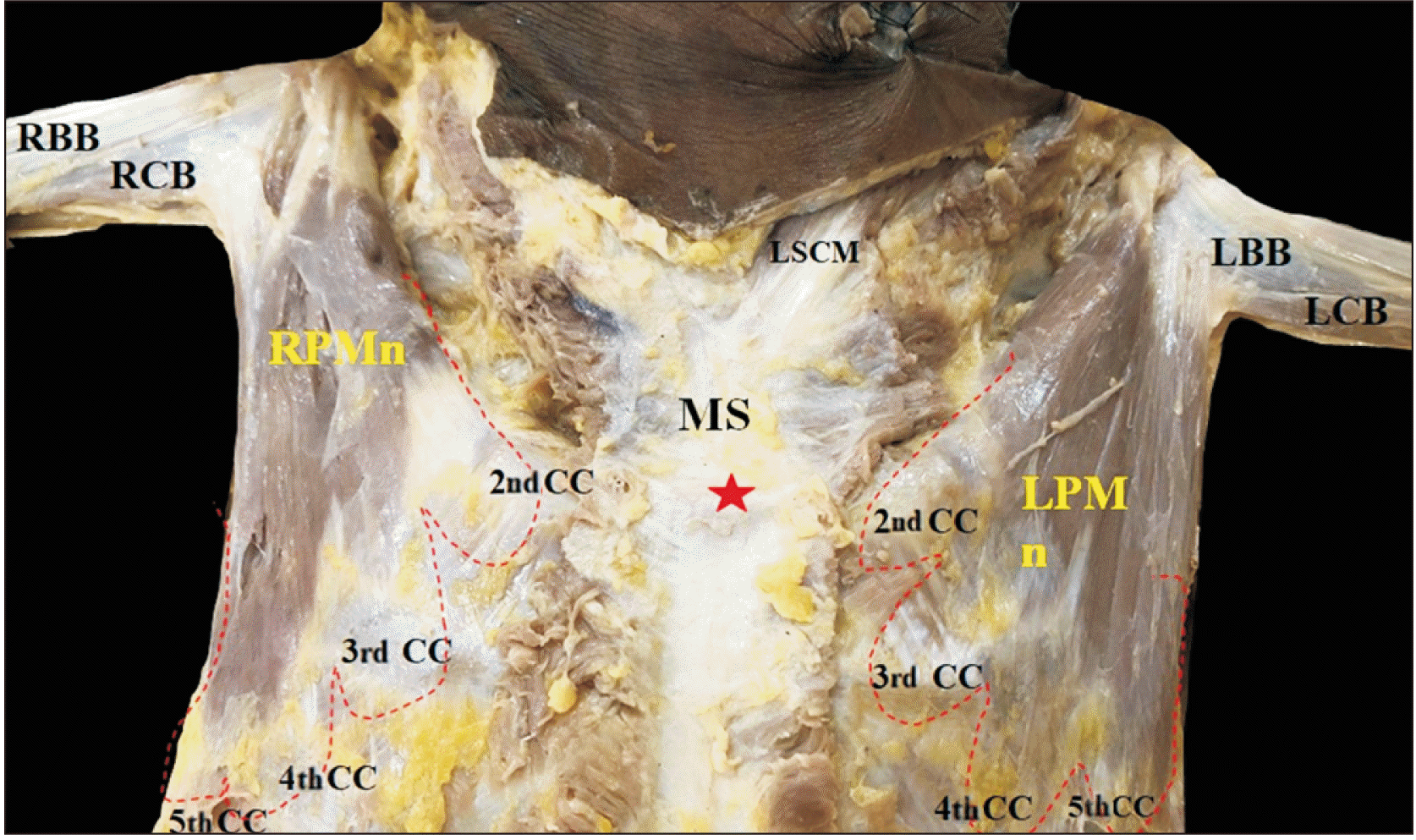
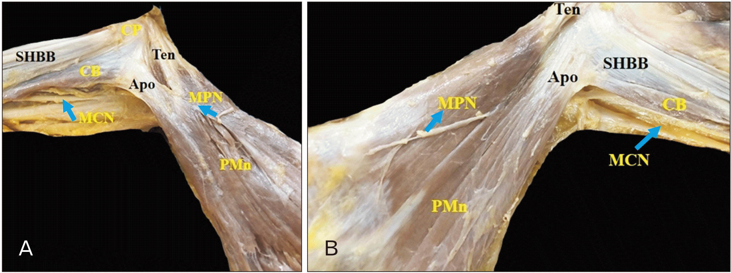
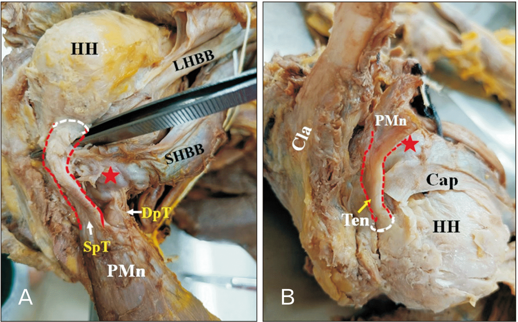
On inspection, the anterior thoracic wall did not appear to have any apparent abnormalities in any of the donated cadavers. Unusual pattern of insertion was observed in 6 limbs (31.6%) out of total 19 limbs. In a female cadaver of 60 year, PMn originated from costal cartilage of 2nd to 5th rib near their costochondral junction on both the sides (Fig. 1). Insertion of the muscle was in an anomalous bilateral dual pattern as aponeurotic and tendinous part. Right PMn had medial tendon of length 3.5 cm and width 1 cm, and lateral aponeurotic part of length 4 cm and width 2 cm approximately. Tendon got inserted onto the medial border and superior surface of coracoid process as usual, whereas aponeurotic part joined coracobrachialis muscle medially (Fig. 2). Left PMn had similar insertion with medial tendon of length 4 cm and width 1 cm, lateral aponeurotic part with length 4 cm and width 2.5 cm (Table 1). Aponeurotic part as on right side got attached to the coracobrachialis muscle on its medial side, whereas tendinous part crossed the coracoid process (Fig. 3A, B) from above on both sides, and passed deep to the CAL and became continuous with the capsule of the shoulder joint in the region of superior part of greater tubercle (Fig. 3).
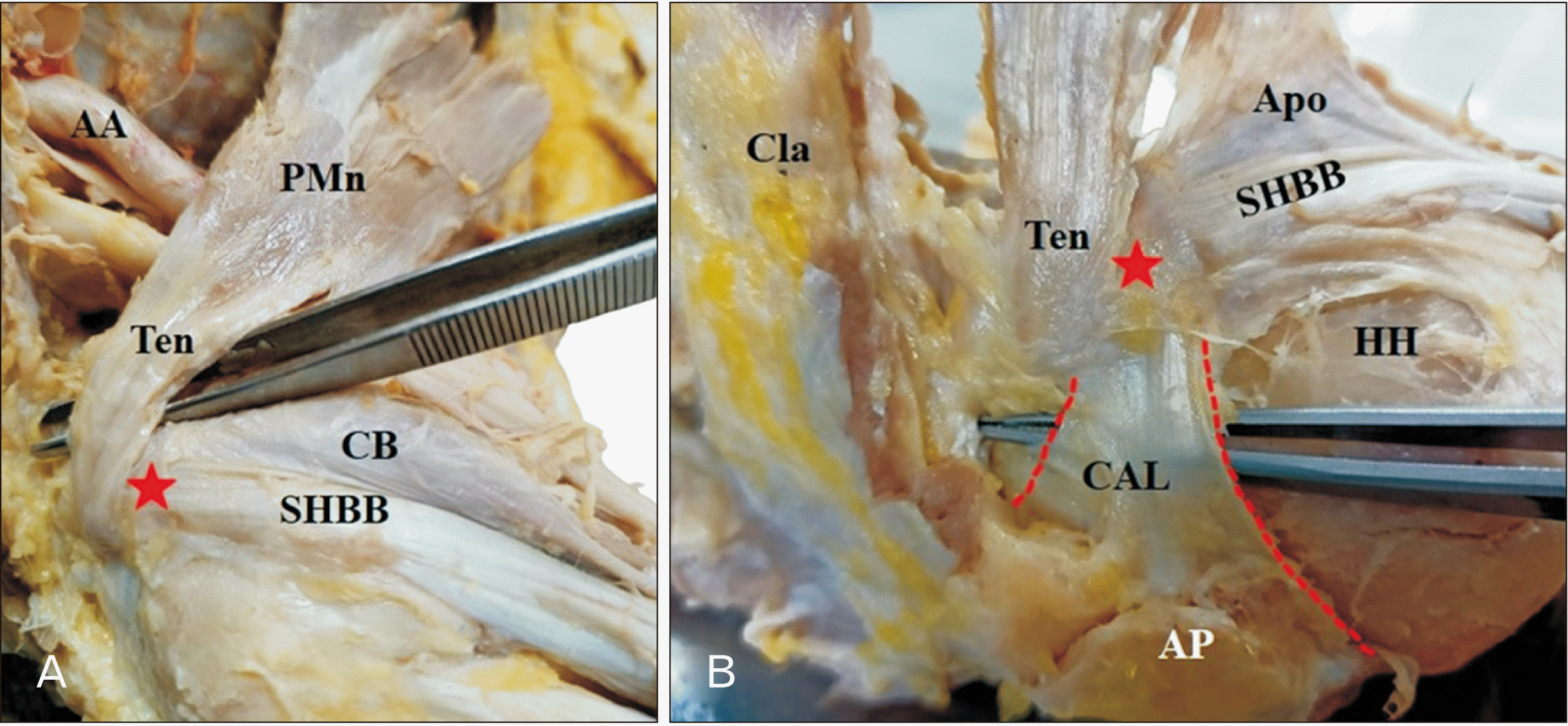
In another 72-year-old female cadaver, PMn originated from 3rd to 5th costal cartilage near costochondral junction on both the sides. Insertion was in the form of superficial and deep tendons (double tendons). Length of right superficial tendon was 3 cm and of left tendon was 3.5 cm, whereas width of right tendon was 1.5 cm and that of left side was 1 cm. Length and width of deep tendon on right side was 2 cm and 2.5 cm respectively, and on left side was 1.5 cm and 2 cm respectively. Deep tendon on both sides was inserted as usual on medial border of the coracoid process, whereas superficial tendon on both sides was found crossing the coracoid process from above, and was inserted in similar fashion as tendons of cadaver 1 (Fig. 4). In the cadaver 3 aged 75-year-old female, PMn took origin from 3rd to 5th costal cartilage near costochondral junction on both the sides. Insertion on both sides was in the form of single tendon which after crossing the coracoid process from above became continuous with the capsule of shoulder joint.
Shoulder pathology is a frequent clinical condition that can be caused by variation in the normal anatomy. PMn origin and insertion variations have been recorded in the literature, with up to 23% of individuals having certain variance of this muscle. Ectopic PMn tendons have shown to contribute to shoulder discomfort and ROM limitations. This has been linked to shoulder stiffness, adhesive capsulitis, SLAP lesions and subacromial-, antero-medial sub coracoid impingement [12]. Surgeons that are aware of such variations are well prepared to adjust techniques and successfully utilize the variation to the patient’s benefit. Thoracic outlet syndrome is also linked to ectopic insertion of PMn [13].
The study on the variation of PMn dates back to 19th century. Krause was the first to describe PMn muscle variations [14]. Le Double et al. proposed three types of variation based on the insertion of PMn. According to him, the deep component of the PMn tendon connects to the coracoid process normally, while the superficial part crosses over it to a more proximal structure in type I variants. In type II variants, the tendon’s majority of fibres adhere to the coracoid process, with only a few passing over it. Tendons of type III variants travel over the coracoid process without attaching, and are frequently separated from it by a bursa. He also emphasized that type 1 variant as the most common and type 3 is the least common [15]. Studies have also reported that the PMn with normal origin getting inserted directly into the shoulder joint capsule and into other muscles [8, 16].
In this study, we describe the existence of three types of variations in the mode of insertion of PMn muscle that was identified during a regular dissection of cadavers. Unusual pattern of insertion was observed in 6 limbs (31.57%) out of total 19 limbs dissected. It was noticed that the PMn towards the insertion splits into two tendons in 4 limbs (21.1%). One of the two tendons in these four limbs got inserted into the coracoid process whereas the other tendon passed more superiorly under the CAL and merged with the capsule of the shoulder joint. The most frequent insertional variation is attachment to the glenohumeral joint capsule, which has been extensively documented in the literature [17, 18]. Lee et al. [6] in an attempt to classify the variation in PMn insertion found that in 29.4% of the dissected samples, the muscle got inserted into the capsule of the shoulder joint. He also described that the PMn inserting into shoulder joint capsule was most of the time associated with an absent CHL. Tubbs et al. [16] described a case in which the left PMn originated as usually from the third to fifth costochondral junction, crossed the coracoid process without connecting, and entered straight into the shoulder joint capsule. Studies have also shown that the ectopic insertion of PMn is often associated with a change in the morphology of CHL [5]. Lee at al. [6] in his Magnetic Resonance arthrogram study found that the PMn when getting inserted into the shoulder joint capsule, in most of the cases was associated with SLAP lesion with the absent CHL. However, in our study there is no associated change in the morphology of CAL or CHL. A meta-analysis on the insertional variation of PMn reported that Le Double type 1 was the most frequently encountered variation [13]. Our study contradicts this statement with Le Double type 3 to be the most commonly encountered variation. Another similar study has documented that Le Double type 3 is the most common type of variation encountered and they proposed that when an atypical PMn tendon is suspected of being a significant reason to shoulder discomfort, the cutting of that tendon is recommended [19]. In this study, one more type of variation was also delineated. In one female cadaver, on both the sides, the PMn was having normal origin from third, fourth and fifth ribs; the fibres were directed upwards and laterally; nearing the insertion, it split into two parts — tendinous and an accessory aponeurotic. On the right side, the PMn had medial tendon of length 3.5 cm and width 1 cm, and lateral aponeurotic part of length 4 cm and width 2 cm approximately. The tendinous part got inserted into the coracoid process of the scapula whereas the aponeurotic part fused with the coracobrachialis muscle. On the left side, the muscle had a medial tendon of length 4 cm and width 1 cm, lateral aponeurotic part with length 4 cm and width 2.5 cm. Aponeurotic part got attached to the coracobrachialis muscle on its medial side, whereas tendinous part crossed the coracoid process from above and got merged shoulder joint capsule. Soni et al. [20] reported a similar anomalous insertion of PMn where the muscle in addition to coracoid process, also got inserted to the coracobrachialis and biceps brachii. Another study reported the insertion of PMn into the tendon of supraspinatus [21]. Studies have also documented the rare insertion of PMn into the clavicle [22]. Observations of some studies in relation to variant insertion of PMn tendon is discussed in Table 2 [6, 15, 16, 20-22].
Furthermore, in one female cadaver, in addition to the insertional abnormality, variation in the origin of the muscle was also noted. Ribs three to five are considered the “typical” origin of the PMn muscle. An origin at ribs two to four, on the other hand, is rather frequent [23, 24]. In this study, the PMn on the left side of a female cadaver was found to have origin from two to five ribs, and towards insertion splits into two tendons — one getting inserted to the coracoid process and the other passes under the CAL to merge with the capsule of the shoulder joint. In the previous studies, there are a few reports of changes in the PMn’s most inferior origin. Turan-Ozdemir and Cankur [24] described a case of a right PMn muscle that came exclusively from the fifth rib. Uncommon origin at the sixth rib have also been reported. Clinically, these variations are likely to be undetectable. Another variation observed bilaterally in a female cadaver in present study was presence of single tendon of insertion of PMn, which crossed the coracoid process superficially without any attachments to the process and inserted into the glenohumeral joint capsule. This type of variation belonged to the type III of Le double classification.
The variations we observed does not fall completely in the classification by Le Double, hence variations we observed can be considered as new and rare variant which to our knowledge is not reported in literature. We propose this new variant to be type 4 of Le Double classification.
We propose a new classification of the insertion of PMn based on our observations (Table 3).
We propose that type 2 and 3 variants of our classification are more vulnerable to shoulder-related diseases, whereas type 4 variant is more susceptible to thoracic outlet syndrome-related symptoms, based on our classification and a thorough literature study.
Anatomical neuromuscular variations can be ascribed to unknown parameters that alter the mechanism of formation of limb muscle. The lateral plate mesoderm forms the limb buds. These buds’ mesenchyme differentiates into the limbs’ muscles. A single muscle mass is created throughout development by fusion of the primary muscle into the various layers, which ultimately fades by apoptosis. Failure to complete this process of primary muscle disappearance during embryonic development might have led to the presence of abnormal aponeurotic part and accessory tendon [5]. The ectopic tendons of the PMn are mainly coursing through or under CHL. According to Bland Sutton’s three basic guidelines concerning the morphology of ligaments [1], the CHL has the same origin and insertion as the PMn tendon; [2] it is possible that CHL is a remnant of PMn muscle; [3] the PMn muscle has anatomical variance in humans; with a humeral insertion of functional significance [25].
Some researchers believe there is a link between shoulder problems and insertion variations of PMn. Rotator cuff diseases are thought to be caused by the proximal insertion of the PMn beyond the coracoid process. The arm’s full abduction (180 degrees) is the consequence of movement at two ‘joints.’ At the glenohumeral joint, there is a total of 120 degrees of abduction. The scapula spinning on the posterior thoracic wall accounts for the remaining 60 degrees (or scapulothoracic joint). Moving the shoulder into full abduction might create discomfort or pain if this 60-degree rotation is restricted or lost entirely. The discomfort is caused by the supraspinatus tendon, subacromial bursa, and joint capsule being squeezed between two bony surfaces: the humerus greater tubercle and the scapula’s acromion process. The ectopic PMn tendon is prone to get squeezed between the greater tubercle of the humerus and the acromion process of the scapula as it is inserted into the shoulder joint capsule, resulting in shoulder discomfort and stiffness [13].
If the atypical insertion of the PMn happens at the same time as lateral rotations and overhead abductions, such as in the throwing movement with the overhand, it could lead to undue strain on the axillary vessels [26]. Tendinopathy and a snapping feeling can also occur when an excessively sliding motion occurs during workouts such as bench presses or weightlifting, leading PMn tendons with unique insertion patterns to move along the coracoid process. In Le Double type 3, this problem will be more prominent [8]. According to Zvijac et al. [27] with regard to variation in PMn insertion, special attention to be given when investigating for costal injuries, sternoclavicular injuries, high rib fractures, tears in the PMj and PMn. Moineau et al. [12] documented that unusual PMn insertion patterns impacted shoulder stiffness. As reported earlier, whenever there is abnormal insertion of PMn, attention should be paid to the morphology of CHL. In a study of 12 patients with coracoid impingement syndrome, Dumontier et al. [28] found that one patient had anterior shoulder discomfort due to an atypical insertion PMn into the rotator interval. The PMn tendon contacted the coracoid process at internal rotation, and the authors were able to successfully treat it using coracoplasty [28]. According to Lee et al. [6] abnormal PMn tendon insertions to the glenohumeral joint associated with absent CHL is a key element in the SLAP lesion development. The CHL is important for supporting the biceps brachii tendon and preventing inferior shoulder translation. Hence, individuals with Le Double type III variants and the absent CHL may be at increased risk for SLAP lesions as both of these acts serve to prevent them [6]. When reconstructing the acromioclavicular joint, the PMn tendon might be employed as a local source of autograft tissue. The tendon that travels across the coracoid process is favourable as a source since the PMn is unaffected [9]. In addition, the abnormal aponeurotic insertion of PMn into the coracobrachialis or other adjacent structures could compress the underlying neurovascular structures (the axillary vein, artery, and brachial plexus) in the axilla resulting in PMn syndrome [29]. Repetitive overhead activity or arm sports or hypertrophied variant PMn might possibly make a patient more vulnerable to this type of lesion [19].
In conclusion, the potential of ectopic PMn tendon should be taken into consideration and tested out, especially in patients with shoulder discomfort and stiffening who have ruled out the more frequent diseases. For proper surgical planning, a preoperative magnetic resonance imaging (MRI) or ultrasound examination examination of the shoulder joint is required considering the prevalence of variation in the insertion pattern of PMn muscle. Preoperative identification of any abnormal PMn insertion can help to reduce the risk of iatrogenic tendon injury and post-operative problems.
Acknowledgements
This study would not have been possible without the body donors and their families who selflessly offered their precious bodies for medical education and research. Our sincere thanks to the donors and their family. We also would like to acknowledge the help of attenders of anatomy department of AIIMS Bibinagar for their efforts in maintaining the cadaveric laboratory.
Notes
References
1. 2020. Gray's anatomy 42nd edition [Internet]. Medicon;Edinburg: Available from: https://topmedicon.com/grays-anatomy-42nd-edition-pdf-free-download/. cited 2022 Apr 4.
2. 2008. Gray's anatomy the anatomical basis of clinical practice, 40th Ed [Internet]. Yumpu;Edinburg: Available from: https://www.yumpu.com/xx/document/view/55652992/grays-anatomy-the-anatomical-basis-of-clinical-practice-40th-ed-pdftahir99-vrg. cited 2022 Apr 4.
3. Chen S, Yang D, Sun Q, Guan Z, Tan P, Zhang K, Mao X. 2022; Effect of pectoralis minor relaxation on the prognosis of rotator cuff injury under arthroscopy. Ann Palliat Med. 11:77–84. DOI: 10.21037/apm-21-3959. PMID: 35144400.
4. Lambert AE. 1925; A rare variation in the pectoralis minor muscle. Anat Rec. 31:193–200. DOI: 10.1002/ar.1090310303.
5. Schwarz GM, Hirtler L. 2019; Ectopic tendons of the pectoralis minor muscle as cause for shoulder pain and motion inhibition-explaining clinically important variabilities through phylogenesis. PLoS One. 14:e0218715. DOI: 10.1371/journal.pone.0218715. PMID: 31226146. PMCID: PMC6588231. PMID: b1657ad813e84b8c9f66b54c093ecc70.
6. Lee SJ, Ha DH, Lee SM. 2010; Unusual variation of the rotator interval: insertional abnormality of the pectoralis minor tendon and absence of the coracohumeral ligament. Skeletal Radiol. 39:1205–9. DOI: 10.1007/s00256-010-0926-0. PMID: 20401480.
7. Lim TK, Koh KH, Yoon YC, Park JH, Yoo JC. 2015; Pectoralis minor tendon in the rotator interval: arthroscopic, magnetic resonance imaging findings, and clinical significance. J Shoulder Elbow Surg. 24:848–53. DOI: 10.1016/j.jse.2015.03.003. PMID: 25979554.
8. Low SC, Tan SC. 2010; Ectopic insertion of the pectoralis minor muscle with tendinosis as a cause of shoulder pain and clicking. Clin Radiol. 65:254–6. DOI: 10.1016/j.crad.2009.11.004. PMID: 20152284.
9. Moinfar AR, Murthi AM. 2007; Anatomy of the pectoralis minor tendon and its use in acromioclavicular joint reconstruction. J Shoulder Elbow Surg. 16:339–46. DOI: 10.1016/j.jse.2006.09.007. PMID: 17408976.
10. Shibata T, Izaki T, Miyake S, Doi N, Arashiro Y, Shibata Y, Irie Y, Tachibana K, Yamamoto T. 2019; Predictors of safety margin for coracoid transfer: a cadaveric morphometric analysis. J Orthop Surg Res. 14:174. DOI: 10.1186/s13018-019-1212-z. PMID: 31182130. PMCID: PMC6558900. PMID: 61da6dc7e0f94f03ba1642c619365b23.
11. Le Double AF. 1897. Treats variations of the muscular system of man and their significance from the point of view of anthropology, zoology [Internet]. Biodiversity Heritage Library;Paris: Available from: https://www.biodiversitylibrary.org/bibliography/44649. cited 2022 Apr 4.
12. Moineau G, Cikes A, Trojani C, Boileau P. 2008; Ectopic insertion of the pectoralis minor: implication in the arthroscopic treatment of shoulder stiffness. Knee Surg Sports Traumatol Arthrosc. 16:869–71. DOI: 10.1007/s00167-008-0535-9. PMID: 18641969.
13. Asghar A, Narayan RK, Satyam A, Naaz S. 2021; Prevalence of anomalous or ectopic insertion of pectoralis minor: a systematic review and meta-analysis of 4146 shoulders. Surg Radiol Anat. 43:631–43. DOI: 10.1007/s00276-020-02610-8. PMID: 33165647.
14. Krause W. 1880. Handbuch der menschlichen Anatomy [Internet]. Hahn'sche Hofbuchhandlung;Halle: Available from: http://archive.org/details/handbuchdermens09kraugoog. cited 2022 Mar 15.
15. Le Double AF. 1897. Prevalence and Le Double classification relative to previous studies. N = number of included shoulders. [Internet]. ResearchGate;Berlin: Available from: https://www.researchgate.net/figure/Prevalence-and-Le-Double-classification-relative-to-previous-studies-N-number-of_tbl1_333937589. cited 2022 Mar 15.
16. Tubbs RS, Oakes WJ, Salter EG. 2005; Unusual attachment of the pectoralis minor muscle. Clin Anat. 18:302–4. DOI: 10.1002/ca.20113. PMID: 15832348.
17. Turgut HB, Anil A, Peker T, Barut C. 2000; Insertion abnormality of bilateral pectoralis minimus. Surg Radiol Anat. 22:55–7. DOI: 10.1007/s00276-000-0055-x. PMID: 10863749.
18. Uzel AP, Bertino R, Caix P, Boileau P. 2008; Bilateral variation of the pectoralis minor muscle discovered during practical dissection. Surg Radiol Anat. 30:679–82. DOI: 10.1007/s00276-008-0382-x. PMID: 18612582.
19. Lee KW, Choi YJ, Lee HJ, Gil YC, Kim HJ, Tansatit T, Hu KS. 2018; Classification of unusual insertion of the pectoralis minor muscle. Surg Radiol Anat. 40:1357–61. DOI: 10.1007/s00276-018-2107-0. PMID: 30306210.
20. Soni S, Rath G, Suri R, Kumar H. 2008; Anomalous pectoral musculature. Anat Sci Int. 83:310–3. DOI: 10.1111/j.1447-073X.2008.00234.x. PMID: 19159367.
21. Musso F, Azeredo R, Tose D, Marchiori JGT. 2004; Pectoralis minor muscle. An unusual insertion. Braz J Morphol Sci. 21:139–40.
22. Taylor AE. 1898; Case of clavicular insertion of the pectoralis minor. J Anat Physiol. 32(Pt 2):218. PMID: 17232299. PMCID: PMC1327880.
23. Shane Tubbs R, Shoja MM, Loukas M. 2016. Bergman's comprehensive encyclopedia of human anatomic variation. John Wiley & Sons;Hoboken: DOI: 10.1002/9781118430309.
24. Turan-Ozdemir S, Cankur NS. 2004; Unusual variation of the inferior attachment of the pectoralis minor muscle. Clin Anat. 17:416–7. DOI: 10.1002/ca.20010. PMID: 15176040.
25. Bland-Sutton J. 1897. Ligaments their nature and morphology [Internet]. AbeBooks;München, London: Available from: https://www.abebooks.co.uk/book-search/title/ligaments-their-nature-and-morphology/author/j-bland-sutton/. cited 2022 Mar 28.
26. Simovitch RW, Bal GK, Basamania CJ. 2006; Thoracic outlet syndrome in a competitive baseball player secondary to the anomalous insertion of an atrophic pectoralis minor muscle: a case report. Am J Sports Med. 34:1016–9. DOI: 10.1177/0363546505284386. PMID: 16476910.
27. Zvijac JE, Zikria B, Botto-van Bemden A. 2009; Isolated tears of pectoralis minor muscle in professional football players: a case series. Am J Orthop (Belle Mead NJ). 38:145–7. PMID: 19377649.
28. Dumontier C, Sautet A, Gagey O, Apoil A. 1999; Rotator interval lesions and their relation to coracoid impingement syndrome. J Shoulder Elbow Surg. 8:130–5. DOI: 10.1016/S1058-2746(99)90005-8. PMID: 10226964.
29. Sanders RJ, Annest SJ. 2017; Pectoralis minor syndrome: subclavicular brachial plexus compression. Diagnostics (Basel). 7:46. DOI: 10.3390/diagnostics7030046. PMID: 28788065. PMCID: PMC5617946.
Fig. 1
Showing dissected pectoral region, with the variation in the origin of pectoralis minor (PMn) muscle bilaterally. Right pectoralis minor (RPMn) and left pectoralis minor (LPMn) muscles taking origin from second to fifth ribs near their costochondral junction. MS, manubrium sternum (red asterisk, sternal angle); LBB, left biceps brachi; LCB, left coracobrachialis; RBB, right biceps brachi; RCB, right coracobrachialis; CC, costal cartilage; LSCM, left sternocleidomastoid muscle.

Fig. 2
Showing variant form of insertion of pectoralis minor (PMn) muscle in cadaver-1. Dual insertion of PMn in right side (A) and in left side (B), in the form of medial tendon (Ten) and lateral aponeurosis (Apo). MCN, musculocutaneous nerve; MPN, medial pectoral nerve; SHBB, short head of biceps brachii; CB, coracobrachialis; CP, coracoid process.

Fig. 3
Showing variant form of insertion of pectoralis minor (PMn) muscle in cadaver-2. (A) Right PMn is with two tendons, superficial tendon (SpT) and deep tendon (DpT). DpT is inserted on the coracoid process (red asterisk) along with coracobrachialis (CB) and short head of biceps brachii (SHBB). SpT got merged with the capsule of the shoulder joint and inserted at upper part of greater tubercle along with supraspinatus muscle. (B) Left PMn muscle (cut) inserted as a single tendon gets merged with the capsule of the shoulder joint and was inserted at upper part of greater tubercle. Cap, capsule of shoulder joint; Cla, clavicle; HH, humeral head; LHBB, long head of biceps brachii.

Fig. 4
Showing variant form of insertion of pectoralis minor (PMn) muscle in cadaver-1. (A) Right PMn where tendon (Ten) part is crossing the coracoid process (red asterisk). (B) Left side PMn Ten crossing the coracoid process (red asterisk) and entering deep to coracoacromial ligament (CAL). SHBB, short head of biceps brachii; CB, coracobrachialis; AA, axillary artery; Cla, clavicle; HH, humeral head; Apo, aponeurosis; AP, acromion process.

Table 1
Showing morphometry of the pectoralis minor muscle at the site of insertion in 6 limbs of 3 donated cadavers
Table 2
Showing observations of other studies in relation to variations of PMn muscle insertion
| No | Author | Insertion of PMn |
|---|---|---|
| 1 | Le Double [15] | Type I: Deep component of the PMn tendon inserted into the coracoid process, while the superficial part crosses over it to a more proximal structure. |
| Type II: The tendon’s majority of fibres adhere to the coracoid process, with only a few passing over it. | ||
| Type III: Tendon travel over the coracoid process without attaching, and is frequently separated from it by a bursa. | ||
| 2 | Lee at al. [6] | Capsule of shoulder joint; mostly associated with absent CHL |
| 3 | Tubbs et al. [16] | Shoulder joint capsule |
| 4 | Soni et al. [20] | Coracoid process; with few muscle fibres blending with coracobrachialis and biceps brachii |
| 5 | Musso et al. [21] | Supraspinatus |
| 6 | Taylor et al. [22] | Clavicle |
Table 3
Classification proposed by the authors


 PDF
PDF Citation
 Print
Print


 XML Download