This article has been
cited by other articles in ScienceCentral.
Abstract
The nomenclature of arteries supplying the thumb and its sources arteries differs between the studies. This makes difficulty in understanding the irrigation pattern to the thumb. The main purpose of this study was to identify the proper digital arteries supplying the thumb on its radial and ulnar side from both the palmar and dorsal aspect. Also this study aimed to trace its source and classify with proper definition with the review of blood supply to thumb. Dissection was carried out in 55 hands form 28 freshly embalmed adult human cadavers of both genders. The proper digital arteries to the thumb and its source arteries were carefully traced and defined. Thumb receives its dominant blood supply mostly from its palmar side. The ulnar palmar digital artery was seen in all the dissected hand (100.0%) whereas; the radial palmar digital artery was present in 53 hands (96.4%). The radial dorsal digital artery and ulnar dorsal digital artery were observed in only 10.0% and 7.3%. The most common source of both the palmar digital arteries to thumb was from first palmar metacarpal artery (FPMA). In majority of the hands, in addition to the radial or ulnar palmar digital arteries from the FPMA, there were also additional palmar digital arteries arising from the superficial palmar arterial system. The universal naming of the proper digital arteries to the thumb as well as its source arteries is mandatory for the proper understanding of normal as well as variant arterial anatomy of thumb.
Go to :

Keywords: Thumb, Radial artery, Anatomy, Ulnar artery
Introduction
Thumb is the most unique and important part of the hand. Loss of thumb by any means may lead to loss of unique functions like pinch, grasp, and fine movements of the hand [
1]. Adequate knowledge of the arterial supply of the thumb is of utmost importance in preventing complications of ischemia of the thumb. Such complications may occur during surgical procedures involving the thumb or the first web space, or during interventional procedures like radial artery harvest, radial artery cannulation, etc [
2]. It also helps to increase the success rate of arterial reconstruction as well as in raising a successful skin flap in various reconstructive surgeries of the injured thumb [
3,
4]. The chief artery supplying the thumb is called arteria princeps pollicis (APP). But there is a huge difference in the literature regarding naming the arteries that supply it and also the source of these arteries which has created confusion in understanding its irrigation pattern [
5-
7].
Standard textbooks define the APP as the largest and main artery to the thumb that arises from the radial artery before it continues as a deep palm arch (DPA). It course along the palmar aspect of the first metacarpal bone and at the level of the base of the proximal phalanx divides into two palmar digital branches to supply the radial and ulnar side of the thumb from its palmar aspect [
3,
6]. There is a controversy in the literature regarding the naming of APP as in most cases it is not the major vessel to supply the thumb [
2,
3,
5,
7-
15]. Parks et al. [
2] and Coleman and Anson [
10] have used the term APP for the first palmar metacarpal artery (FPMA) as it arises from the deep branch of the radial artery and followed close to the palmar aspect of the first metacarpal bone similar to the other palmar metacarpal arteries. Still, others have considered that the term FPMA should be used for a common trunk from the deep branch of radial artery that gives both the radialis indicis artery (RIA) and APP [
3].
Few authors have considered that in a typical superficial palmar arch (SPA) formed by the continuity of ulnar artery (UA) and the superficial palmar branch of radial artery (SPBRA), a common stump from the SPA giving major arteries to the thumb as well as RIA as the first common palmar digital artery (CPDA) and the rest of the common palmar digital arteries were numbered from second to fourth respectively [
3,
8,
9,
11]. Similarly, Bilge et al. [
9] observed that in nearly 66% the major arteries supplying the thumb and the RIA arise as a common trunk from either the terminal part of the superficial branch of the UA or palmar continuity of the first dorsal metacarpal artery (FDMA) and hence named the common trunk as the first CPDA. In the remaining 34%, the major artery to the thumb and RIA originated separately from the SPBRA [
9].
Ramírez and Gonzalez [
15] have used both the terms APP and FPMA as two separate entities. In the above-mentioned study, the term FPMA was used instead of the term first CPDA as described by Bilge et al. [
9]. This further adds on to the existing controversies in naming the source arteries supplying the thumb. Apart from these arteries mentioned above, the other sources of the dominant artery to the thumb reported in the literature are the FDMA and the second palmar metacarpal artery [
2]. Sometimes the chief artery to the thumb arises as a direct branch from the SPA instead of the radial artery, or in addition to APP, significant contributions were from the SPA, median artery (MA), and UA [
2,
4]. Hence accordingly these arteries should also be called as APP. On the other hand, there is also a difference in opinion regarding naming the digital arteries of the thumb from its palmar and dorsal aspects. For many decades and even recently, many authors have recommended naming the thumb arteries using systemic anatomical terms to avoid confusion [
2,
5,
15,
16]. So our study aimed at identifying the digital arteries supplying the thumb on both its radial and ulnar side and from both the palmar and dorsal aspects. To trace the source of these proper digital arteries to the thumb and give an acceptable definition to these source arteries.
Go to :
Materials and Methods
The present study was performed on 28 embalmed adult human cadavers (18, male; 10, female) that have been donated to the Department of Anatomy, following the ethical guidelines through the institutional body donation program. The research protocol for conducting this study was approved by the Institute Ethics Committee of Jawaharlal Institute of Postgraduate Medical Education and Research, Puducherry, project no–JIP/IEC/SC/2016/29/889. The study excluded cadavers with evidence of damage or loss of tissues in the region of the thumb and hand. In one female cadaver the right hand was amputated previously and thus was not included in the study. Dissection was carried out in 55 upper limbs. The hand was dissected according to Cunningham’s dissection manual and the digital arteries supplying the thumb on the radial and ulnar side from both the palmar and dorsal aspects were noted [
17]. The names given to the digital arteries supplying the thumb on the palmar aspect are radial palmar digital artery (RPDA), ulnar palmar digital artery (UPDA) and on the dorsal aspect radial dorsal digital artery (RDDA), ulnar dorsal digital artery (UDDA) respectively [
16]. The sources of these digital arteries were traced backward and noted accordingly. If they were found to arise from SPA it was also noted whether it was from complete or incomplete SPA according to our previous study on SPA [
18]. The surfaces of the arteries were painted carefully with Nerolac universal stainer (red color) using a paintbrush instead of injecting latex into the arteries and photographs were taken. All relevant data were recorded and analyzed using IBM SPSS Statistics for Windows, Version 19.0 (IBM Co., Armonk, NY, USA).
Go to :
Results
Out of 55 dissected hand specimens, the RPDA was present in 53 hands (96.4%) and UPDA in all 55 hands (100.0%). But dorsally, the RDDA and UDDA were seen in only 6 hands (10.9%) and 4 hands (7.3%) respectively.
Source of the radial palmar digital artery and ulnar palmar digital artery to thumb
The most common source of RPDA to the thumb was the FPMA in 81.8% (45 hands) (
Figs. 1–
3). Other sources of RPDA include the terminal branch of the UA in 9% (
Fig. 4), the SPBRA in 9% (
Fig. 5), and the MA in 2% (
Fig. 6). In about 13% the RPDA arouse separately from the typical SPA that is formed by the continuation of the UA and completed by SPBRA in the hand. Out of seven hands, where RPDA was from the typical SPA, in one case the RPDA had its origin from the first CPDA from SPA that also gave UPDA and RIA (
Fig. 7). More than one RPDA was observed in 35 hands, where in addition to RPDA from the FPMA significant RPDA to the thumb came from one of the other superficial palmar arterial systems (
Tables 1 and
2) [
7,
12].
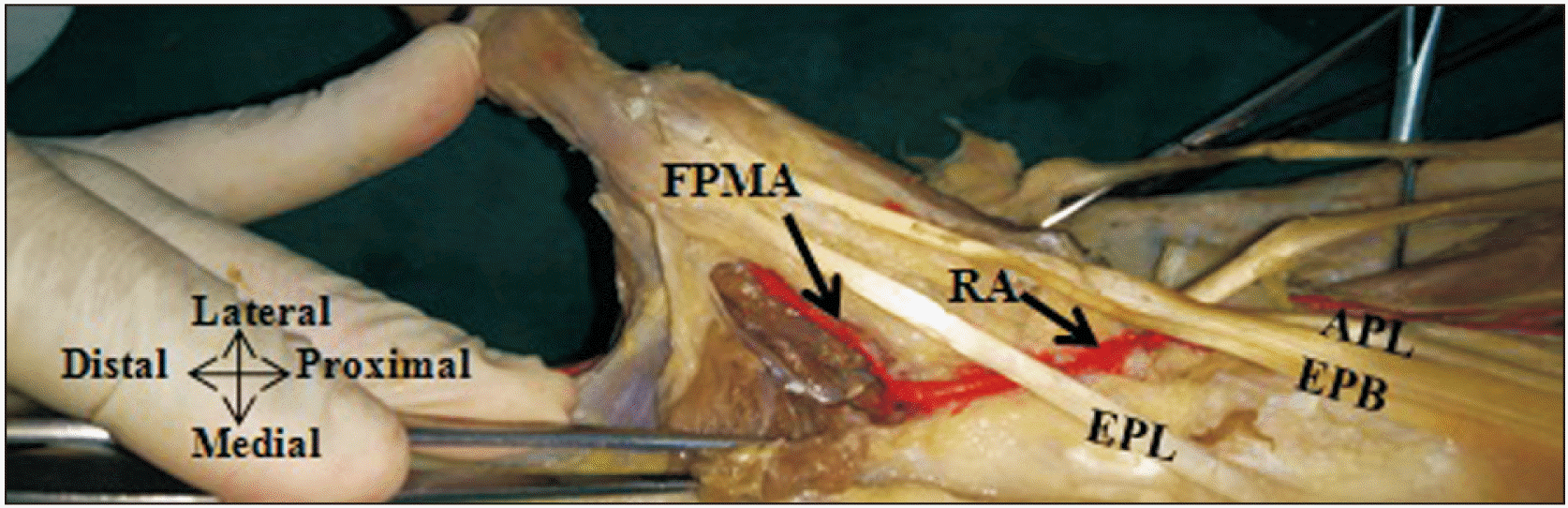 | Fig. 1FPMA arises from the deep branch of radial artery after it pierces the first dorsal interosseous muscle to continue as deep palmar arch. In this figure the first dorsal interosseous muscle is incised to show the course of FPMA running between the first dorsal interosseous and adductor pollicis muscles. APL, abductor pollicis longus; EPB, extensor pollicis brevis; EPL, extensor pollicis longus; FPMA, first palmar metacarpal artery; RA, radial artery. 
|
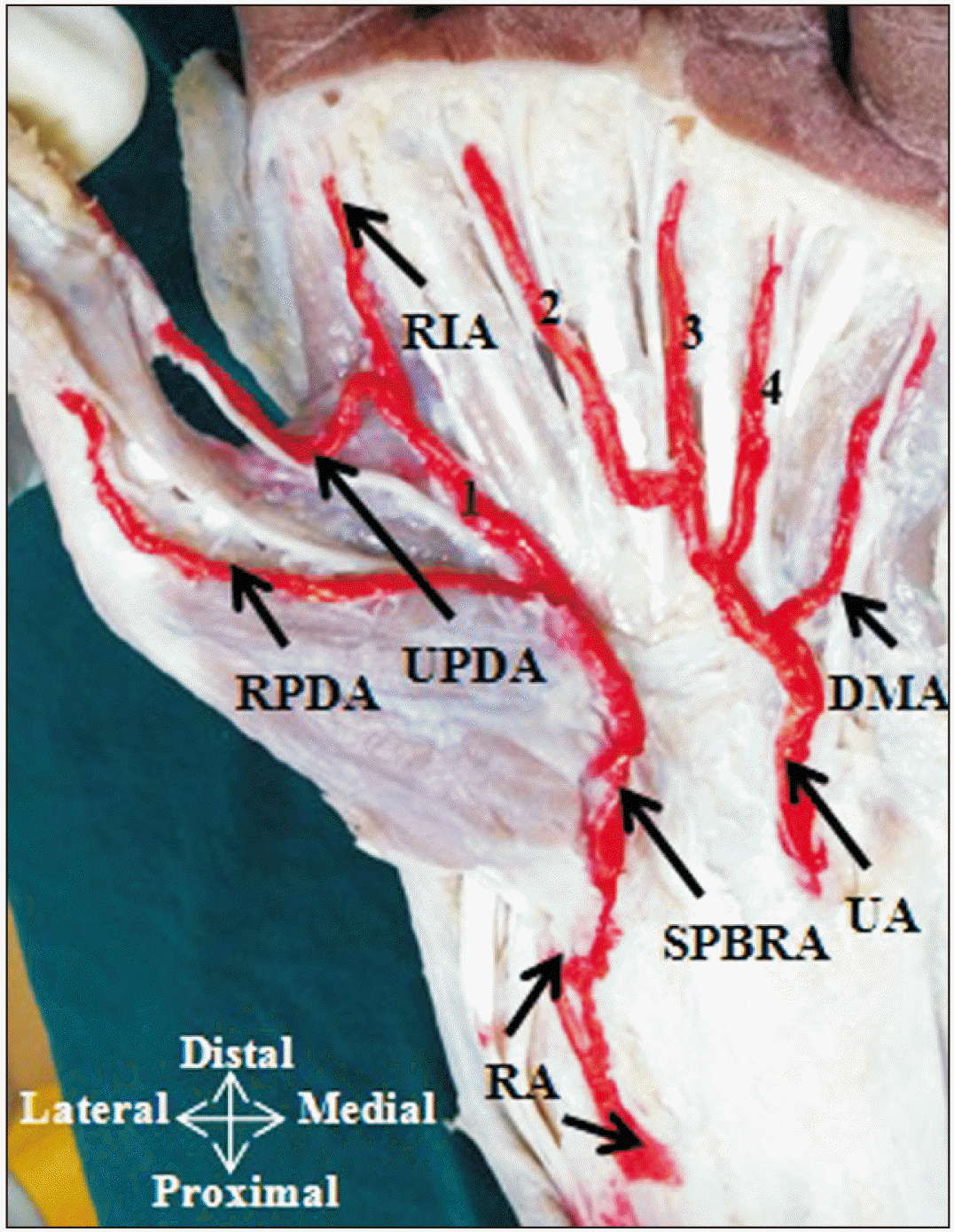
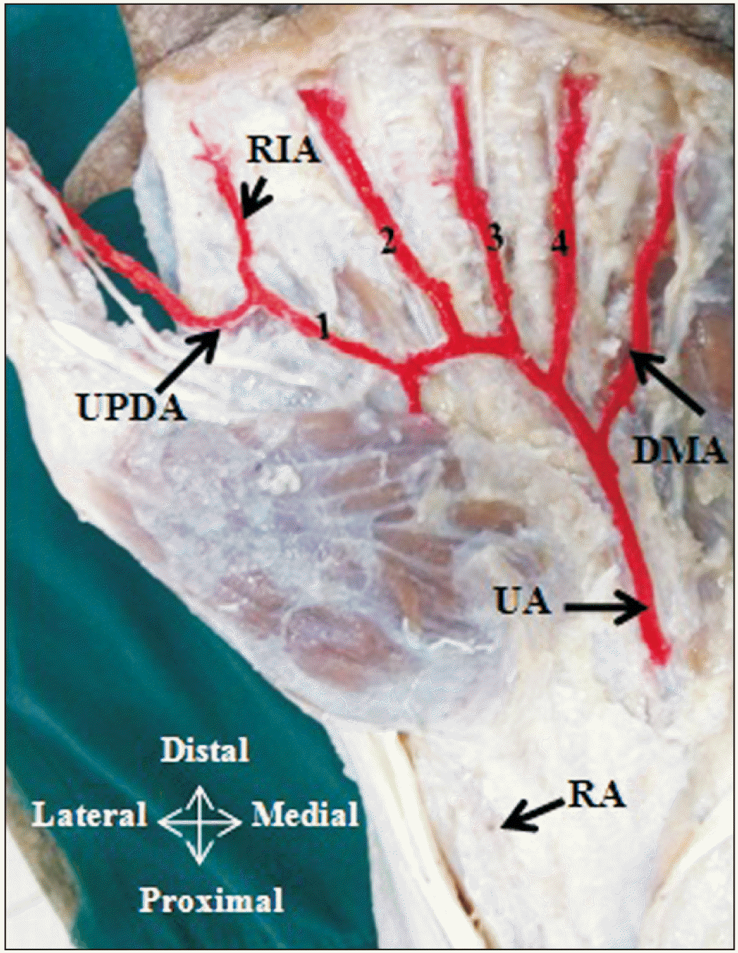
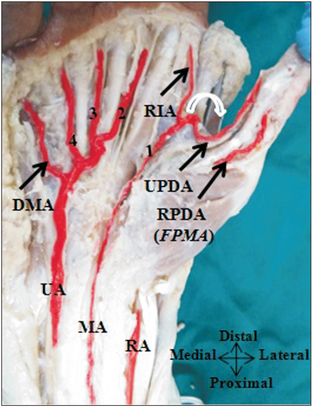
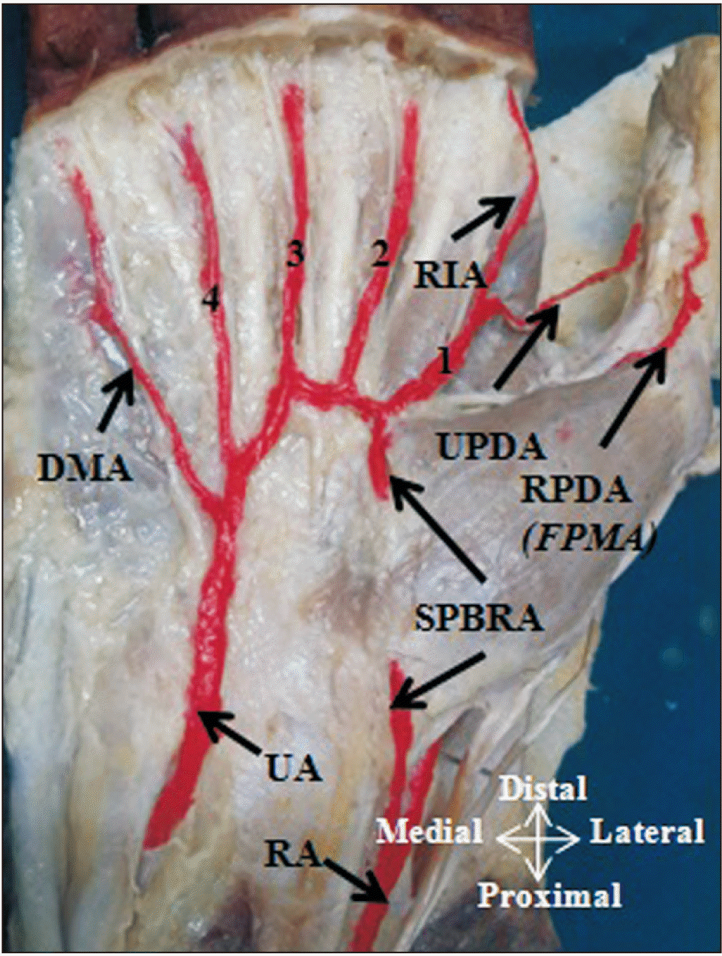 | Fig. 2RPDA from the FPMA and UPDA from the first CPDA from typical SPA. CPDA, common palmar digital artery; 1, 2, 3, 4, CPDA; DMA, digiti minimi artery; FPMA, first palmar metacarpal artery; RA, radial artery; RIA, radialis indicis artery; RPDA, radial palmar digital artery; SPA, superficial palmar arch; SPBRA, superficial palmar branch of radial artery; UA, ulnar artery; UPDA, ulnar palmar digital artery.
|
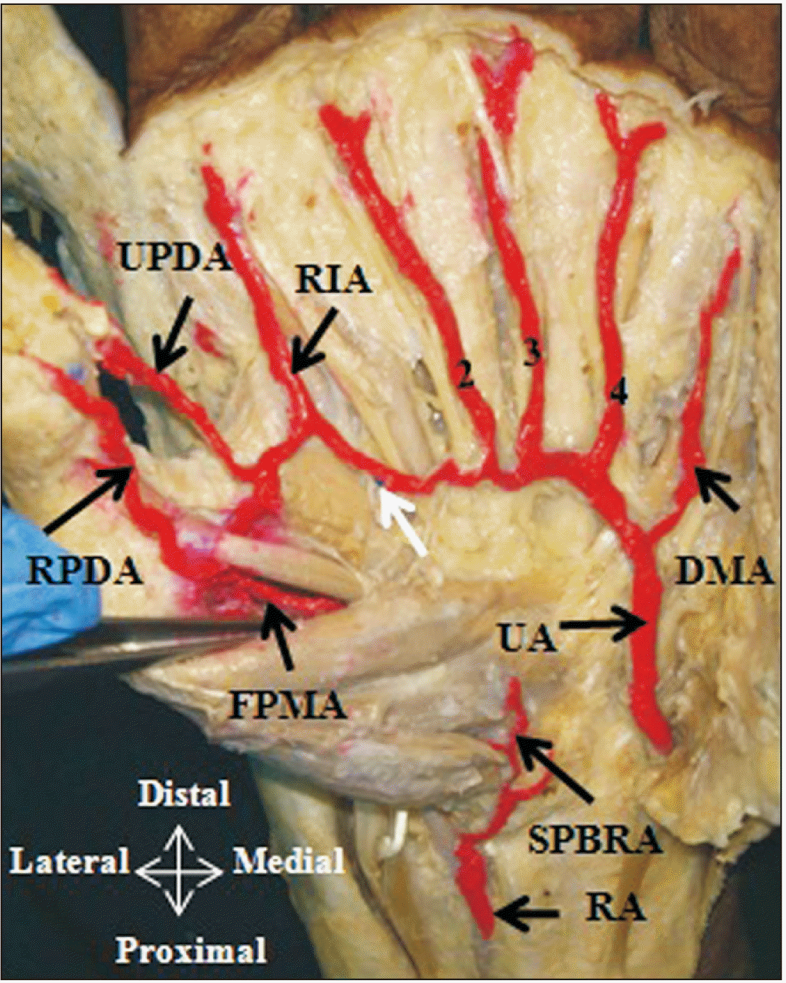 | Fig. 3RPDA and UPDA from the FPMA. CPDA, common palmar digital artery; 1, 2, 3, 4, CPDA; DMA, digiti minimi artery; FPMA, first palmar metacarpal artery; RA, radial artery; RIA, radialis indicis artery; RPDA, radial palmar digital artery; SPBRA, superficial palmar branch of radial artery; UA, ulnar artery; UPDA, ulnar palmar digital artery; white arrow, anastomosis of UA with RIA.
|
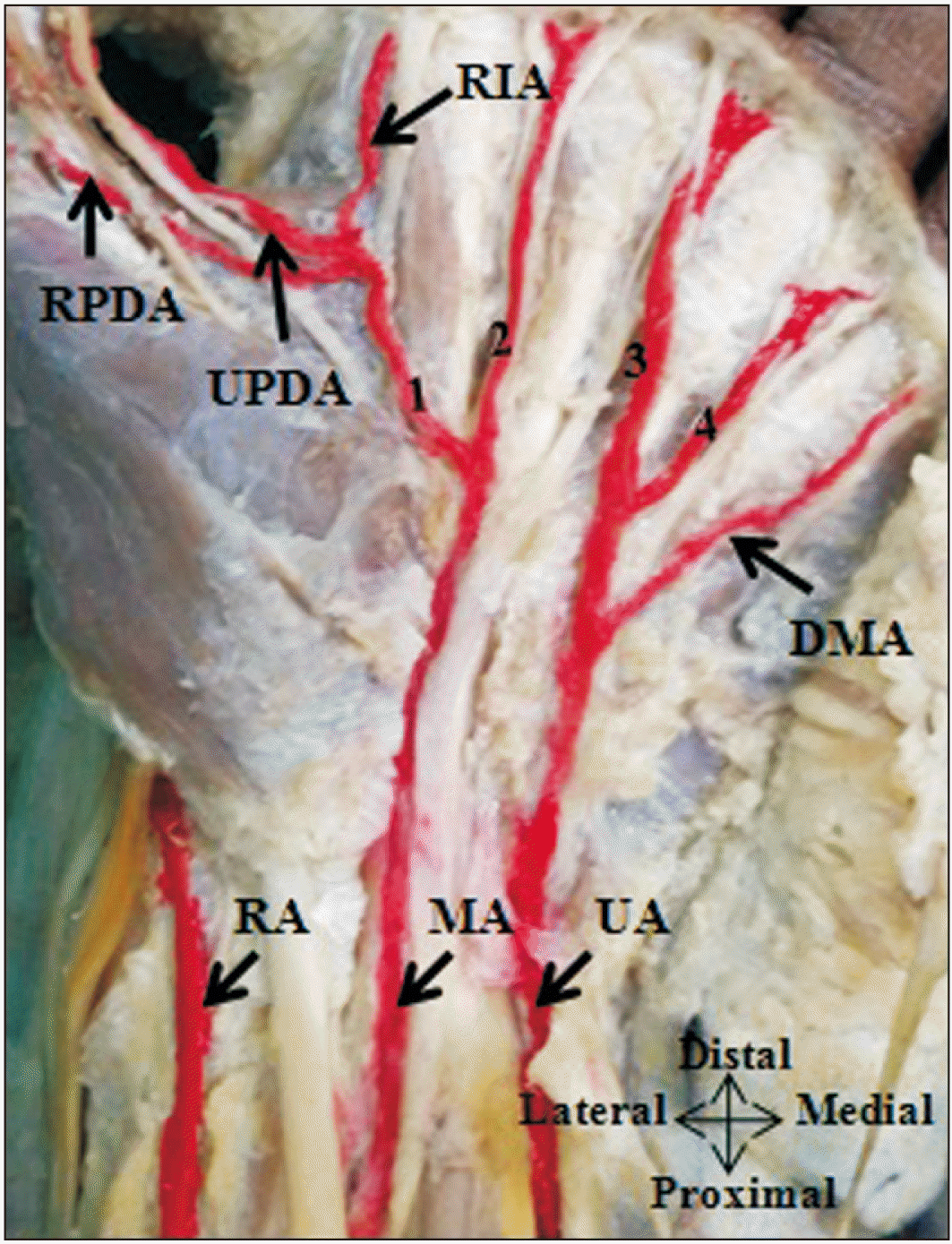
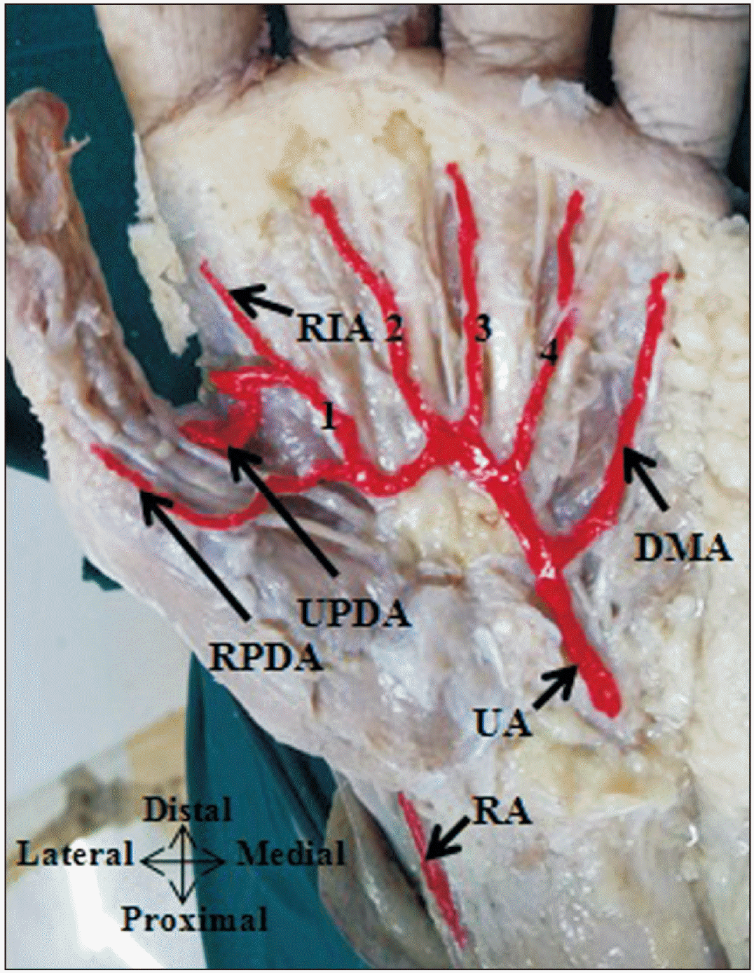 | Fig. 4RPDA arise directly from the UA and UPDA from the UA through first CPDA. CPDA, common palmar digital artery; 1, 2, 3, 4, CPDA; DMA, digiti minimi artery; RA, radial artery; RIA, radialis indicis artery; RPDA, radial palmar digital artery; UA, ulnar artery; UPDA, ulnar palmar digital artery.
|
 | Fig. 5RPDA arises directly from the SPBRA whereas; the UPDA and radialis indicis artery arise from the SPBRA through the first CPDA. CPDA, common palmar digital artery; 1, 2, 3, 4, CPDA; DMA, digiti minimi artery; RA, radial artery; RIA, radialis indicis artery; RPDA, radial palmar digital artery; SPBRA, superficial palmar branch of radial artery; UA, ulnar artery; UPDA, ulnar palmar digital artery.
|
 | Fig. 6UPDA and RPDA from the MA through first CPDA. CPDA, common palmar digital artery; 1, 2, 3, 4, CPDA; DMA, digiti minimi artery; MA, median artery; RA, radial artery; RIA, radialis indicis artery; RPDA, radial palmar digital artery; SPBRA, superficial palmar branch of radial artery; UA, ulnar artery; UPDA, ulnar palmar digital artery.
|
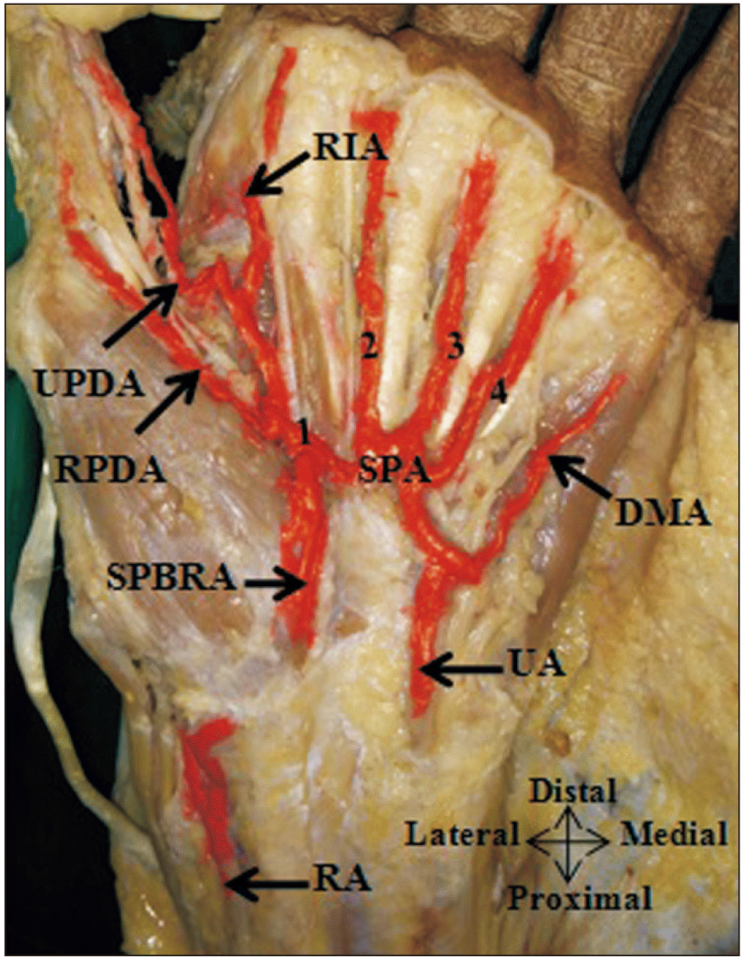 | Fig. 7RPDA, UPDA from the typical SPA through the first CPDA. CPDA, common palmar digital artery; 1, 2, 3, 4, CPDA; DMA, digiti minimi artery; RA, radial artery; RIA, radialis indicis artery; RPDA, radial palmar digital artery; SPA, superficial palmar arch; SPBRA, superficial palmar branch of radial artery; UA, ulnar artery; UPDA, ulnar palmar digital artery.
|
Table 1
Frequency of source of RPDA and UPDA to thumb
|
No. |
Source artery |
RPDA (n=55) |
UPDA (n=55) |
|
1 |
FPMA |
45 (81.8) |
38 (69.1) |
|
2 |
UA |
5 (9.1) |
11 (20.0) |
|
3 |
SPA |
7 (12.7) |
20 (36.4) |
|
4 |
SPBRA |
5 (9.1) |
3 (5.5) |
|
5 |
FDMA |
0 |
11 (20.0) |
|
6 |
MA |
1 (1.8) |
4 (7.3) |

Table 2
Comparison of source of RPDA to the thumb with other studies
|
No. |
Source of RPDA |
Studies, sample size |
|
Earley [7], n=20 |
Ikeda et al. [12], n=220 |
Present study, n=55 |
|
1 |
FPMA |
18 (90.0) |
167 (75.9) |
45 (81.8) |
|
2 |
UA |
0 |
- |
5 (9.1) |
|
3 |
SPA |
0 |
41 (18.6) |
7 (12.7) |
|
4 |
SPBRA |
2 (10.0) |
- |
5 (9.1) |
|
5 |
FDMA |
0 |
10 (4.5) |
0 |
|
6 |
MA |
0 |
- |
1 (1.8) |
|
7 |
Absent |
0 |
2 (0.9) |
2 (3.6) |
|
Total RPDA |
20 |
220 |
65 |
The most common source of UPDA to the thumb was also the FPMA, which was seen in 69.1% (38 hands) (
Figs. 3,
8). Other sources of UPDA include the terminal branch of the UA in 20% (
Figs. 4,
9), the typical SPA in 36% (
Fig. 10), SPBRA in 6% (
Fig. 5), FDMA in 20% (
Fig. 11) and MA in 7% (
Fig. 6). More than one UPDA was observed in 32 hands where in addition to UPDA from the FPMA significant UPDA to the thumb came from one of the other superficial palmar arterial systems (
Tables 1 and
3) [
7,
12].
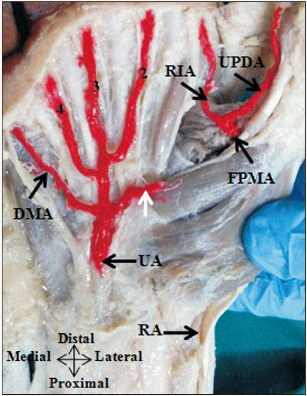 | Fig. 8UPDA and radialis indicis artery arising from the FPMA. CPDA, common palmar digital artery; 2, 3, 4, CPDA; DMA, digiti minimi artery; FPMA, first palmar metacarpal artery; RA, radial artery; RIA, radialis indicis artery; UPDA, ulnar palmar digital artery; UA, ulnar artery; white arrow muscular branch to thenar muscles from UA, according to Gnanasekaran and Veeramani [ 18]. Adapted from Gnanasekaran and Veeramani. Surg Radiol Anat 2019;41:791-9 [ 18].
|
 | Fig. 9UPDA arise from the UA through the first common palmar digital. CPDA, common palmar digital artery; 1, 2, 3, 4, CPDA; DMA, digiti minimi artery; RA, radial artery; RIA, radialis indicis artery; UA, ulnar artery; UPDA, ulnar palmar digital artery, according to Gnanasekaran and Veeramani [ 18]. Adapted from Gnanasekaran and Veeramani. Surg Radiol Anat 2019;41:791-9 [ 18].
|
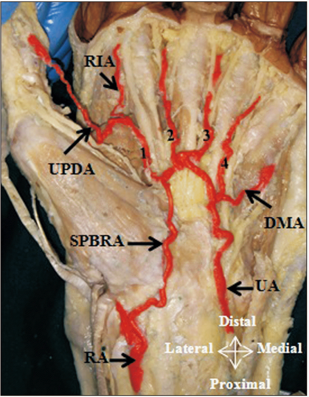 | Fig. 10UPDA arise from typical SPA through the first CPDA. CPDA, common palmar digital artery; 1, 2, 3, 4, CPDA; DMA, digiti minimi artery; RA, radial artery; RIA, radialis indicis artery; SPA, superficial palmar arch; SPBRA, superficial palmar branch of radial artery; UA, ulnar artery; UPDA, ulnar palmar digital artery, according to Gnanasekaran and Veeramani [ 18]. Adapted from Gnanasekaran and Veeramani. Surg Radiol Anat 2019;41:791-9 [ 18].
|
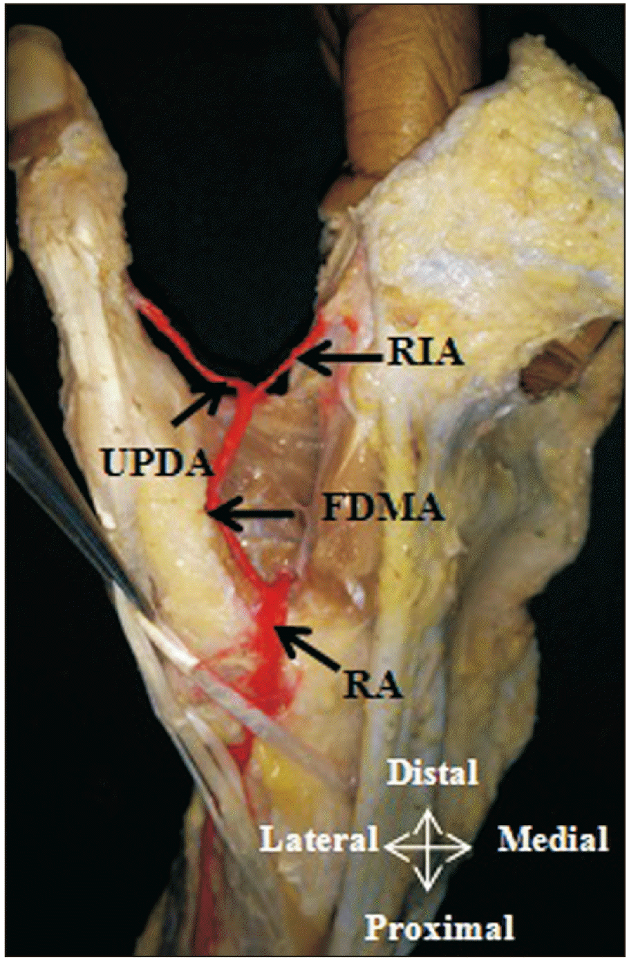 | Fig. 11UPDA and radialis indicis artery arise from the FDMA, a direct branch of radial artery. FDMA, first dorsal metacarpal artery; RA, radial artery; RIA, radialis indicis artery; UPDA, ulnar palmar digital artery, according to Gnanasekaran and Veeramani [ 18]. Adapted from Gnanasekaran and Veeramani. Surg Radiol Anat 2019;41:791-9 [ 18].
|
Table 3
Comparison of source of UPDA to the thumb with other studies
|
No. |
Source of UPDA |
Studies, Sample size |
|
Earley [7], n=20 |
Ikeda et al. [12], n=220 |
Present study, n=55 |
|
1 |
FPMA |
11 (55.0) |
158 (71.8) |
38 (69.1) |
|
2 |
UA |
3 (15.0) |
- |
11 (20.0) |
|
3 |
SPA |
3 (15.0) |
32 (14.5) |
20 (36.4) |
|
4 |
SPBRA |
1 (5.0) |
- |
3 (5.5) |
|
5 |
FDMA |
1 (5.0) |
30 (13.6) |
11 (20.0) |
|
6 |
MA |
0 |
- |
4 (7.3) |
|
7 |
UDDA |
1 (5.0) |
0 |
0 |
|
Total UPDA |
20 |
220 |
87 |
When there is an extra RPDA or UPDA to the thumb in addition to the FPMA, the RPDA and UPDA branching from the FPMA terminate at the level of the metacarpophalangeal joint or base of proximal phalanx itself. And, the RPDA or UPDA from the superficial palmar arterial system extends up to the tip of the thumb and seems to be the dominant vessel.
Source of the radial dorsal digital artery and ulnar dorsal digital artery to thumb
The RDDA to the thumb was observed in 10.9% (6 hands) and UDDA in 7.3% (4 hands). Both of these arteries were found to arise either directly from the radial artery (
Fig. 12) or sometimes as the continuation of the FDMA that arises from the radial artery before it pierces the first dorsal interosseous muscle. Out of 10.9% (6 hands) in 2 hands the RDDA was found to be the sole artery to supply the thumb on its radial side, as RPDA was absent here. In the remaining 4 hands, the RDDA was present in addition to RPDA. Similarly, UDDA seen in 4 hands was found to be supplementary to UPDA, as UPDA was observed in 100.0% of the dissected hands.
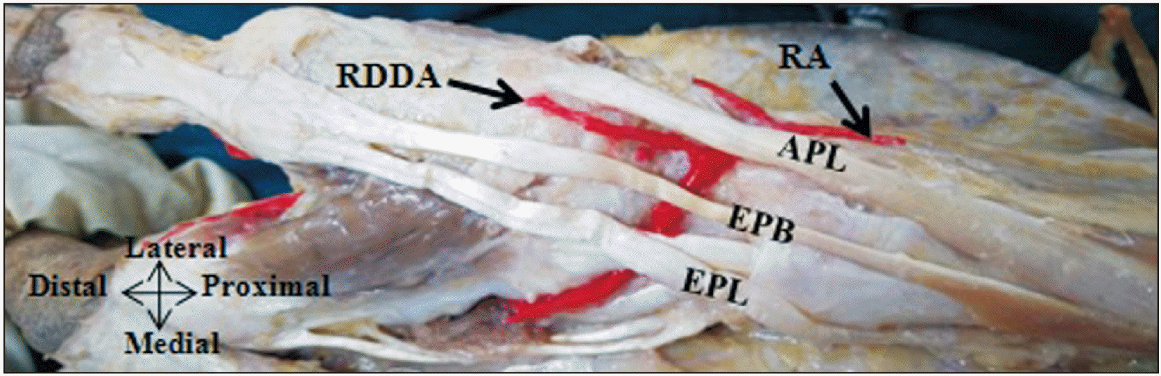 | Fig. 12RDDA arise as a direct branch from the radial artery. APL, abductor pollicis longus; EPB, extensor pollicis brevis; EPL, extensor pollicis longus; RA, radial artery; RDDA, radial dorsal digital artery.
|
Palmar arteries to the thumb either RPDA or UPDA or both the RPDA and UPDA arise from the complete SPA in 80.0% (44 hands) according to our previous study [
18]. Out of these, in 8 hands the arch was completely formed by the UA alone (
Fig. 9). In 7 out of 8 hands, the UPDA was from the first CPDA (from the terminal branch of the UA). In the remaining one hand, both the RPDA and the UPDA were from the first CPDA (
Fig. 4). In the 11 hands (20.0%) both palmar arteries to the thumb originated from incomplete SPA (
Figs. 5,
6,
8). Out of these 11 cases, in 2 hands the FDMA anastomosed with the UPDA provided by the MA around the first web space (
Fig. 13).
 | Fig. 13FDMA anastomosed with the UPDA provided by the MA around the first web space. CPDA, common palmar digital artery; 1, 2, 3, 4, CPDA; DMA, digiti minimi artery; FDMA, first dorsal metacarpal artery; FPMA, first palmar metacarpal artery; MA, median artery; RA, radial artery; RPDA, radial palmar digital artery; RIA, radialis indicis artery; UA, ulnar artery; UPDA, ulnar palmar digital artery, according to Gnanasekaran and Veeramani [ 18]. Adapted from Gnanasekaran and Veeramani. Surg Radiol Anat 2019;41:791-9 [ 18].
|
Go to :
Discussion
The knowledge of arterial anatomy of the thumb and associated variation is crucial for the surgeon performing reconstructive surgeries on the thumb. In the current study, the RPDA and UPDA were present in 96% and 100% which is similar to Earley [
7] Ikeda et al. [
12] Ramírez and Gonzalez [
15] and, Tan and Lahiri [
19] The UPDA was seen in 100% of the dissected limbs similar to the above studies. On the other hand, the RDDA and UDDA were present in 11% and 7% only, which contradicts the observation by previous studies (
Table 4) [
7,
12,
15]. The reason for such a low incidence of RDDA and UDDA reported in the present may because we have taken into consideration only the major arteries to the thumb that was visible by gross dissection and probably the finer dorsal branches could have been missed. Authors from many studies also have commented that the dorsal digital arteries to the thumb were very small and had a variable origin and the mean diameter of these branches was less than 1 mm. They could do so because they used either a radio-opaque dye, latex, or resin to see these tiny arteries [
7,
12,
15]. According to Omokawa among the dorsal metacarpal arteries of the hand, the FDMA was constantly present and irrigates the thumb [
20]. But Ramírez and Gonzalez [
15] reported that even though the FDMA was seen in all of the 30 specimens none was found irrigating the thumb. So based on these it is inferred from the present study that the dominant blood supply to the thumb is mostly from the RPDA and UPDA (
Table 4) [
7,
12,
15]. In case, if any one side of the major palmar digital arteries to the thumb is absent then only the thumb is supplied by the corresponding side major dorsal digital artery.
Table 4
Comparison of incidence of digital arteries to the thumb with other studies
|
Studies (sample size) |
Type of cadaveric study |
Thumb arteries (%) |
|
RPDA |
UPDA |
RDDA |
UDDA |
|
Earley [7] (n=20) |
Intra arterial radio opaque dye with two-dimensional arterio graph & latex injection with dissection |
100 |
100 |
70 |
70 |
|
Ikeda et al. [12] (n=220) |
Intra arterial radio-opaque dye with three-dimensional arterio graph |
99 |
100 |
64 |
80 |
|
Ramírez and Gonzalez [15] (n=30) |
Intra arterial synthetic resin with dissection |
100 |
100 |
93 |
100 |
|
Present study (n=55) |
Dissection |
96 |
100 |
11 |
7 |
The source of UPDA to the thumb was mainly observed arising from the FDMA in 69% in the present study which is in concordance with the study by Ikeda et al. [
12]. In 5% it was from SPBRA similar to the study by Earley [
7]. The terminal branch of UA was found to be the UPDA in 20% which is similar to the observation by Earley [
7]. However, Ikeda et al. [
12] considered though UPDA was from the terminal branch of UA, it was kept under the classification of the branch of SPA predominantly of ulnar type. But we strictly placed the palmar digital branches to thumb under SPA only when it comes from the typical SPA to avoid confusion. In the current study, the UPDA was found to arise for the typical SPA formed by SPBRA and the superficial branch of UA in 36%. The UPDA arose separately from the typical SPA or the first common palmar digital arteries from the typical SPA. The MA was also found to give the UPDA to the thumb in 7% and such variation was not observed by Earley [
7] and Ikeda et al. [
12]. The UPDA from the FDMA was present in 20% in the present study whereas, Ikeda et al. [
12] reported that it was seen in 14%. The source of UPDA and RPDA to the thumb from various studies is summarized in
Tables 2 and
3 [
7,
12].
The most common source of RPDA to the thumb is the FPMA which was seen in 82%. Earley [
7] and Ikeda et al. [
12] also found that RPDA to the thumb was mainly from the FPMA in most of the cases. The RPDA was from SPBRA in 10% of dissected cases similar to Earley [
7]. In the present study, MA was found to provide the RPDA in only 2%. In the case of absent RPDA, the dominant RDDA from the radial artery was found supplying the thumb. In the present study, the RDDA and UDDA were found to arise either directly from the radial artery or it continues from the FDMA which is a branch of radial artery. Other sources of the RDDA and UDDA arteries reported by Ikeda et al. [
12] were the FPMA and SPA. In contrast to the low incidence of RDDA and UDDA reported in the present study, other studies have proved its significant presence and their utilization as flaps in various reconstruction procedures of hand (
Table 4) [
7,
12,
15].
When the radial artery or UA providing the main blood supply to the thumb does not form anastomosis with each other, the obstruction or injury to either of those may lead to the compromised blood supply to the thumb [
1]. In the present study, the palmar digital arteries to the thumb originating from SPA were found to arise from the complete SPA in 80% and incomplete SPA in 20 % according to our previous study on SPA [
18]. In the case of SPA formed entirely by the UA, obstruction, or damage to this artery may lead to thumb ischemia. On the other hand, the radial artery can be harvested safely, as the sole blood supply was from the UA. This study proposes a single acceptable nomenclature taking into consideration the merits & demerits of the already existing nomenclature mentioned in the literature (
Table 5) [
2,
5,
6,
7,
9-
12,
15,
16].
Table 5
Description of various sources of digital arteries to the thumb
|
FPMA |
It arise from the RA soon after it enters between the two heads of first dorsal interosseous to form the DPA and passes along the first metacarpal bone either palmar to the adductor pollicis or between the first dorsal interosseous muscle and adductor pollicis. At the level of the metacarpal phalangeal joint, and deep to the flexor pollicis longus tendon, it usually terminates in two palmar digital arteries namely RPDA and UPDA to the thumb [2, 5, 16]. Fig. 1 Or FPMA trifurcates into RPDA and UPDA to the thumb and FDMA [5]. Or FPMA gives RPDA and UPDA as separate branch and the terminal end of FPMA continues as RIA [Present study]. Fig. 3 Or the FPMA divides into UPDA to the thumb and RIA [Present study]. Fig. 8 Or the FPMA provides only RPDA [Present study]. Fig. 2
|
|
FDMA |
It arise from the RA just before it passes between the two heads of the first dorsal interosseous. In most cases, FDMA follows a fascial course overlying the first dorsal interosseous and parallel to the second metacarpal bone. Occasionally, it may follow an intramuscular course. It divides, into UDDA and RIA [6]. Fig.11 Sometimes the FDMA continues as UPDA [7, 12].
|
|
Typical/classical complete SPA |
Formed by the terminal part of UA and completed by SPBRA [2, 5, 6, 9]. Figs. 2, 7, 10 Sometimes RPDA and UPDA arise separately from the typical SPA [6]. Or RPDA, UPDA and RIA arise from the common trunk called first CPDA from this arch [Present study]. Fig. 7 Or only UPDA and RIA arise from the first CPDA from this arch [Present study]. Fig. 10 The parent sources of the RPDA and UPDA to the thumb that arise from first CPDA from the SPA were classified under branches from SPA.
|
|
SPBRA |
It arises from the RA just before it curves round the carpus and passes superficial to flexor retinaculum. In the hand it passes either superficial to or through the thenar muscles. It gives both the RPDA and UPDA to thumb and also the RIA [5, 6, Present study]. Fig. 5 Sometimes it continues as only RPDA to the thumb [5].
|
|
First CPDA |
First CPDA from the SPA
The classical/ typical complete SPA gives a common trunk called first CPDA to the first web space which gives three branches namely RPDA and UPDA to the thumb and RIA [Present study]. Fig. 7 Or the first CPDA divides into UPDA and RIA [5, 10, Present study]. Fig. 10 Or this common trunk gives only UPDA and RPDA to thumb [5]. To avoid confusion palmar digital arteries to the thumb arising either through first CPDA from typical SPA or directly from the typical SPA were classified under branches of SPA.
First CPDA from MA
Sometimes MA gives first CPDA that trifurcates into RPDA, UPDA to the thumb and RIA [Present study]. Fig. 6 Or first CPDA from the MA gives only UPDA and RIA [5, 15, 16]. In the above two conditions the parent source of palmar digital arteries to thumb were classified under branch from MA only.
First CPDA from the SPBRA
First CPDA from the terminal part of UA
The terminal part of UA continues as first CPDA to the first web space and divides into UPDA to the thumb and RIA [11, Present study]. Fig. 9 Or the terminal part of UA provides first CPDA that later divides into UPDA to the thumb and RIA then it continues as RPDA [Present study]. Fig. 4 The parent source of palmar digital arteries to the thumb from the terminal part UA and through the first CPDA was classified under branch of UA only. Note: When palmar continuity of FDMA provides UPDA to the thumb and RIA Bilge et al. [9] considered the continuity of FDMA as first CPDA. But we didn’t agree with this naming as the so called first CPDA didn’t arise from any of superficial palmar arterial system and it came from the dorsal side of the hand.
|
Conclusion
Universal naming of the proper digital arteries to the thumb as well as its source arteries is mandatory for proper understanding of the normal as well as variant arterial anatomy of the thumb. We recommend that the term APP should not be used as it creates mere confusion only. Our study described the incidence of thumb vasculature on both radial and ulnar sides from its dorsal and palmar aspects in addition to it traced the source of these arteries. This study may be of paramount importance to vascular surgeons operating on the thumb for reimplantation and reconstructive surgeries. Adequate anatomical knowledge about the arteries on the radial and ulnar side and their source is also important in preservative surgeries of the thumb that involve multiple traumatic injuries.
Go to :
Acknowledgements
We sincerely thank the relatives of the cadavers who have donated the bodies for the research purpose.
Go to :
Notes
Go to :
References
1. Cho SH, Bahar-Moni AS, Park HC. 2016; Thumb replantation using the superficial palmar branch of the radial artery. J Hand Microsurg. 8:106–8. DOI:
10.1055/s-0036-1585058. PMID:
27625540. PMCID:
PMC5018973.
2. Parks BJ, Arbelaez J, Horner RL. 1978; Medical and surgical importance of the arterial blood supply of the thumb. J Hand Surg Am. 3:383–5. DOI:
10.1016/S0363-5023(78)80044-6. PMID:
681725.
3. Hierner R, Putz R, Bishop AT, Shen ZL, Wilhelm K, Brugger U, Kellner L, Rintelen H. 2013. Flaps in hand and upper limb reconstruction: surgical anatomy, operative techniques and differential therapy. Urban & Fischer;Munchen: DOI:
10.1016/s0363-5023(78)80044-6.
4. Schlenker JD, Kleinert HE, Tsai TM. 1980; Methods and results of replantation following traumatic amputation of the thumb in sixty-four patients. J Hand Surg Am. 5:63–70. DOI:
10.1016/S0363-5023(80)80046-3. PMID:
7365219.
6. Biant LC. Standring S, editor. 2016. Elbow and forearm. Gray's Anatomy: The Anatomical Basis of Clinical Practice. 41st ed. Elsevier Limited;Philadelphia: p. 837–61. DOI:
10.1201/b13469-22.
7. Earley MJ. 1986; The arterial supply of the thumb, first web and index finger and its surgical application. J Hand Surg Br. 11:163–74. DOI:
10.1016/0266-7681_86_90253-6. PMID:
3734551.
8. Al-Turk M, Metcalf WK. 1984; A study of the superficial palmar arteries using the Doppler ultrasonic flowmeter. J Anat. 138(Pt 1):27–32. PMID:
6706837. PMCID:
PMC1164307.
9. Bilge O, Pinar Y, Ozer MA, Gövsa F. 2006; A morphometric study on the superficial palmar arch of the hand. Surg Radiol Anat. 28:343–50. DOI:
10.1007/s00276-006-0109-9. PMID:
16642281.
12. Ikeda A, Ugawa A, Kazihara Y, Hamada N. 1988; Arterial patterns in the hand based on a three-dimensional analysis of 220 cadaver hands. J Hand Surg Am. 13:501–9. DOI:
10.1016/S0363-5023(88)80085-6. PMID:
3418051.
13. Murakami T, Takaya K, Outi H. 1969; The origin, course and distribution of arteries to the thumb, with special reference to the so-called A. princeps pollicis. Okajimas Folia Anat Jpn. 46:123–37. DOI:
10.2535/ofaj1936.46.2-3_123. PMID:
5820050.
15. Ramírez AR, Gonzalez SM. 2012; Arteries of the thumb: description of anatomical variations and review of the literature. Plast Reconstr Surg. 129:468e–76e. DOI:
10.1097/PRS.0b013e3182402d43. PMID:
22373995.
16. Miletin J, Sukop A, Baca V, Kachlik D. 2017; Arterial supply of the thumb: systemic review. Clin Anat. 30:963–73. DOI:
10.1002/ca.22973. PMID:
28791730.
17. Cunningham DJ. 2005. Cunningham's manual of practical anatomy. 15th ed. Vol 1:Oxford University Press;Oxford: DOI:
10.1002/ca.22973.
18. Gnanasekaran D, Veeramani R. 2019; Newer insights in the anatomy of superficial palmar arch. Surg Radiol Anat. 41:791–9. DOI:
10.1007/s00276-019-02223-w. PMID:
30923841.
20. Omokawa S, Tanaka Y, Ryu J, Kish VL. 2005; The anatomical basis for reverse first to fifth dorsal metacarpal arterial flaps. J Hand Surg Br. 30:40–4. DOI:
10.1016/J.JHSB.2004.09.006. PMID:
15620490.
Go to :


 PDF
PDF Citation
 Print
Print


 XML Download