Introduction
Computed tomography (CT) of the nose and paranasal sinuses is currently considered the imaging modality of choice for radiological diagnosis of nasal and paranasal disorders [
1]. Unlike plain radiography, CT provides an excellent idea about the soft tissue and bony anatomical details. Understanding the details of sinonasal anatomy helps to reach the proper diagnosis and hence the guidance for safe surgery [
2].
The nasal septum is a crucial supportive component of the nasal cavity which is composed of bony and cartilaginous parts and divides the nasal cavity into two sides [
3]. The bony component is made up of the vomer and perpendicular plate of ethmoid bones, contributing more than 70% of the whole nasal septum [
4]. Although a perfectly straight nasal septum is extremely rare, some degree of deviation can be clinically accepted [
5]. In contrast, a higher percentage of nasal septal deviation (NSD) is a potent risk factor for nasal cavity obstruction and sinusitis [
6]. Moreover, significant NSD can produce compensatory hypertrophy of inferior turbinate and concha bullosa of the middle turbinate at the contralateral side, aggravating the obstruction, or additionally causing hypoplasia of the ipsilateral turbinates. According to the side, NSD can be classified as right-sided, left-sided or S-shaped variants [
7].
Nasal septal spur (NSS) is a popular anatomical variation that is frequently associated with NSD. If prominent, NSS may interfere with the surgical access to the nasal cavity and also could narrow the middle meatus or ethmoid infundibulum [
8]. In addition, the pressure between mucosal surfaces in the region of NSS can be a source of excruciating pain [
7]. Another anatomical variation of the nasal septum is its pneumatization which could block the osteomeatal complex and thus potentially predispose to various sinonasal mucosal diseases [
9]. Commonly the nasal septal pneumatization is produced as an extension of the air cells from the sphenoid sinus or crista galli to the nasal septum. Nevertheless, it usually does not cause any clinical relevance but may be contributed to some sort of narrowing of the sphenoethmoidal recess [
8]. Thus, most anatomical nasal septal variations could finally lead to chronic or recurrent rhino-sinusitis due to obstruction of osteomeatal complex and affection of the mucocilliary clearance [
10].
Hence, this study aimed to use coronal CT scan imaging of the nasal cavity and paranasal sinuses to investigate the prevalence of NSD, NSS and nasal septal pneumatization (NSP) among adult Saudi population, and their possible association with the incidence of sinusitis.
Go to :

Results
The current study included subjects aged between 20 and 71 years old with a mean of 44.7±14.3 years old. NSD was present in 512 cases (75.2%), while normal septum was present only in 169 subjects (24.8%). NSD was more frequent in males than females (80.0% vs. 67.4% respectively), with a significant difference in-between (
P<0.001). Regarding the side of NSD, there was no statistical difference between the frequencies of right and left sides according to gender (
Table 1). In addition, the degrees of NSD in both males and females showed no statistical difference regarding right-sided and S-shaped NSD (9.6° vs. 10.4° and 9.1° and 8.7° respectively) (
P=0.163 and
P=0.751 respectively), while was with significantly higher septal deviation degrees in males than females regarding left-sided NSD (14.1° vs. 10.6° respectively) (
P<0.001) (
Fig. 2). According to the correlation between NSD and sinusitis, sinusitis was detected to be more frequent in subjects with NSD (57.0% of septal deviation subjects vs. 29.0% of subjects with no septal deviation) with a statistically significant difference in-between (
P<0.001) (
Table 2) (
Fig. 3A–D). Furthermore, mild, moderate and severe NSDs were associated with nearly similar incidence percentages of sinusitis (58.7%, 56.2%, and 57.0% respectively) (
Fig. 4A).
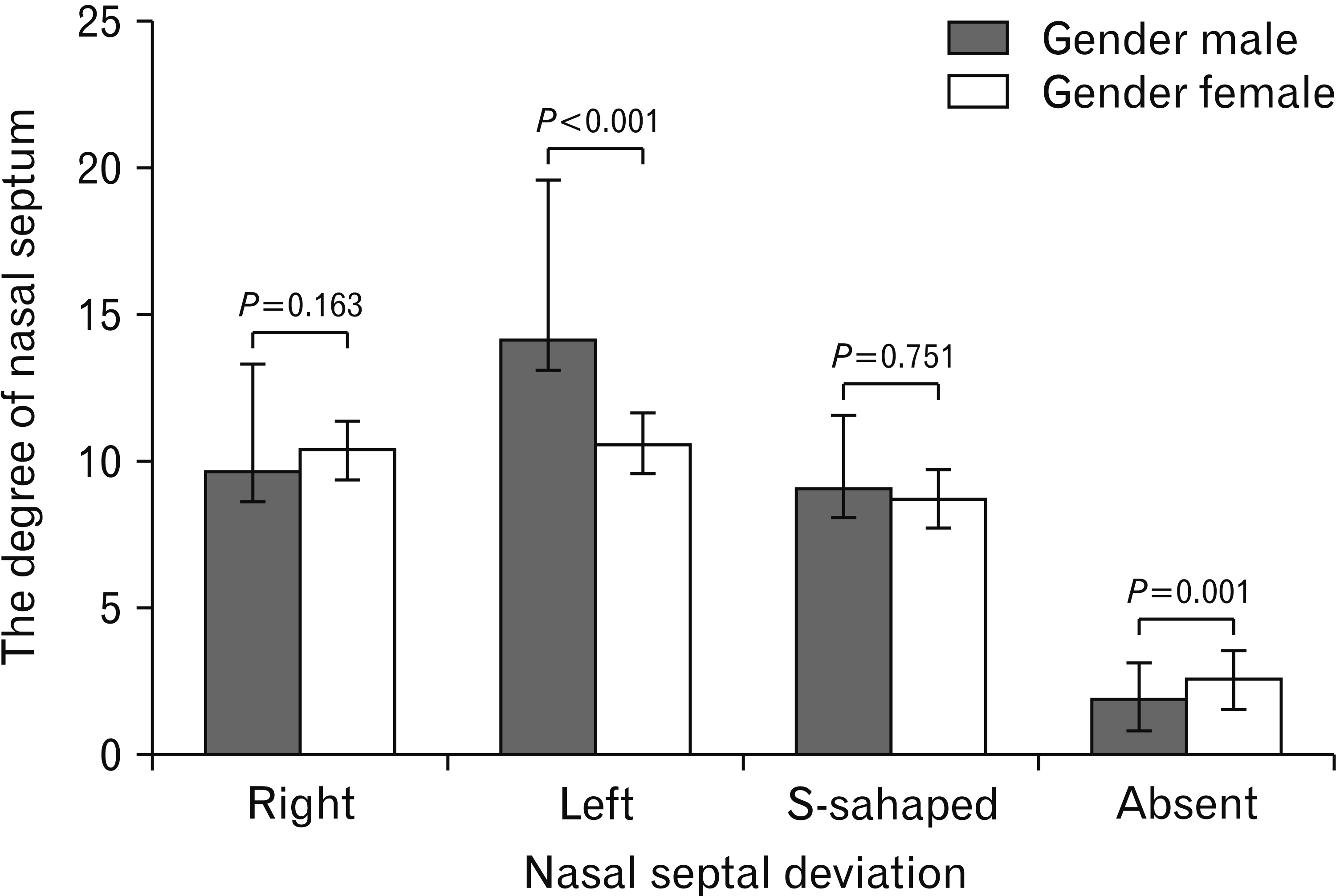 | Fig. 2The degree of NSD in male and female subjects. Values are presented as means with the minimal and maximal values. Statistical analysis was performed by unpaired Student’s t-test. NSD, nasal septal deviation. 
|
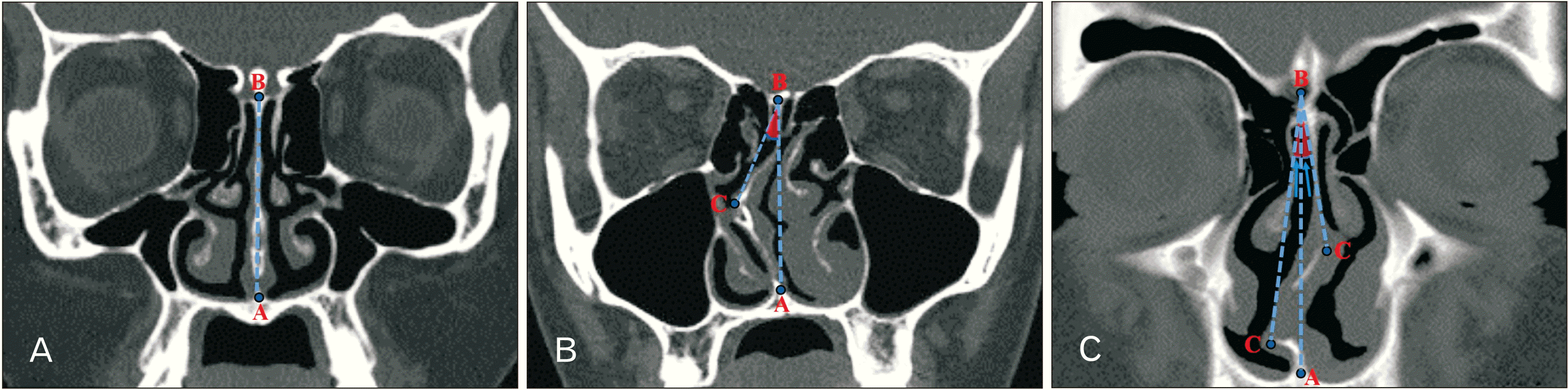
 | Fig. 3Coronal CT images. (A) Left-sided NSD (arrow) with bilateral normal mucosa of both maxillary (asterisks) and ethmoid (arrowheads) sinuses. (B) Left-sided NSD (arrow) with mild sinusitis of both maxillary sinuses (asterisks). (C) S-shaped NSD (arrows) with bilateral normal mucosa of both maxillary (asterisks) and ethmoid (arrowheads) sinuses. (D) S-shaped NSD (arrows) with bilateral maxillary (asterisks) and ethmoid (arrowheads) sinusitis. (E) Left-sided NSD with ipsilateral NSS (arrow), and normal mucosa of both maxillary (asterisks) and ethmoid (arrowheads) sinuses. (F) Centralized nasal septum with left NSS (arrow) and mucosal thickening of both maxillary sinuses (asterisks). (G) Pneumatization (arrow) of a centralized nasal septum with normal mucosa of both maxillary (asterisks) and ethmoid (arrowheads) sinuses. (H) Pneumatization (arrow) of a centralized nasal septum with left maxillary sinusitis (asterisk). CT, computed tomography; NSD, nasal septal deviation; NSS, nasal septal spur.
|
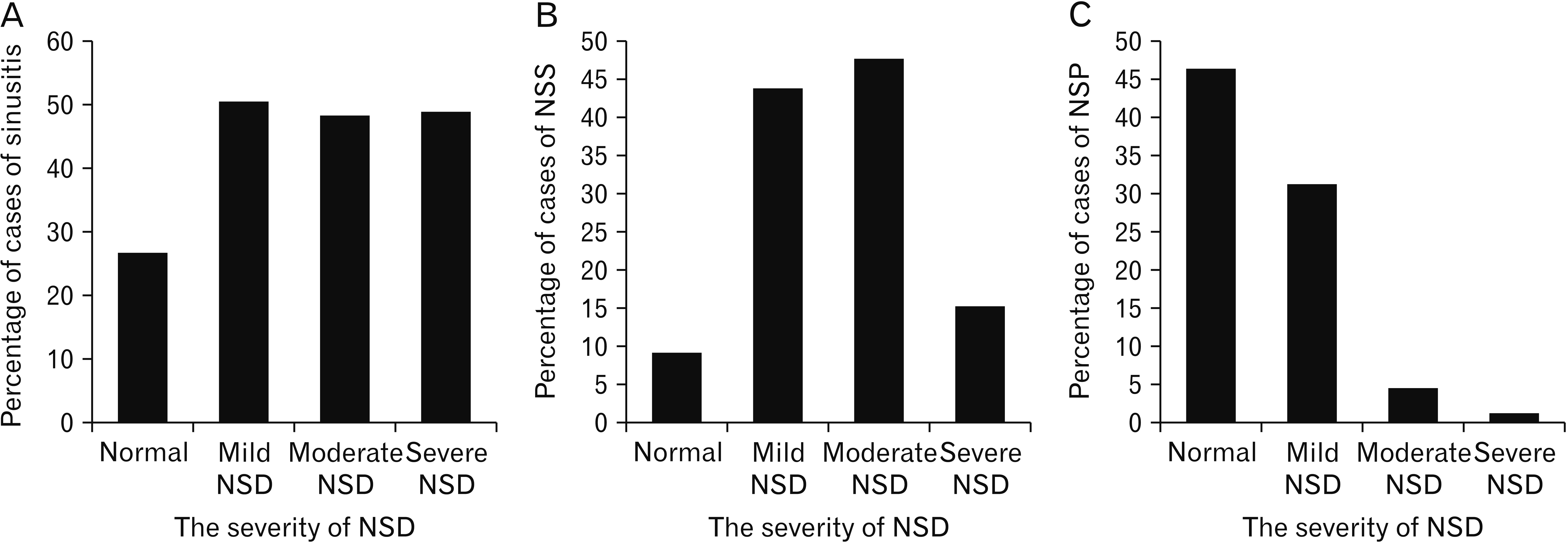 | Fig. 4Percentages of incidence of sinusitis according to the severity of NSD. (B) Percentages of incidence of NSS according to the severity of NSD. (C) Percentages of incidence of NSP according to the severity of NSD. NSD, nasal septal deviation; NSS, nasal septal spur.
|
Table 1
Relation between the incidence of different types of NSD and gender
|
Gender |
NSD |
Total |
|
Right |
Left |
S-shaped |
Total cases of NSD |
Absent |
|
Male |
151 (36.0) |
154 (36.7) |
31 (7.4) |
336 (80.0) |
84 (20.0) |
420 (100.0) |
|
Female |
74 (28.4) |
81 (31.0) |
21 (8.1) |
176 (67.4) |
85 (32.6) |
261 (100.0) |
|
Total |
225 (33.0) |
235 (34.5) |
52 (7.6) |
512 (75.2) |
169 (24.8) |
681 (100.0) |

Table 2
Relation between the incidence of NSD and sinusitis (mucosal thickening)
|
Sinusitis |
NSD |
|
Present |
Sinusitis cases relative to total NSD cases (512 cases) |
Absent |
Sinusitis cases relative to total absent NSD cases (169 cases) |
Total |
P-value |
|
Present |
292 (85.6) |
57.0% |
49 (14.4) |
29.0% |
341 (100.0) |
<0.001 |
|
Absent |
220 (64.7) |
- |
120 (35.3) |
- |
340 (100.0) |
|
|
Total |
512 (75.2) |
|
169 (24.8) |
|
681 (100.0) |
|
The frequency of NSS was significantly higher in males than females (34.5% vs. 24.9% respectively) (
P=0.008), with no statistical difference between frequencies of both right- and left-sided NSS incidence according to gender (
P=0.861) (
Table 3). NSS cases were present only in 5.3% of subjects with no NSD. All cases of NSS that were associated with NSD followed the same side of the deviation. So, the presence of NSS was significantly associated with the incidence of NSD (
P<0.001) (
Table 4). In addition, mild and moderate NSDs were associated with relatively high prevalence percentages of NSS (43.7 and 47.6% respectively) compared to subjects with normal nasal septa and those with severe NSDs (9.0 and 15.0% respectively) (
Fig. 4B). Furthermore, the angles of the nasal septum in subjects with left spur were significantly higher than those with right spur (
P=0.015) (
Table 5). On the other hand, the presence of NSS was significantly linked with the incidence of sinusitis (
P<0.001), affecting 77.1% of subjects having NSS versus 38.0% of those with no spur. Also, the side of sinusitis significantly followed the side of NSS (
P<0.001) (
Table 6,
Fig. 3E, F).
Table 3
Relation between the side of NSS and gender
|
Gender |
NSS |
Total |
|
Right |
Left |
Absent |
|
Male |
67 (16.0) |
78 (18.6) |
275 (65.5) |
420 (100.0) |
|
Female |
31 (11.9) |
34 (13.0) |
196 (75.1) |
261 (100.0) |
|
Total |
98 (14.4) |
112 (16.5) |
471 (69.2) |
681 (100.0) |
Table 4
Relation between the incidence of NSD and incidence of NSS
|
NSS |
NSD |
Total |
P-value |
|
Right |
Left |
S-shaped |
Absent |
|
Right |
82 (83.7) |
0 (0.0) |
12 (12.3) |
4 (4.1) |
98 (100.0) |
P<0.001 |
|
Left |
0 (0.0) |
94 (83.9) |
13 (11.6) |
5 (4.5) |
112 (100.0) |
|
|
Absent |
143 (30.4) |
141 (29.9) |
27 (5.7) |
160 (34.0) |
471 (100.0) |
|
|
Total |
225 (33.0) |
235 (34.5) |
52 (7.6) |
169 (24.8) |
681 (100.0) |
|
Table 5
Nasal septal angles in subjects with NSS
|
Gender |
NSS |
|
Right |
Left |
|
Male |
9.3±3.1 |
10.6±3.7 |
|
Female |
8.4±5.1 |
9.8±4.7 |
|
Total |
9.0±3.9 |
10.4±4.0 |
|
P-value |
P=0.015 |
Table 6
Relation between the incidence of side of NSS and sinusitis (mucosal thickening)
|
Sinusitis |
NSS |
P-value |
|
Present |
Absent |
Total |
|
Present |
162 (47.5) |
179 (52.5) |
341 (100.0) |
<0.001 |
|
Absent |
48 (14.1) |
292 (85.9) |
340 (100.0) |
|
|
Total |
210 (30.8) |
471 (69.2) |
681 (100.0) |
|
There was no statistical difference between the frequency of NSP regarding gender (
P=0.670), nevertheless, the presence of NSP was linked with lower nasal septal angles (
Fig. 4C). However, there was no difference between incidences of presence or absence of NSP and sinusitis (
P=0.131). Even so, sinusitis was less frequent in nasal septa with no pneumatization (48.5% of subjects) than that with pneumatization (55.3% of cases) (
Fig. 3G, H). Moreover, the frequency of NSP was significantly lower in cases of NSD (15.6% of cases) compared to subjects without NSD (47.9% of subjects) (
P<0.001). On the other hand, the incidence of NSP was significantly higher in s-shaped than unilateral NSD (36.5% of s-shaped NSD cases vs. 13.3% of unilateral NSD cases) (
P<0.001) (
Table 7). Moreover, the presence of NSS was associated with a significant increase in the incidence of NSP (
P<0.001) (
Table 8).
Table 7
Relation between the incidence of NSD and NSP
|
NSP |
NSD |
Total |
|
Right |
Left |
S-shaped |
Absent |
|
Present |
32 (19.9) |
29 (18.0) |
19 (11.8) |
81 (50.3) |
161 (100.0) |
|
Absent |
193 (37.1) |
206 (39.6) |
33 (6.4) |
88 (16.9) |
520 (100.0) |
|
Total |
225 (33.0) |
235 (34.5) |
52 (7.6) |
169 (24.8) |
681 (100.0) |
Table 8
Relation between the incidence of NSS and NSP
|
NSP |
NSS |
Total |
|
Right |
Left |
Absent |
|
Present |
37 (23.0) |
31 (19.3) |
93 (57.8) |
161 (100.0) |
|
Absent |
61 (11.7) |
81 (15.6) |
378 (72.7) |
520 (100.0) |
|
Total |
98 (13.7) |
112 (15.7) |
471 (70.6) |
681 (100.0) |
Go to :
Discussion
In the current study, coronal CT scans of 681 adult Saudi subjects (420 males and 261 females) were used to evaluate their nasal septa and paranasal sinuses. Our results revealed that the most common nasal septal variation was NSD (in 75.2% of cases), followed by NSS (in 30.8% of cases) and then NSP (in 23.6% of cases). Both NSD and NSS were significantly correlated to the incidence of sinusitis (P<0.001 for both), while sinusitis was not significantly linked with NSP (P=0.131).
Regarding the present study, the NSD incidence was significantly higher in males than in females (80.0% vs. 67.4%) which is in contrast to the results of Smith et al. [
13] who revealed that NSD was more prevalent in females. Moreover, Bora et al. [
14] and Shrestha et al. [
15] found that there is no statistical difference between frequencies of septal deviation in both genders (
Table 9).
Table 9
Comparison between the results of present study and previous ones
|
Author |
Country |
Method of assessment |
Number of cases |
Studied variation |
Frequency |
Link with sinusitis |
|
Present study |
KSA |
CT scan |
681 |
NSD |
Male: 74.7% |
Significant correlation (P<0.001) |
|
Female: 67.4% |
|
Right: 33.0% |
|
Left: 34.5% |
|
S-shaped: 7.6% |
|
NSS |
Male: 34.5% |
Significant correlation (P<0.001) |
|
Female: 24.9% |
|
Right: 14.4% |
|
Left: 16.5% |
|
NSP |
Male: 23.1% |
No significant correlation (P=0.131) |
|
Female: 24.5% |
|
Chandel et al. [2] |
India |
Single slice spiral CT scan |
180 |
NSD |
Male: 78.6% |
- |
|
Female: 79.3% |
|
NSS |
Male: 13.3% |
- |
|
Female: 15.9% |
|
Biswas et al. [9] |
India |
CT scan |
50 |
NSD |
78.0% |
- |
|
NSP |
12.0% |
- |
|
Janovic et al. [11] |
Serbia |
CT scan |
386 |
NSD |
92.7% |
- |
|
Smith et al. [13] |
United States of America |
Cone beam CT scan |
883 |
NSD |
Male: 18.9% |
Only 19.7% of cases had maxillary sinusitis |
|
Female: 19.9% |
|
Bora et al. [14] |
Turkey |
Multi-detector CT scan |
1,567 |
NSD |
Male: 44.0% |
- |
|
Female: 56.0% |
|
NSP |
Male: 53.7% |
- |
|
F: 46.3% |
|
Shrestha et al. [15] |
Nepal |
CT scan |
76 |
NSD |
Male: 39.5% |
- |
|
Female: 25.0% |
|
Right: 34.2% |
|
Left: 26.3% |
|
NSP |
Male: 3.9% |
- |
|
Female: 2.6% |
|
Turna et al. [19] |
Turkey |
Multi-detector CT |
6,224 |
NSD |
Right: 26.5% |
- |
|
Left: 25.0% |
|
S-shaped: 7.5% |
|
NSS |
19.9% |
- |
|
NSP |
34.8% |
- |
|
Qureshi and Usmani [20] |
Pakistan |
CT scan |
50 |
NSD |
56.0% |
No significant correlation with sinusitis |
|
Onwuchekwa and Alazigha [22] |
Nigeria |
CT scan |
365 |
NSD |
20.9% |
- |
|
NSP |
20.2% |
- |
|
Pérez-Piñas et al. [24] |
Spain |
CT scan |
110 |
NSD |
80% |
- |
|
NSS |
18% |
- |
|
Earwaker [30] |
Australia |
CT scan |
800 |
NSD |
Unilateral: 79.0% |
- |
|
S-shaped: 21.0% |
|
Alsubael and Hegazy [35] |
KSA |
CT scan |
100 |
NSD |
Male: 76.0% |
- |
|
Female: 80.0% |
|
NSS |
Male: 6.0% |
- |
|
Female: 8.0% |
|
NSP |
Male: 20.0% |
- |
|
Female: 20.0% |
|
Al-Qudah [39] |
Jordan |
CT scan |
110 |
NSD |
Right: 23.6% |
- |
|
Left: 19.1% |
|
S-shaped: 0% |
|
NSP |
27.0% |
- |
The previously reported prevalence of NSD varies widely due to the differences in the applied criteria to consider that the nasal septum is deviated, the used populations and study samples. In the present study, the prevalence rate of NSD was 75.2% in the Saudi population in which we considered that septal deviation was defined when the septal angle is 5° or more. Our result was higher than the results of Badia et al. [
16] and Smith et al. [
13] who considered NSD is defined when the septal angle is more than 4°. So, Badia et al. [
16] found that NSD frequencies are 13%–20% in Caucasians and 7%–8% in Chinese, while, Smith et al. [
13] found that NSD prevalence is 19.4% of the American population (
Table 9). Furthermore, a lower prevalence was reported by Sazgar et al. [
17] (62.9% in Iranians), Devareddy and Devakar [
18] (62.0% in Indians), Turna et al. [
19] (59.1% in Turkish), Qureshi and Usmani [
20] (56.0% in Pakistanis), Adeel et al. [
21] (26.0% in Pakistanis), Onwuchekwa and Alazigha [
22] (20.2% in Nigerians), and Espinosa et al. [
23] (20.0% in Filipinos) (
Table 9).
In contrast, others reported a higher prevalence of NSD than in the current study. For example, NSD frequency in the Spanish population was 80.0% as revealed by Pérez-Piñas et al. [
24] when diagnosed NSD with any detected angle of deviation. In addition, the prevalence of septal deviation was higher than our results according to Clark et al. [
25] (76.0% in Americans), Janovic et al. [
11] (92.7% in Serbians), Bora et al. [
14] (79.7% in Turkish) and Chandel et al. [
2] (78.9% in Indians) (
Table 9).
In this study, the most frequent type of NSD was left-sided then right-sided and followed by the S-shaped one (45.9, 44.0, and 10.2% respectively). These results were matched with the results of both Bagri et al. [
26], Madani et al. [
27], and Poorey and Gupta [
28] who found that left-sided NSD is more prevalent than right. Whereas, Stallman et al. [
29], Shrestha et al. [
15] and Turna et al. [
19] revealed that the right NSD is more frequent than the left one. In addition, Shrestha et al. [
15] and Earwaker [
30] showed that the rate of unilateral NSD is significantly higher than the S-shaped one (
Table 9).
In the present results, the presence or absence of sinusitis was significantly linked to the presence or absence of NSD (<0.001). This was in accordance with studies conducted by Elahi et al. [
31] and Hatipoglu et al. [
32] who reported that an increased incidence of sinusitis was noted with the existence of NSD. Conversely, Mohibbi et al. [
33], Bagari et al. [
26], and Smith et al. [
13] reported that there is no significant association between NSD and sinusitis. In addition, our study revealed that there was an increased incidence of sinusitis associated with mild and moderate NSDs which may be due to their high association with the presence of NSS providing an additional risk factor for sinusitis.
NSS is frequently concomitant with the NSD that is, if prominent, can make any surgical access difficult and also narrows the middle meatus or ethmoid infundibulum [
8]. In the present study NSS was found in 30.8% of subjects which closely matches the prevalence of 32.0% and 29.0% reported by Alshaikh and Aldhurais [
34] in Saudi population, and Bagari et al. [
26] in Indians respectively. Whereas, these results are higher than those found by other authors such as Alsubael and Hegazy [
35], Perez-Pinas et al. [
24], Turna et al. [
19] and Chandel et al. [
2] where the prevalence was 7.0% in Saudi population, 18.0% in Spanish, 19.9% in Turkish and 11.7% in Indian populations respectively (
Table 9). Furthermore, our results showed a significantly higher prevalence of NSS in males compared to females (
P=0.008) and left spurs were more frequent than the right ones. These results are in accordance with the results of Bagari et al. [
26] in Indians. In the harmony with the results of the present study, there is a significant relationship between the presence of NSS and sinusitis, as reported by Dua et al. [
36] in Indians.
NSP is an important anatomical variation occurring mainly at the bony part of the nasal septum due to air extension from the sphenoid sinus or crista galli. Usually, it has no health effect but it may cause narrowing in the sphenoethmoidal recess [
8]. Nevertheless, it is not widely described in the literature because of its low prevalence rates and its weak clinical relevance. In the same context, we found that there is no significant link between the presence or absence of NSP and the presence or absence of sinusitis (
P=0.131), indicating that the presence of NSP mostly does not affect the paranasal sinus integrity. These results are in the harmony with the results of Azila et al. [
37] and Zinreich et al. [
38].
The prevalence of NSP in the present study was 23.6% which closely matches the prevalence reported by Al-Qudah [
39] as 27.0% in Jordanians. On the other hand, higher frequencies were reported by Turna et al. [
19] as 34.8% in Turkish, while lower prevalence rates were observed by Biswas et al. [
9] as 12.0% in Indians, Alshaikh and Aldhurais [
34] as 15.0% in Saudi, Onwuchekwa and Alazigha [
22] as 8.2% in Nigerians, Shrestha et al. [
15] as 6.5% in Nepali, and Bora et al. [
14] as 1.3% in Turkish. Moreover, no case with NSP was revealed by Adeel et al. [
21] in Pakistanis (
Table 9). Chandel et al. [
2] and Shrestha et al. [
15] noted that NSP is more in the males compared to females in Indians and Nepali respectively which is concomitant with our results in the Saudi population.
As noted, the wide variability of percentages of the incidence of nasal septal variations and their link with the incidence of sinusitis among various studies could be due to different types of used CT for assessment, the different number of involved subjects or different included populations according to their age, gender and ethnicity.
As our study was a retrospective one, many limitations were related, including the absence of data concerning the occupation, lifestyle, height, weight and body mass index of the subjects that may be correlated to the findings concerning the conditions of nasal septum and sinuses.
In conclusion, the findings of the current study revealed that NSD and NSS were frequently prevalent among the Saudi population and commonly associated with sinusitis. Nevertheless, NSP was less prevalent and not related to the incidence of sinusitis. Knowledge of the details of these anatomical variations and their relation with the incidence of sinusitis may provide useful radiological data for otolaryngologists that help them in the management of nasal diseases.
Go to :


 PDF
PDF Citation
 Print
Print


 XML Download