

 PDF
PDF Citation
 Print
Print


INTRODUCTION
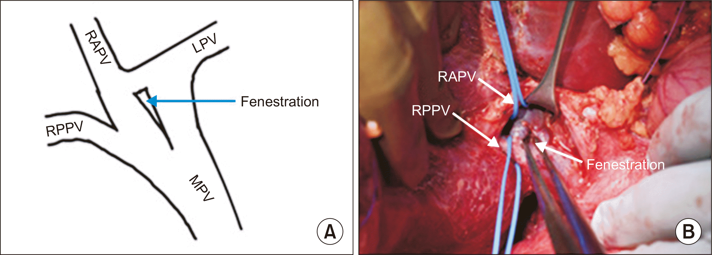
Anatomical variations of the portal vein (PV) occur during embryonic life, while the vitelline vein evolves into the PV [1]. Precise knowledge of these variations is necessary before planning donor liver hepatectomy. Many classifications of PV anatomical variants have been described, but PV fenestration has never been reported in the literature. Nakamura et al. [2] classified PV anatomy into five types A–E. Type A (92.5%) is the usual bifurcation of the right PV (RPV) and left PV (LPV). Type B (2.5%) is the trifurcation of the right anterior PV (RAPV), right posterior PV (RPPV), and LPV. Type C (2.5%) represents extra-parenchymal branching and type D (1.7%) intra-parenchymal branching of the RAPV from the LPV. In type E, the branches of segments 8 and 5 originate separately from the LPV. PV fenestration is an unreported anatomic variation in which a segment of a vessel divides into at least two channels that reunite into a single distal lumen. We report a case of PV fenestration incidentally detected during standard right donor hepatectomy (Fig. 1).
CASE REPORT
The study was approved by the Ethics Committee of Max Super Specialty Hospital, New Delhi, India (approval No. MHILDA00017). Informed consent from the patient was taken.
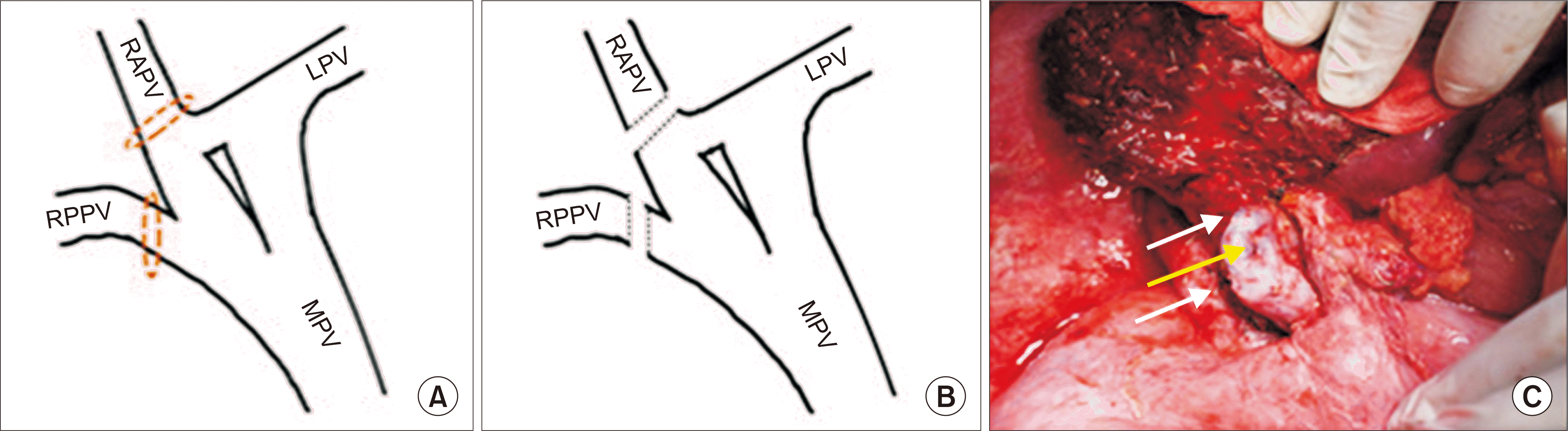
A 25-year-old male patient with no comorbidities planned for liver donation and underwent a routine preoperative evaluation before hepatectomy for a liver transplant. Computed tomography (CT) angiography and volumetry were performed to visualize the vascular anatomy of the hepatobiliary system. The PV anatomy appeared to be type C according to the classification by Nakamura et al. [2]. Intraoperatively, while performing RPV looping, we were able to obtain a single stump of the RPV (Fig. 2A). Since this finding was not correlated with the CT angiography images, we dissected further proximally and were able to see the confluence of the LPV with the RAPV and fenestration in the common trunk of the PV (Fig. 1A). The RPPV and RAPV were looped separately and clamped temporarily, and the ischemic line was marked (Fig. 1A). The donor underwent an unremarkable standard right lobe hepatectomy, with no adverse intraoperative events. We obtained a standard right lobe graft of 718 g, with two PVs, a single right hepatic artery, and a single right hepatic duct. The RAPV and RPPV were divided separately, and the stumps were closed without compromising the lumen of the PV trunk and retaining the PV fenestration (Fig. 3).
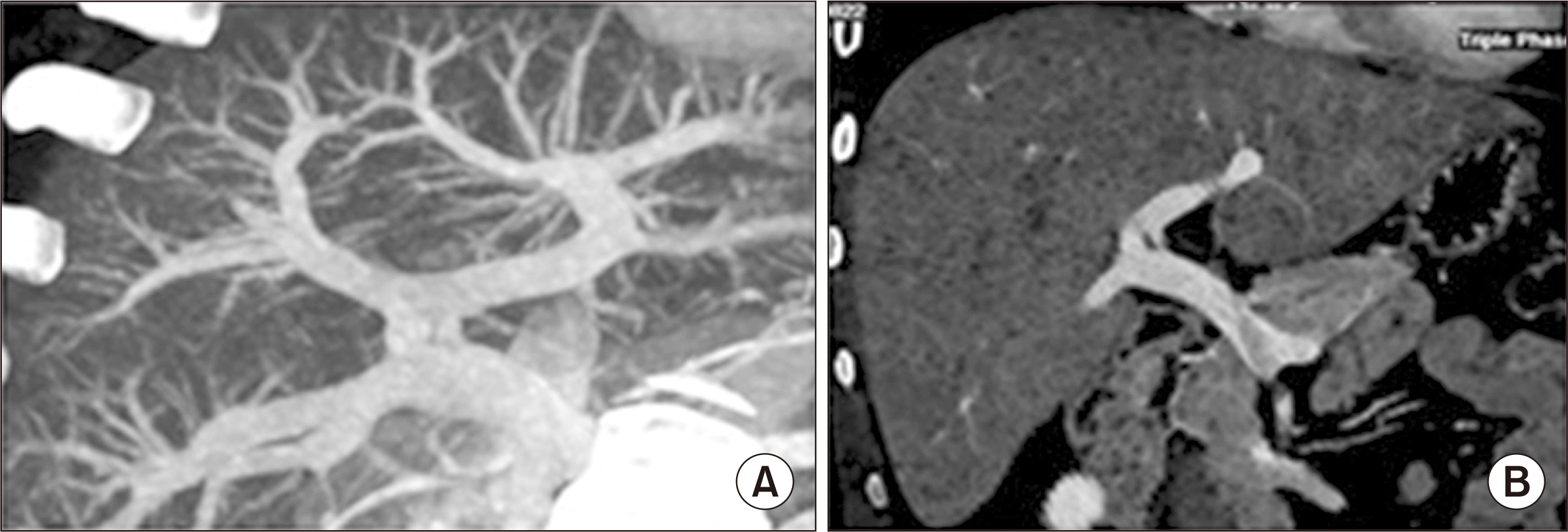
Quilt venoplasty was performed between the two PV orifices and circumferential fencing using a recipient liver PV graft on a back table bench [3]. PV reconstruction was performed in an end-to-end fashion using the recipient’s PV trunk. Both the donor and recipient remained stable during the postoperative period, without any complications. The donor and recipient were discharged on postoperative days 8 and 15, respectively. On reviewing the CT angiography images retrospectively, we found the fenestration in the PV trunk (Fig. 4).
DISCUSSION
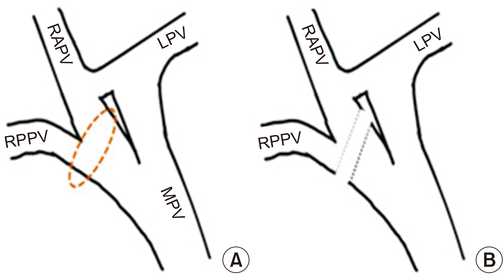
Vascular fenestration is an uncommon anatomic variation in which a segment of a vessel divides into at least two channels that reunite into a single distal lumen [4]. PV duplication is defined as two separated PVs that course upward to the porta hepatis and divide into segmental branches, whereas PV fenestration refers to a bifurcation that reunites distally [5]. Although duplication of the PV may present with portal hypertension, variceal bleeding, or duodenal obstruction, fenestration of the PV has no clinical consequences in healthy individuals [5]. However, in patients undergoing hepatectomy, this developmental anomaly can lead to catastrophic intraoperative events. A wrongly looped RPV through the fenestration below the confluence of the RAPV and LPV will lead to incorrect marking of the ischemic line, and if further divided, will compromise the portal flow to the remnant liver (Fig. 2). Looping the RAPV higher and RPPV separately will prevent these catastrophic events.
Anatomical variations of the PV were classified by Nakamura et al. [2] and Cheng et al. [6], but fenestration of PV has never been described in the literature. Fenestration of the internal jugular vein and its clinical consequences in neck dissection have been described in a few case reports [4]. Failure to identify PV fenestration can have devastating effects on the vascularity of the remaining liver parenchyma in a liver donor.
This case report depicts the importance of the correlation between intraoperative findings on CT angiography in liver donor hepatectomy and the existence of PV fenestration, which can lead to catastrophic events if not carefully observed.
 XML Download