

 PDF
PDF Citation
 Print
Print


Go to : 
INTRODUCTION
Liver transplantation (LT) is considered the most effective treatment for end-stage liver disease [1], and living-donor LT (LDLT) has been increasingly performed due to a shortage of liver grafts from deceased donors [2]. LDLT is performed based on the assumption that the liver has a rapid and abundant regenerative capacity [3]. In adult LDLT, liver regeneration is a crucial process to ensure donor safety and avoid hepatic dysfunction in recipients [4]. The success of LDLT depends on the capacity for remnant and graft liver regeneration in donors and recipients, respectively [5]. Studies have demonstrated that early liver regeneration occurs with a peak in DNA synthesis around postoperative day (POD) 7–10, and that future liver remnant regeneration has the strongest potential within 7 days after surgery [3,6]. Previous studies have suggested that presurgical factors including age, body mass index (BMI), and future remnant liver volume (FRLV) may influence liver regeneration [7,8].
It is well known that sarcopenia, defined as a reduction of skeletal muscle mass, is a clinical feature of metabolic dysfunction related to end-stage liver disease, and skeletal muscle mass has been found to be an important predictor of LT outcomes in recipients [9]. Additionally, a previous study reported that skeletal muscle mass, as measured on preoperative computed tomography (CT), had a significant positive correlation with the graft regeneration rate in LT recipients, and that low pretransplant skeletal muscle mass was associated with impaired graft regeneration after LDLT [10]. However, to the best of our knowledge, the association between skeletal muscle mass and remnant liver regeneration volume has not been studied in living donors.
Therefore, the purpose of the present study was to investigate the correlation between the preoperative skeletal muscle index (SMI) and remnant liver regeneration in living donors 7 days after right hemihepatectomy for LDLT, and to identify preoperative predictors of greater early remnant liver regeneration.
Go to :
METHODS
This retrospective study was approved by the Institutional Review Board of Asan Medical Center (IRB No. S2021-2223), which waived the requirement for informed consent.
Study Population
We retrospectively searched the LT database and identified 654 live donors who underwent right hemihepatectomy for LDLT between January 2017 and December 2018 at Asan Medical Center. The donors underwent CT of the liver as part of the preoperative work-up for the evaluation of anatomical variations in the hepatic vasculature and the estimation of liver volume, and postoperative CT at or around POD 7 to screen for complications and evaluate remnant liver regeneration. We excluded 129 subjects in whom the time between LT surgery and postoperative CT was shorter or longer than 7 days.
Computed Tomography Acquisition
Preoperative CT scans were performed using 64-slice (SOMATOM Definition; Siemens, Erlangen, Germany) or 128-slice (SOMATOM Definition AS+ or SOMATOM Definition Edge, Siemens) multidetector scanners. Unenhanced CT scans were obtained, followed by biphasic (hepatic arterial and portal venous phases) contrast-enhanced scans after the administration of 150 mL of iopromide (Ultravist 370, Bayer HealthCare, Berlin, Germany) for the anatomical mapping of the hepatic vasculature and CT volumetry. The scanning parameters were as follows: beam collimation of 64 or 128 slices by 0.6 mm; spiral pitch of 1; gantry rotation time of 0.5 seconds; 200 effective mAs with automatic exposure control (Care Dose 4D, Siemens); and 100 kVp. Images were reconstructed using a section thickness of 5 mm at 5-mm intervals. Postoperative CT scans for the screening of surgical complications and evaluation of remnant liver regeneration in living liver donors were obtained using monophasic (portal venous phase) contrast-enhanced CT with an ultra-low-dose protocol (40 effective mAs and 100 kVp). The other parameters were the same as those used for the preoperative scans.
Computed Tomography Volumetry
A semi-automated volumetric analysis of the donor liver was performed on CT images prior to surgery and on POD 7 using computer-aided in-house liver volumetry software. Volume measurements were performed by a board-certified abdominal radiologist (KWK) with 20 years of experience in hepatobiliary imaging. The preoperative volumetric analysis was performed sequentially in three steps. First, the initial liver outline was detected through sequential application of seeded region growing onto level-set speed images, which were generated as a map inversely proportional to the gradient magnitude [11]. Second, the level-set method was used to perform liver segmentation based on the initially detected liver contour [11], with Malladi’s level-set method being used for the level-set propagation [12]. After the manual correction of mis-segmentation, if any, the total liver volume (TLV) was obtained. Third, and finally, the radiologist repeatedly defined a resection line dividing the liver into the right hemiliver and the left hemiliver plus segment 1 on a few representative slices based on Cantlie's line or the long axis of the middle hepatic vein. FRLV was defined as the volumetric sum of the left hemiliver plus segment 1 under the assumption of right hemiliver donation [8]. The postoperative liver volume analysis was performed using the same techniques described above, except there was no need for the final step. The remnant liver regeneration volume on POD 7 was calculated by subtracting the FRLV measured on the preoperative CT from the remnant liver volume measured on the POD 7 CT.
Skeletal Muscle Measurement
A single axial preoperative CT image at the level of the inferior endplate of the third lumbar vertebra was processed for each patient. Abdominal CT image analyses were performed with a fully convolutional, network-based, automatic segmentation technique using a deep-learning system [13]. The skeletal muscle was assessed using artificial intelligence software (AID-U; iAID Inc., Seoul, Korea) [13]. CT images were automatically segmented to generate boundaries, and the total abdominal muscle area was measured. The skeletal muscle area (SMA; cm2), including all muscles on the selected axial image (i.e., psoas, paraspinal, transversus abdominis, rectus abdominis, quadratus lumborum, and internal and external oblique muscles) was demarcated using predetermined thresholds (–29 to 150 Hounsfield units). The SMI was normalized to stature by dividing the muscle area by the height squared, as follows: SMA (cm2)/height (m2).
Data Collection
Preoperative anthropometric measurements (body weight and height) and laboratory parameters (platelet count, prothrombin time, and serum levels of aspartate aminotransferase, alanine aminotransferase, total bilirubin, and albumin) were collected preoperatively. BMI was calculated as body weight (kg) divided by the square of the height (m2). Perioperative findings included operation time and blood loss. Information on the hospital stay duration and complications after donor hepatectomy was collected postoperatively.
Statistical Analysis
Continuous variables were expressed as mean±standard deviation, and categorical variables were expressed as frequencies with percentages. Correlations between remnant liver regeneration volume on POD 7 and other variables were analyzed using Pearson correlation coefficients. Stepwise multiple regression analysis was performed using the backward selection method, including all significant variables from the aforementioned analysis. Statistical significance was set at P<0.05, and statistical analyses were performed using IBM SPSS ver. 23 (IBM Corp., Armonk, NY, USA).
Go to :
RESULTS
Characteristics of the Study Population
A total of 525 donors (mean age, 28.9±8.3 years; 345 men and 180 women) were included in the analysis. The characteristics of the study population are summarized in Table 1. The mean FRLV, TLV, and FRLV-to-TLV ratio (FRLV/TLV) on preoperative CT images were 412.1±96.3 cm3, 1,164.5±239.4 cm3, and 35.3%±3.6%, respectively. The remnant liver regeneration volume on POD 7 CT images was 374.2±117.3 cm3. The mean SMI on preoperative CT images was 48.2±8.9 cm2.
Table 1
Characteristics of the study population
![]()
Correlation between Regenerated Liver Volume and Clinical Parameters
The remnant liver regeneration volume on POD 7 CT images was positively correlated with BMI (r=0.280, P<0.001), alanine aminotransferase (r=0.137, P=0.002), and SMI (r=0.322, P<0.001), and negatively correlated with age (r=–0.154, P<0.001) and the FRLV/TLV ratio (r=–0.261, P<0.001) (Table 2).
Table 2
Correlation between preoperative variables and remnant liver regeneration volume on POD 7
| Variable | Remnant liver regeneration volume on POD 7 (cm3) | |
|---|---|---|
|
|
||
| Correlation coefficient (r) | P-value | |
| Age (yr) | –0.154 | <0.001a) |
| Body mass index (kg/m2) | 0.280 | <0.001a) |
| Platelet count (×109/L) | 0.053 | 0.354 |
| Prothrombin time (INR) | 0.047 | 0.442 |
| AST (IU/L) | 0.067 | 0.125 |
| ALT (IU/L) | 0.137 | 0.002a) |
| Total bilirubin (mg/dL) | 0.061 | 0.166 |
| Albumin (g/dL) | 0.081 | 0.065 |
| FRLV/TLV ratio (%) | –0.261 | <0.001a) |
| Skeletal muscle index (cm2/m2) | 0.322 | <0.001a) |
![]()
Preoperative Predictors of Remnant Liver Regeneration
Stepwise multiple regression analysis showed that a high BMI (β=0.146, P=0.001), high SMI (β=0.228, P<0.001), young age (β=−0.091, P=0.025) and low FRLV/TLV ratio (β=−0.225, P<0.001) were predictors of greater remnant liver regeneration volume at POD 7 (Table 3, Fig. 1).
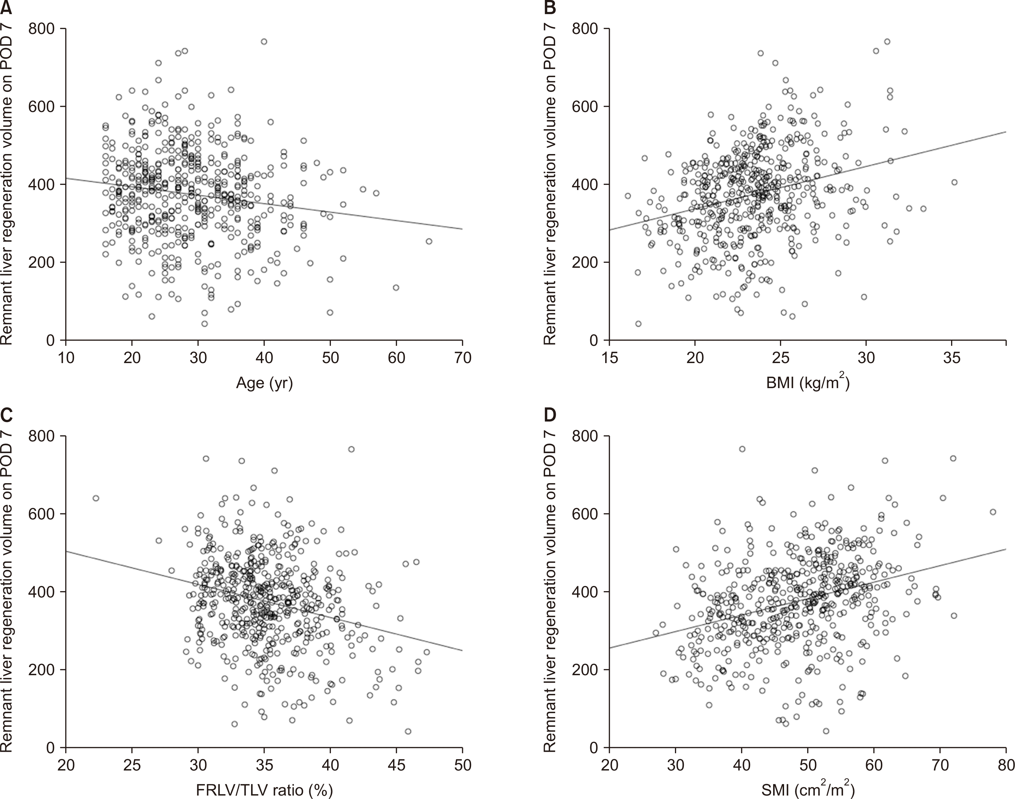 | Fig. 1Relationships between remnant liver regeneration volume on postoperative day (POD) 7 and preoperative variables, including age (A), body mass index (BMI; B), future remnant liver volume to total liver volume (FRLV/TLV) ratio (C), and skeletal muscle index (SMI; D) in living liver donors after right hemihepatectomy.
|
Table 3
Preoperative factors predictive of remnant liver regeneration volume on POD 7 by multiple regression analysis
![]()
Perioperative and Postoperative Outcomes of Living Donors
The remnant liver regeneration volume on POD 7 was not significantly correlated with the operation time (r=0.008, P=0.851), blood loss (r=0.042, P=0.335), or length of hospital stay (r=–0.017, P=0.699). None of the donors experienced post-hepatectomy hepatic failure or postoperative complications.
Go to :
DISCUSSION
In the present study, the early remnant liver regeneration at POD 7 of LT donors after right hemihepatectomy showed significant negative correlations with age and the FRLV/TLV ratio, while it was positively correlated with preoperative BMI and SMI. Additionally, the results of the present study indicated that high BMI and SMI, young age, and a low FRLV/TLV ratio were significant predictors of greater early remnant liver regeneration in living donors after LT.
Several studies have shown that advanced donor age may have a significant negative influence on early graft regeneration in LDLT recipients [7,10,14]. In a previous study by Ikegami et al. [14], early graft regeneration was significantly lower at POD 7 in older donors (≥50 years) than in younger donors (<30 years), in which an extended left hemiliver graft was used. Pravisani et al. [10] also reported that donor age showed a significant negative correlation with the graft regeneration rate in LDLT patients who received an extended left hemiliver graft. Additionally, Tanemura et al. [7] identified that donor age (≥50 years) was an independent factor correlated with impaired graft regeneration at POD 7 in both right and left hemiliver LDLT recipients. Meanwhile, the effects of donor age on remnant liver regeneration in living liver donors remains undetermined. Previous studies did not show significant differences in remnant liver regeneration volumes between younger and older donor groups [15,16]. However, Ono et al. [17] revealed that the remnant liver regeneration rate at POD 7 in living donors was impaired with increased age, especially after right hemihepatectomy. Similarly, the results of the present study showed that the remnant liver regeneration volume at POD 7 after right hemihepatectomy was negatively correlated with age in living donors. Aging is accompanied by a gradual decline in the regenerative capacity of hepatocytes, with a decrease in the cell cycle and an increase in autophagy and apoptosis [3].
In the present study, the remnant liver regeneration volume in LDLT donors after right hemihepatectomy showed a negative correlation with the preoperatively estimated FRLV/TLV ratio. This result is consistent with a previous study in Japan that included living donors who underwent left lateral sectionectomy or left hemihepatectomy [18]. In previous studies, lower FRLV or FRLV/TLV ratio were independent predictors of greater liver regeneration in living donors [4,8]. The results of the present study also demonstrated that a lower FRLV/TLV ratio was a positive predictor of early remnant liver regeneration after LDLT. The release of proinflammatory cytokines, such as interleukin-6 or tumor necrosis factor-α, initiates the regenerative process after resection [3,8]. Larger hepatic resections may lead to a greater concentration of cytokines, thereby promoting greater remnant liver regeneration [3,8]. Additionally, Gruttadauria et al. [8] reported that higher BMI was a predictor of greater remnant liver regeneration in living donors after right hemihepatectomy, which is in line with the results of the present study.
A limited number of studies have evaluated the influence of pretransplant skeletal muscle mass on liver grafts after LDLT [10,19]. Pravisani et al. [10] evaluated the correlation between recipients’ pretransplant skeletal muscle mass and liver graft regeneration in left hemiliver LDLT. In that study, decreased skeletal muscle mass was associated with a significantly lower graft regeneration rate in both sexes, and recipients’ pretransplant skeletal muscle indices showed a significant positive correlation with graft regeneration rates at 1 month after LDLT in men [10]. Miyachi et al. [19] investigated the effect of donors’ skeletal muscle mass and quality on recipients’ graft survival after LDLT. They found that high muscle mass and quality in male donors were independent protective factors for graft loss in recipients [19]. Unlike these previous studies, the present study focused on the association of donors’ preoperative skeletal muscle mass with remnant liver regeneration after right hemihepatectomy for LDLT. The results of the present study demonstrated a significant positive correlation between preoperative SMI and remnant liver regeneration, and identified high SMI as a significant predictor of early remnant liver regeneration in LT donors.
The present study has several limitations. First, this was a single-center retrospective study, and the results need to be prospectively validated in a multicenter trial. Second, because of the ethnic homogeneity of Koreans, the impact of race on remnant liver regeneration after LT remains unclear. Third, we did not evaluate muscle quality or strength. Because sarcopenia is defined as low muscle strength and low muscle quantity or quality [20], we could not analyze the relationship between the presence of sarcopenia and liver regeneration. Further studies may be required to assess the associations of muscle quality, muscle strength, and sarcopenia with liver regeneration. Fourth, since the present study focused on early remnant liver regeneration in donors after LDLT, we did not evaluate graft liver regeneration or graft survival outcomes in LT recipients.
In conclusion, high SMI and BMI, young age, and a low FRLV/TLV ratio may be predictors of greater early remnant liver regeneration in living donors after LDLT.
Go to :
 XML Download