

 PDF
PDF Citation
 Print
Print


INTRODUCTION
Lumbosacral transforaminal epidural steroid injection is a valuable therapeutic option for various lumbosacral pathologies, including radiculopathy caused by spinal stenosis or a herniated intervertebral disc. It is a target-specific approach in which a steroid can be intensively injected into the pathological site and around the dorsal root ganglion [1–4]. Because lumbosacral pathologies such as intervertebral disc herniation develop most frequently at the L4-5 and L5-S1 regions [5], 1st sacral transforaminal epidural steroid injection (S1 TFESI) may be a promising therapeutic option for treating the pain associated with compression of inflammation of the S1 nerve root [6].
S1 TFESI is generally performed under fluoroscopy or computed tomography (CT) guidance, but these techniques have some disadvantages, such as radiation exposure, space limitation, and high costs. Moreover, identification of the S1 posterior foramen is often difficult because it is smaller than the anterior foramen in the anteroposterior fluoroscopic view [7–9]. It may be more challenging in patients with intestinal gas overlap, obesity, severe spinal degeneration, osteoporosis, or anatomic variations [10,11]. Several researchers have suggested practical methods that may help predict the location of the S1 posterior foramen using the alignment of the lumbar facet joint [12,13]. Such methods may narrow down the possible location of the S1 posterior foramen in difficult cases, but they cannot facilitate its visualization.
Ultrasound-guide S1 TFESI is a practical and easily applicable bedside alternative to CT and fluoroscopy that can be performed without exposure to radiation. Lumbar transforaminal injections under fluoroscopy- and ultrasound-guided techniques have been shown to be comparable with reliable accuracy and feasibility in clinical settings [14–17]. Furthermore, the S1 posterior foramen being placed relatively close to the skin, can be easily detected via ultrasound [18,19]. However, poor visualization of the needle tip may lead to unwanted drug leakage from the epidural space or a visceral injury during injection [10,20]. Therefore, further research for a better understanding of the surface anatomy and sonographic features of the S1 foramen is necessary to conduct the procedure effectively and safely.
In this retrospective study, the authors aimed to evaluate the anatomical information in terms of the depth, width, length, and angle of the S1 posterior foramen, using three-dimensional CT (3D-CT) to improve ultrasound-guided S1 TFESI performance. And the demographic factors associated with imaging parameters were also identified.
Go to : 
MATERIALS AND METHODS
This study retrospectively reviewed 3D-CT images of the pelvis of 670 consecutive adult patients who underwent imaging between January 2019 and February 2021. This retrospective study was approved by the Institutional Review Board of Jeonbuk National University Hospital (No. CUH 2021-04-028) and the requirement for informed consent was waived.
The inclusion criteria were being aged 19–99 years and having underwent CT for disease assessment.
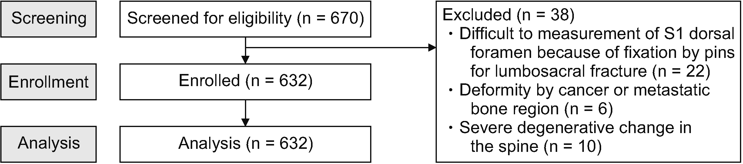
The exclusion criteria were the presence of S1 posterior foramen parameters that were difficult to measure owing to lumbosacral bone surgery, sacral deformity caused by primary or metastatic bone cancer, and presence of severe degenerative changes in the lumbosacral spine. The patient selection process is presented as a flow diagram (Fig. 1).
1. CT machines
Multi-slice Computed Tomography scanning was performed on a 128-slice dual-source CT scanner (Somatom definition flash, Siemens, Germany). Images of the pelvis were obtained by automatically selecting the patient’s optimal kV setting in 10 kV increments by balancing the radiation dose and the image contrast using Siemens’ CARE kV function.
2. Radiological measurement of the first sacral foramen
All parameters were measured using axial 3D-CT images of the pelvis. The vertebral level was determined by counting from the 5th lumbar vertebra in the caudal direction, and the largest diameter of the S1 posterior foramen was determined after identifying the first and second sacral bones. According to a previous study, lumbarization of the first sacral bone has a prevalence of 4.1%, which can result in incorrect counting of the vertebra resulting in misidentification of the first sacral foramen [21]. Therefore, the position of the 5th lumbar vertebra was confirmed with 3D-CT imaging.
Radiological measurements included the following (Fig. 2): S1 posterior foramen depth (S1D, mm), defined as the length from the posterior surface of the sacrum to the margin of the sacral canal; S1 posterior foramen width (S1W, mm), defined as the largest diameter of the S1 posterior foramen; S1 posterior foramen angle (S1A, °), defined as the angle between the S1 posterior foramen and the midsagittal line of the sacrum; and S1 posterior foramen distance (S1ds, mm), defined as the distance between the midsagittal line of the sacrum and the parallel line passing through the center of the S1 posterior foramen. To minimize measurement errors, two trained anesthesiologists measured each parameter, and the obtained values were averaged.
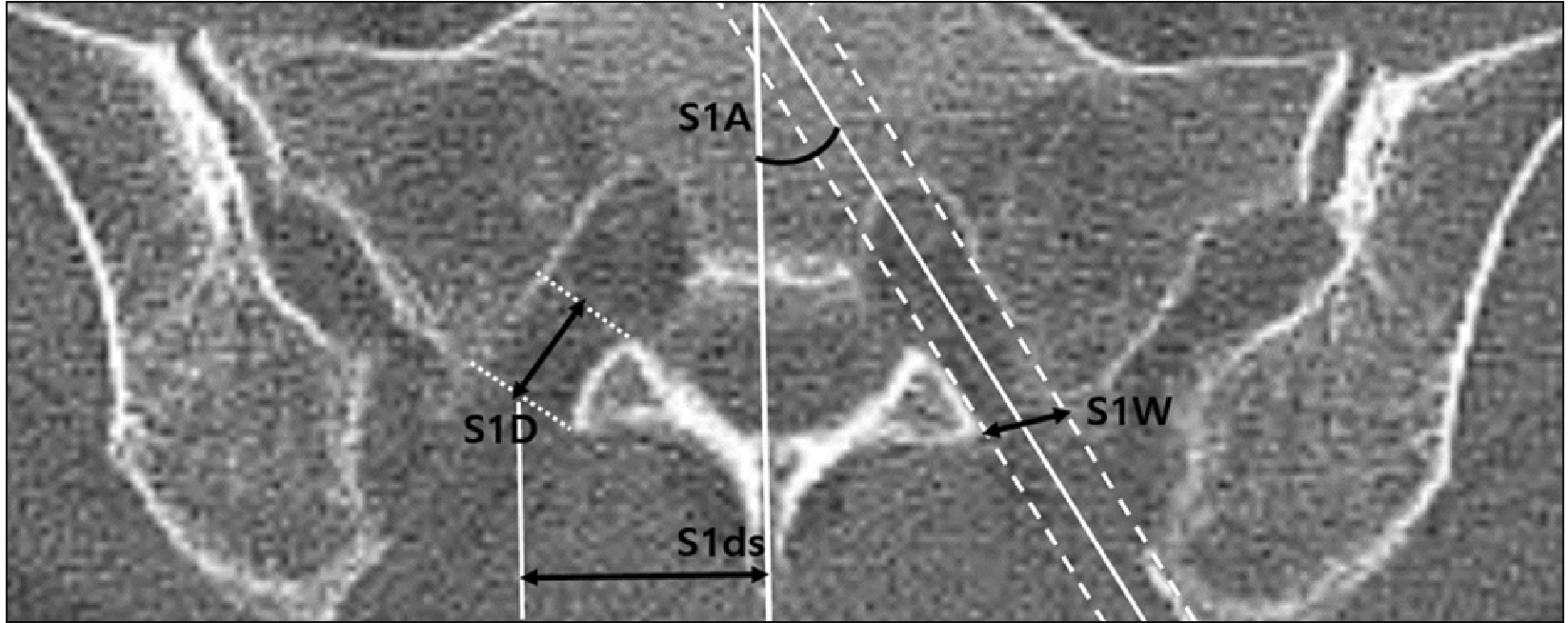 | Fig. 2Measured parameters using axial computed tomography at the level of the 1st posterior sacral foramen. S1D: 1st posterior sacral foramen depth defined as the length from the posterior surface of the sacrum to the margin of the sacral canal, S1W: 1st posterior sacral foramen width defined as the largest diameter of the S1 posterior foramen, S1A: angle of the 1st posterior sacral foramen defined as the angle between the S1 posterior foramen and the midsagittal line of the sacrum, S1ds: 1st posterior sacral foramen distance defined as the distance between the midsagittal line of the sacrum and the parallel line passing through the center of the S1 posterior foramen.
|
Demographic data, such as age, sex, height, weight, and body mass index (BMI), were evaluated to verify their effects on the measured values. The primary outcome assessed the S1D (mm), while the secondary outcomes included other measurements of the S1 posterior foramen features. Furthermore, we evaluated the correlation between the demographic data and S1 posterior foramen parameters was evaluated.
3. Statistical analysis
An independent sample t-test was performed to analyze continuous variables after the normality test. A linear regression model was used to analyze the potential demographic factors in relation to the measured values. Based on the regression analysis, the Bland–Altman method was used to identify the 95% limits of agreement. One-way analysis of variance was used to determine whether S1D was different among stratified groups according to patients’ heights. All statistical analyses were performed using IBM SPSS statistics for Windows, version 27 (IBM Co., Armonk, NY). All descriptive statistics are expressed as mean ± standard deviations. Statistical significance was set at P < 0.050.
Go to :
RESULTS
A total of 632 patients, 287 male and 345 female, were examined. Demographic data are shown in Table 1, and measurements of parameters are presented in Table 2.
Table 1
Demographic data
![]()
Table 2
S1 posterior sacral foramen parameter values
| Parameters | Total (n = 632) | Male (n = 287) | Female (n = 345) | P value |
|---|---|---|---|---|
| S1D (mm) | 11.0 ± 1.9 (4.9–17.5) | 11.5 ± 1.9 (7.1–17.5) | 10.6 ± 1.8 (4.9–17.4) | < 0.001* |
| S1W (mm) | 8.5 ± 1.6 (3.7–19.1) | 8.3 ± 1.6 (3.7–19.1) | 8.6 ± 1.6 (4.7–15.1) | 0.066 |
| S1A (°) | 29.2 ± 4.9 (12.5–45.8) | 28.2 ± 4.8 (12.5–44.7) | 30.1 ± 4.9 (18.5–45.8) | < 0.001* |
| S1ds (mm) | 23.5 ± 2.8 (16.2–32.2) | 24.1 ± 2.9 (17.8–32.2) | 22.9 ± 2.6 (16.2–32.2) | < 0.001* |
![]()
The mean S1D of the males was significantly greater than that of the females (11.5 ± 1.9 mm vs. 10.6 ± 1.8 mm; P < 0.001, two-tailed t-test). S1A and S1ds values also significantly differed from each other. The mean S1A values of males were smaller than those of females (28.2 ± 4.8° vs. 30.1 ± 4.9°; P < 0.001), whereas the mean S1ds values of the males were larger than those of the females (24.1 ± 2.9 mm vs. 22.9 ± 2.6 mm; P < 0.001). However, no difference regarding S1W values was observed.
Univariate linear regression analysis revealed a significant correlation between demographic variables and S1D (Fig. 3). The Bland–Altman plots are presented in Fig. 4. Age, height, and weight were significantly correlated, but no significant correlation with BMI was identified. The significantly associated variables were integrated into a multivariate linear regression analysis; height was the only significant predictor for S1D (β = 0.318; 95% confidence interval [CI], 0.020 to 0.101; P = 0.004) (Table 3).
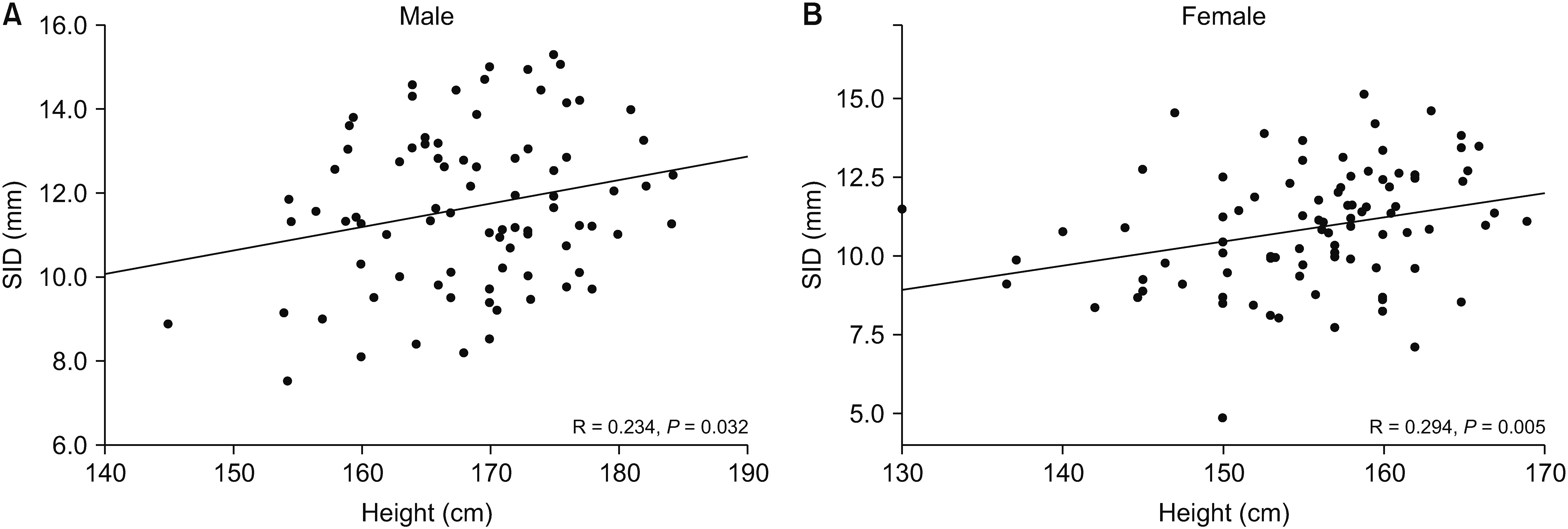 | Fig. 3Relationships between S1D and the patients’ height. (A) Male. (B) Female. S1D: 1st posterior sacral foramen depth.
|
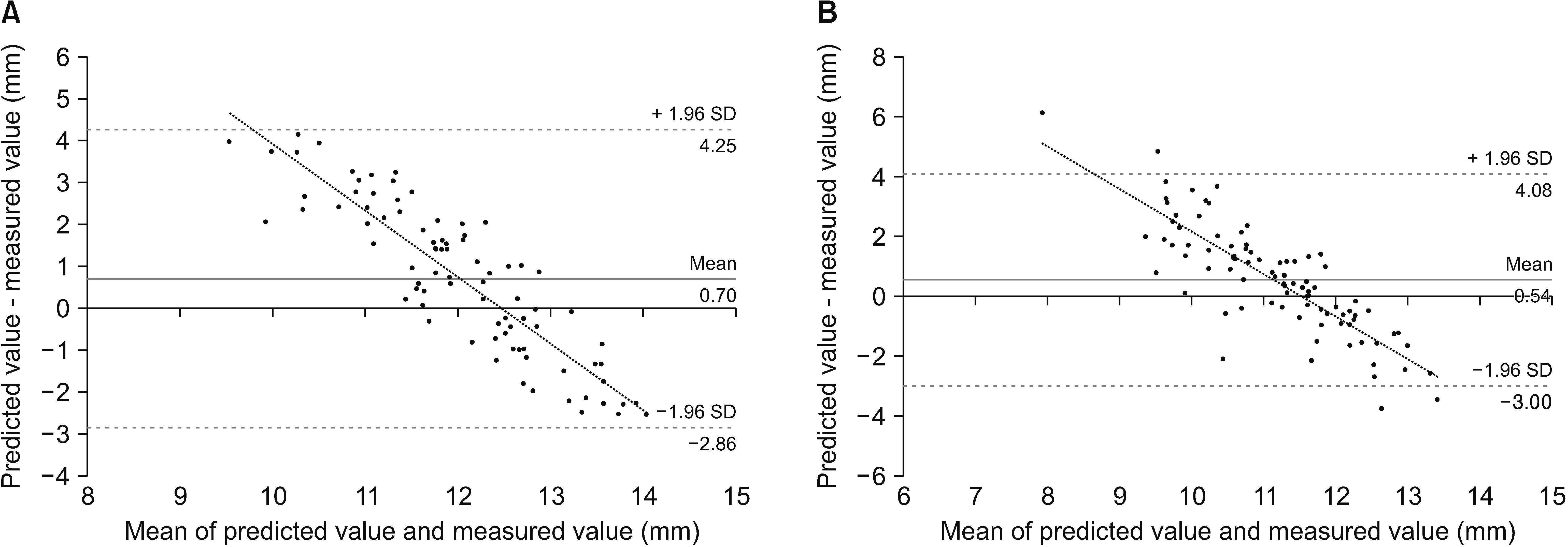 | Fig. 4Bland–Altman plot comparing predicted and measured values of S1D. (A) Male. (B) Female. S1D: 1st posterior sacral foramen depth, SD: standard deviation.
|
Table 3
Correlation between the depth of the S1 posterior sacral foramen and demographic variables on linear regression analysis
| Variable | Univariate analysis | Multivariate analysis | |||||
|---|---|---|---|---|---|---|---|
| β | 95% CI | P value | β | 95% CI | P value | ||
| Age (yr) | –0.191 | –0.029 to –0.012 | < 0.001* | –0.058 | –0.023 to 0.011 | 0.489 | |
| Height (mm) | 0.333 | 0.037 to 0.091 | < 0.001* | 0.318 | 0.020 to 0.101 | 0.004* | |
| Weight (kg) | 0.222 | 0.012 to 0.058 | 0.003* | –0.018 | –0.034 to 0.028 | 0.855 | |
| BMI (kg/m2) | 0.007 | –0.081 to 0.089 | 0.927 | ||||
![]()
We further investigated whether height influences S1D. In the males, S1D was significantly reduced in participants with a height < 155 cm compared with those with a height ≥ 175 cm (9.8 ± 1.8 vs. 12.2 ± 1.6 mm, P = 0.006). In the females, S1D was significantly reduced in participants with a height < 155 cm compared with those with a height ≥ 165 cm tall (10.1 ± 1.9 vs. 12.0 ± 1.0 mm, P = 0.019) (Table 4).
Table 4
Depth of the S1 posterior sacral foramen stratified by height
![]()
Other parameters and demographic variables were analyzed by linear regression analysis. S1ds showed a significant correlation with height (β = 0.278; 95% CI, 0.035 to 0.112; P < 0.001), but no significant correlation with age, weight, or BMI. S1W and S1A showed no significant relationship.
Go to :
DISCUSSION
Ultrasound imaging has been suggested as an important alternative to fluoroscopy for various spinal interventions in that it is easy to apply without space limitations and free from radiation exposure. Moreover, it can visualize almost all spinal structures, including the musculature, bony surfaces, facet joints, and intervertebral foramen. Ultrasound imaging also helps in visualization of S1 posterior foramen in those with a relatively clear sonoanatomy [22], and its feasibility compared with that of conventional fluoroscopy has been validated [6,14–17]. However, the main drawback of ultrasound-guided S1 TFESI is that the needle tip may not be well visualized inside the sacral foramen [20,23]. Besides, excessive needle advancement into the sacral foramen may cause drug leakage through the anterior sacral foramen or a pelvic organ injury, resulting in a failed procedure [10,20].
For minimizing these kinds of unexpected side effects, many studies have suggested anatomical information via direct measurement using cadavers [7,24] or imaging studies [11,25]. Watanabe et al. [25] analyzed the relationship between the S1 posterior foramen and S1 nerve root by 3D-CT to prevent nerve root damage during a sacral surgical approach. They reported that the average distance from the posterior surface of the sacrum to the S1 nerve root was 13.9 mm. Although direct comparison of those results with the current study is limited due to the methodological discrepancy, the measured parameter approximately corresponds to the S1D of the current study. This study revealed similar results, in which S1D is 11.0 mm. However, the wide range of S1D, as presented in the current study (min–max; 4.9–17.5), should be taken into consideration to prevent accidental nerve and organ injury in clinical practice. Another study investigated various parameters such as the largest diameter of the S1 foramen as well as the angle of the S1 foramen [11]. The comparison of the measured parameters between the present study and previous studies are shown in Table 5.
Table 5
Comparison of morphological parameters
| Reference | S1D (mm) | S1W (mm) | S1A (°) | S1ds (mm) |
|---|---|---|---|---|
| The current study |
Male (n = 287) 11.5 ± 1.9 Female (n = 345) 10.6 ± 1.8 |
Male (n = 287) 8.3 ± 1.6 Female (n = 345) 8.6 ± 1.6 |
Male (n = 287) 28.2 ± 4.8 Female (n = 345) 30.1 ± 4.9 |
Male (n = 287) 24.1 ± 2.9 Female (n = 345) 22.9 ± 2.6 |
| Arman et al. [7] | N/A |
Adult (n = 100) 7.97 ± 1.89 |
N/A | N/A |
| Hwang et al. [11] | N/A |
Male (n = 202) 7.1 ± 0.7 Female (n = 214) 7.1 ± 0.7 |
Male (n = 202) 26.4 ± 3.4 Female (n = 214) 26.2 ± 3.3 |
N/A |
| Bagheri and Govsa [24] | N/A |
Adult (n = 87) 7.97 ± 1.89 |
N/A | N/A |
![]()
The measured values of the present study suggest some interesting facts for pain physicians, which are as follows. (1) The patient’s height is an independent predictor of S1D, indicating the need to adjust needle insertion depth congruently to patient’s height during ultrasound-guided S1 TFESI. In both male and female with a height < 155 cm, the needle tip should not be inserted more than approximately 10 mm from the posterior margin of the S1 posterior foramen to prevent potential risk to the patients. Moreover, the Bland–Altman plot showed a negative correlation, suggesting that S1D can potentially be overestimated in shorter patients but underestimated in taller ones. (2) S1ds show significant correlation with height. This suggests that S1ds will also increase with the patients’ heights, as the vertebral cross-section area has a positive correlation with the height as well as study by Oura et al. [26]. In this respect, the depth of the posterior foramen is also expected to increase with the patient’s height due to bone growth because it is an estimate of the length of a bone structure (3). However, S1A has shown no significant relationship which would explain that the growth ratio of width and depth in vertebral body growth is almost constant and therefore has barely any effect on the angle.
Fluoroscopy remains the gold standard for spinal interventions, and ultrasound imaging has some technical limitations, including difficulty in identifying the correct epidural spread of drugs and negative intravascular uptake. Park et al. [10] suggested a novel method that combines both ultrasound imaging and fluoroscopy for S1 TFESI. They used ultrasound to guide the initial needle placement and subsequently used fluoroscopy to confirm correct needle placement and drug spread. However, further investigations are urgent for improving the accuracy, safety, and applicability of ultrasound-guided procedures. Thus, to the best of the authors’ knowledge, this is the first study to measure depth of the S1 posterior foramen using 3D-CT. The data of this study will provide useful information for minimizing the disadvantage of ultrasound-guided S1 TFESI by facilitating the prediction of an appropriate needle insertion depth. However, this study has a few limitations. First, the measured parameters may not reflect the actual values as they depend on the angulation and rotation axis of the CT scan, which may not always focus adequately on the sacral foramen. The 3D-CT scan can be used to prevent such errors only to some extent. Second, the possibility of selection bias cannot be excluded because the sample consisted of mostly elderly patients, meaning it did not adequately reflect all age groups. In addition, all patients included in this study were Koreans, and racial differences are possible. Third, the data obtained in this study are from analysis of radiographically measured data. Therefore, additional follow-up studies applying the data are needed for clinical practice. To obtain more clinically valuable data, multi-center studies of patients of different age groups and races are needed.
In conclusion, this study evaluated various anatomical parameters of the S1 posterior foramen, whereby the results indicated the importance of needle insertion depth adjustment corresponding to a patient’s height for a successful ultrasound-guided S1 TFESI.
Go to :
 XML Download