

 PDF
PDF Citation
 Print
Print


INTRODUCTION
Fibromyalgia (FM) is a chronic pain condition that comprises a widespread pain index (WPI) and emotional distress, fatigue, sleep disorders, depression, catastrophizing thinking related to pain, and cognitive compromise. Although its pathophysiology remains unclear, several studies have found altered activations of several brain regions, such as the thalamic nuclei, somatosensory cortices, anterior cingulate, and the insular and prefrontal cortices [1–3]. A review of structural and functional rearrangements in chronic pain indicates more active neural networks in pain, including the thalamus, anterior cingulate cortex (ACC), primary and secondary somatosensory cortices (S1 and S2, respectively), and the insular cortex [4].
According to functional magnetic resonance imaging (fMRI) data, the ventrolateral-periaqueductal gray matter (PAG) has indirect connections with central lateral and medial pathways, the ACC, and upper pons/medulla [5]. The PAG relates to somatosensory and affective-attentional pain components [2,6], with downward projections to spinal dorsal horn neurons to modulate pain transmission information [7]. These pathways constitute the descending pain modulatory system (DPMS), whose dysfunction is frequent in chronic pain syndromes [8].
The conditioned pain modulation test (CPM-test) assesses the function of the DPMS based on the "pain suppresses pain" phenomenon [9]. The CPM-test activates a cortical spinal-bulb-spinal loop responsible for diffuse noxious inhibitory control. The stimulation parameters, test sites, and study population influence the reliability of the CPM-test and conditioning stimuli [10]. A systematic review found fifty percent of intersession reliability measured by intraclass correlation coefficient (ICC) rated as good (ICC 0.60 to 0.75) or excellent (ICC > 0.75) [10]. The CPM-test permits the evaluation of the DPMS function. Its dysfunction in FM has been correlated with the serum biomarkers of neuroplasticity, such as the brain-derived neurotrophic factor and S100-B protein [11]. Furthermore, the dysfunction of DPMS is related to hyperinhibition at the cortical level and increased scores in the Central Sensitization Inventory (CSI) [12].
Resting-state functional magnetic resonance imaging (rs-fMRI) allows the evaluation of the resting-state functional connectivity (rs-FC) between different brain regions. This is a practical approach when investigating the existence of a specific brain’s functional signature and coherence patterns associated with specific patient groups across chronic pain conditions [13]. Regions with synchronous activity tend to correlate when blood oxygen level-dependent (BOLD) activity is measured. However, studies have found no information on the intrinsic FC of pain processing areas with the severity of dysfunction of the DPMS.
FM is a disease associated with central sensitization syndrome (CSS), likely related to a maladaptive function of the neural networks involved in pain processing. However, a gap in the literature persists regarding the understanding about the connection between pain processing brain networks and the modulation of the pain by the DPMS. Thus, the authors aimed to answer the following questions: (i) to examine whether there are differences in the rs-FC pattern between the primary somatosensory cortex and pain processing areas in patients who do/do not respond to the CPM-test, and (ii) to evaluate whether the differences in primary somatosensory cortex rs-FC patterns in patients who do/do not respond to the CPM-test are related to pain, sleep quality, central sensitization, and the impact of FM on quality of life. It was hypothesized that the connection between the networks involved in the pain neuromatrix and the DPMS might be a suitable marker for identifying patients with more severe clinical symptoms.
MATERIALS AND METHODS
The Institutional Review Board (IRB, CAAE 2018-0353) at the Hospital de Clínicas de Porto Alegre (HCPA) approved the protocol of this cross-sectional study, which was conducted according to the Declaration of Helsinki. Participants provided verbal and written informed consent before participating and did not receive payment in exchange for their participation. Recruitment was undertaken from January 2018 to December 2019.
1. Recruitment, inclusion, and exclusion criteria
We included 33 adult females, all right-handed, ages 18 to 65, who could read and write. Patients from the Pain Outpatient Clinic of the HCPA were invited to the study. Other participants became aware of the study through local newspaper publicity. All included subjects met the diagnostic criteria of FM by the standard assessment protocol criteria for the diagnosis of FM—according to the American College of Rheumatology (ACR) 2016 criteria [14]—which was applied by senior physicians with more than fifteen years of experience in chronic pain care. Furthermore, they had to demonstrate daily disability for routine activities due to FM for the three months preceding enrollment. In addition, they needed to report a score of at least six on the numerical pain scale (NPS, 0–10).
We did not include people who had a history of rheumatoid arthritis, lupus, or any other autoimmune, neurologic, or oncological disease, any uncompensated clinical disease (e.g., ischemic heart disease, chronic kidney disease, or hepatic disease), pregnancy, or illegal drug use in the last six months. People with contraindications to fMRI due to the presence of a metal brain implant (e.g., aneurysm clip), a cardiac pacemaker, a cochlear prosthesis, metal fragments or prostheses, or claustrophobia were also excluded.
2. Sample size justification and power of analysis
A prior sample size was estimated, expecting a large effect size (ES, f squared, f2 0.35) for multiple regression analysis, allowing for five predictors. For error types I and II, which had values respectively of 0.05 and 0.20, the estimation showed that the sample size should be 31 patients (Post-hoc Statistical Power Calculator for Hierarchical Multiple Regression: https://www.danielsoper.com/statcalc/calculator.aspx?id=17) [15]. To guarantee the study’s power, it was decided to include 33 patients. However, the outcome variable (i.e., left S1-PAG rs-FC) showed a skewed distribution. So, a generalized linear model (GLM) was used, and the power of the study was calculated using post-hoc analysis. It revealed a power of 89.562% in a sample of 33 patients based on an adjusted ES of the GLM of 0.56 for (χ2 = 10.41) for a type I error of 0.05.
3. Instruments and assessments
All psychological tests used in this study have been validated for the target population. The following instruments were used to assess psychological symptoms and sleep quality: the Central Sensitization Inventory [12], the Beck Depression Inventory-II [16], the Pain Catastrophizing Scale [17], the State-Trait Anxiety Inventory [18], and the Pittsburgh Sleep Quality Index [19].
4. Outcomes assessment
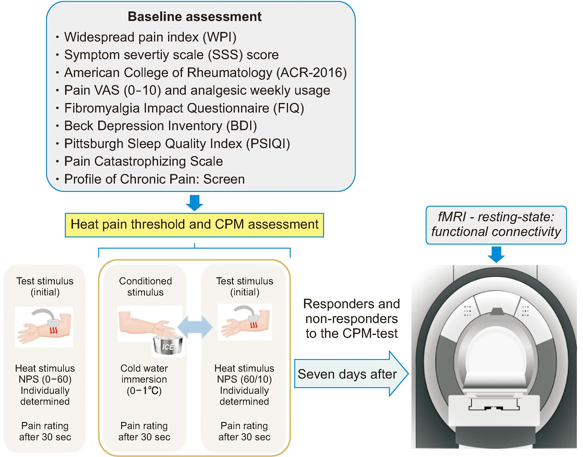
The dependent variables were the rs-FC in the left S1 and the PAG. The main interest factor was the DPMS function, which was assessed through a CPM-test—expressed by the difference in score on an NPS (0–10) produced by the Quantitative Sensory Testing (QST) before and concurrently with the CPM-test induced by immersion of the dominant hand in cold water (zero to 1°C). The CPM-test permits the classification of non-responders and responders. Independent variables included age, years of education, pain catastrophizing, pain-related disability, quality of sleep, pain scores, anxiety, number of psychiatric diagnoses, use of analgesics, and heat pain threshold. The timeline of assessments is shown in Fig. 1.
5. fMRI acquisition, processing, and analysis
1) Imaging acquisition
Structural and functional images were acquired with a 3T scanner (Ingenia 3.0T; Phillips, Best, Netherlands) using a 15-channel head coil. The rs-fMRI was a single-shot T2*-weighted fast-field echo, echo-planar imaging sequence (repetition time [TR] = 2,000 ms, repetition time [TE] = 30 ms, Matrix = 80 × 80, field of view [FOV] = 240 mm, flip angle = 90, 3 × 3 × 3.5 mm voxel size, 36 slices in ascending order with 0.35 mm gap) with 300 volumes, totaling 10 minutes. Anatomical references were acquired using a T1-weighted gradient-echo sequence (TR/TE = 8.5/3.9 sec, Matrix/FOV = 240/240 mm, flip angle = 8°, 0.94 mm isotropic voxel size, 200 sagittal slices).
All rs-fMRI processing was done using CONN18 (www.nitrc.org/projects/conn) [20], which is a toolbox that uses SPM12 (www.fil.ion.ucl.ac.uk/spm) commands over MATLAB (MathWorks, Natick, MA) [21]. The pipeline starts with preprocessing steps (segmenting anatomical volumes in gray matter, white matter, and cerebrospinal fluid, realigning and unwarping, normalizing, and smoothing the functional volumes). CONN’s default denoising pipeline combines two general steps: linear regression of potential confounding effects in the BOLD signal and temporal band-pass filtering. The BOLD signal was extracted from each region of interest (ROI) based on the Harvard-Oxford Atlas predefined within CONN standard settings. Other ROIs were added based on the relevant areas related to the pain connectivity literature (described in more detail below). Factors identified as potential confounding effects on the estimated BOLD signal were estimated and removed separately for each voxel, subject, and functional run/session using Ordinary Least Squares regression to project each BOLD signal time series to the subspace orthogonal to all potential confounding effects. The temporal preprocessing started with a regression out of the BOLD signal to control motion artifacts and residual physiological noise, then controlling the confounding variables (first-level covariates, the BOLD signal from white matter, cerebrospinal fluid), realignment parameters, scrubbing, and the CompCor approach [22]. Twelve potential noise components were defined from the estimated subject-motion parameters to minimize motion-related BOLD variability, including three translation parameters and three rotation parameters plus their associated first-order derivatives. A variable number of noise components (one for each identified outlier scan during the outlier identification preprocessing step) were used as potential confounding effects to remove any influence of these outlier scans on the BOLD signal. Temporal frequencies below 0.008 Hz or above 0.09 Hz were removed from the BOLD signal to focus on slow-frequency fluctuations while minimizing the influence of physiological noise, noise from head motion, and other noise sources [20].
Then, the BOLD signal from each individual was filtered (0.008–0.09 Hz). Filtering was implemented using a discrete cosine transform windowing operation to minimize border effects, having been performed after regression to avoid any frequency mismatch in the nuisance regression procedure. Each rs-FC correlation measure was calculated from all ROIs concerning each other in an FC matrix. Finally, after extracting the average time course from each ROI, rs-FC was estimated from Fisher Z scores.
3) rs-FC analysis
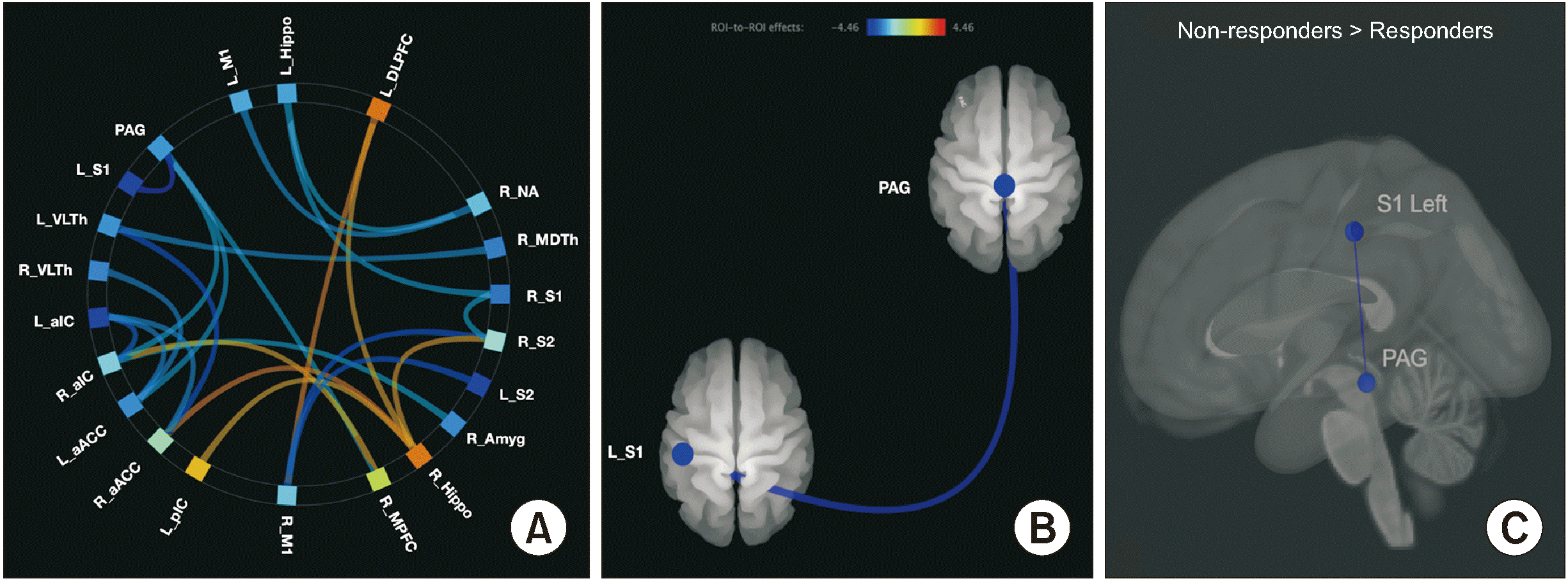
Spherical ROIs with MNI (Montreal Neurological Institute) coordinates [23] were chosen as predefined ROIs based on the Harvard-Oxford Atlas using the CONN-fMRI Functional connectivity toolbox [24]. The radius used was 6 mm for the PAG and 10 mm for the remaining areas. ROIs were acquired from prior studies as seeds. Together, they were used to create rs-FC maps of specific networks related to chronic pain and FM for each subject, with rs-FC being measured using the following source seeds (see Table 1 for coordinates): PAG [6], left (L) and right (R) primary and secondary somatosensory cortex (L_S1, R_S1, L_S2, R_S2) [25], left and right primary motor cortex (L_M1, R_M1) [26], left and right dorsolateral and ventromedial prefrontal cortexes (L_DLPFC, R_DPLFC, L_MPFC, R_MPFC) [27,28], left and right anterior and posterior insular cortexes (L_aIC, L_pIC, R_aIC, R_pIC), left and right ACC (L_ACC, R_ACC) [25], left and right ventrolateral and mediodorsal thalamus (L_VLTh, R_VLTh, L_MDTh, R_MDTh) [29,30], left and right hippocampus (L_Hippo, R_Hippo) [31], amygdalae (L_Amyg, R_Amyg) [32], and nucleus accumbens (L_NA, R_NA) [33]. After calculating the subjects’ FC map, the second-level comparison was performed through an ROI-to-ROI analysis. The threshold for bidirectional explorations of FC was P ≤ 0.001 (i.e., positive and negative associations). The exploratory analysis results were considered significant if they remained after correction for multiple comparisons (false discovery rate [FDR] ≤ 0.05).
6. Assessment of widespread pain index, pain catastrophizing, disability due to pain
1) For the FM diagnosis, the ACR [14] criteria were applied by a physician. The scale for FM symptoms ranges from 0 to 3 and is composed of the WPI and the modified severity scale (SS scale) [14].
2) The Fibromyalgia Impact Questionnaire (FIQ) [34] was used to evaluate the impact of symptoms on quality of life. The FIQ consists of 10 domains and items comprising questions to assess the patient’s ability to perform routine activities of daily living, fatigue, morning, stiffness, mood, anxiety, and depression. Higher scores indicate the worst conditions, and the maximum score is 100 [34].
3) The Pain Catastrophizing Scale (BP-PCS) [17] is a self-administered questionnaire comprising thirteen items to evaluate the presence of pain catastrophizing thoughts. It is composed of a 0–4 score (zero being "not at all" and four being "all the time"). Furthermore, it has subscales divided into the following dominions: magnification, rumination, and helplessness. The sum of all items computes its total score, ranging from 0 to 52 points [17].
4) The Central Sensitization Inventory (CSI-BP) [12] assesses central sensitization symptoms (CS). Higher scores are indicative of severe symptoms. It has 25 items and a total score that ranges from 0 to 100, evaluating the presence of physical signs, emotional distress, headache, and urological symptoms. Part B of the test assesses psychiatric and neurological disorders related to CS [12].
5) The Pittsburgh Sleep Quality Index (PSIQI) was used to measure the quality and pattern of sleep over the previous month [19].
6) The Beck Depression Inventory (BDI-II): this tool was used to evaluate the severity of depressive symptoms [16].
7) The State-Trait Anxiety Inventory (STAI) is a short and adapted version that was applied to evaluate state/trait anxiety. The score ranges from 13 to 52 on the state-anxiety scale and from 12 to 36 for the trait-anxiety scale. Higher scores denote higher levels of anxiety [18].
7. Psychophysical measures
1) The heat pain threshold test (HPT) was assessed by QST. The QST device was developed by the Biomedical Engineering Department at the Hospital de Clínicas de Porto Alegre (Porto Alegre, Brazil) in partnership with the Laboratory of Pain & Neuromodulation. This device has been validated and used in previous studies [35]. The QST-test is based on the method of limits with a computer Peltier-based contact thermode (30 × 30 mm2) attached to the skin on the ventral aspect of the subject’s non-dominant forearm. A computerized version of the thermode determines the HPT on the volar side of the non-dominant forearm. The heat is set at 32°C. The thermode heats up at a rate of 1.0°C/second to 52°C. The HPT is the minimum heat stimulus to induce pain, and it was obtained by the average of three assessments performed with an inter-stimulus interval of 40 seconds [35]. The thermode (Heat Pain Stimulator–1.1.10, Brazil) was manufactured by the Biomedical Engineering Department at the authors’ institution and validated in a previous study [35].
2) The CPM-test evaluates patients’ endogenous DPMS by the psychophysical paradigm of conditioned pain modulation. It was assessed by the same computerized version of the thermode described to determine the HPT. On the non-dominant forearm, three QST assessments were conducted with a 40-second interstimulus gap. Firstly, the temperature at the thermode site was recorded for patients’ reported scores of 6/10 (NPS, 0–10) by the QST. Thus, the temperature used in the test stimulus was calculated as the temperature average (T0). Secondly, the QST was introduced in the non-dominant arm 30 seconds after patients immersed their dominant hand into the water at a temperature of around 1°C (Cold Pressor Test, CPT). So, they reported the pain score on the NPS (0–10) in the non-dominant arm produced by the set temperature to a score of 6/10 on the NPS (T1). Thirdly, the CPM-test index was calculated, consisting of the difference between the pain score on the NPS (0–10) during T1 and the score of 6/10 on the NPS (T0). Non-responders would have a difference in the count on the NPS (T1 minus T0) equal to zero or higher, whereas responders would have a difference below zero [36]. The magnitude of the CPM effect depends on the sensory modality used to deliver the conditioning, test stimuli, and the body area tested [10]. According to a meta-analysis, the CPM-test with a thermal test stimulus showed reliability for repeatability by an ICC ranging from fair to excellent (ICC 5 0.53; ICC 5 0.64; and ICC 5 0.83) [10]. The CPM-test has been proposed as a reliable prognostic factor in experimental and clinical pain studies [10].
8. Sociodemographic characteristics, health status, medicine use, psychiatric diagnoses, and pain score
1) Demographic data and medical comorbidities were evaluated using a standardized questionnaire. Psychiatric diagnoses were assessed by the Mini-International Neuropsychiatric Interview (MINI) [37]. Weekly analgesic intake in the last three months was recorded. They were dichotomized as < 4 times per week or > 4 times per week.
2) The visual analog scale (VAS) is a visual scale for pain score assessment based on millimeters, which ranges from "no pain" (zero) to “the worst possible pain" (100 mm). Patients had to score their worst pain during the last 24 hours.
9. Efforts to address potential sources of bias
Subjects were chosen based on pre-set criteria evaluated by two researchers with clinical experience in outpatient treatment for chronic pain. Two evaluators specifically trained for performing all assessments applied standardized protocols (e.g., the CPM-test) and questionnaires.
10. Statistical analysis
Categorical and continuous variables were summarized using conventional descriptive statistics. Continuous variables were compared with t-tests for independent samples, while chi-square or Fisher’s exact tests were used for categorical variables. The Shapiro–Wilk test was used to assess normality. The univariate analysis presented in Table 2 and the Spearman correlation analysis (Table 3) were used to identify potential confounding variables in the relationship between the left S1-PAG rs-FC according to responders and non-responders to the CMP-test. The criterion for a variable to be included and retained in the GLM was a P value equal to or less than 0.05. The multivariate GLM included the following variables: psychiatric diagnosis, central nervous system medication (i.e., antidepressant and anticonvulsant), central sensitization, catastrophizing pain thinking, sleep quality, and the impact of FM symptoms on quality of life. One variable was included at a time in the model to select a minimum number of variables, considering that the sample size was small. However, the final GLM model comprises all variables retained in the final model. The forwarding method was used to generate the best model GLM based on the lower Akaike’s Information Criteria (AIC). This choice was based on the idea that the AIC method might penalize the model when additional parameters are added. In this case, several possible models were created, and AIC was used to compare them. So, the models were run one by one, and thus the authors compared their AIC: "regress response on predictor A", "regress response on predictor B", "regress response on predictor C", etc. The model’s goodness of fit, according to the lower AIC scores, was used to define the final model. When the AIC of models were compared with psychiatric diagnosis or psychotropic medications, these variables did not show a statistically significant association; they increased the AIC score and produced a worse fit model. Hence, they were excluded from the final model. The following approaches were used to detect potential multicollinearity: (i) the magnitude of the change in standard errors with each additional factor added to the model, (ii) the changes in the regression coefficients when a factor was added or removed, and (iii) the authors compared whether the beta changed the coefficients’ signs when the relationship between the factor and the outcome was examined separately and in combination with other factors. The Cramer’s V measured ES for chi-square tests. The authors wanted to differentiate properties based on the CPM-test division between non-responders and responders. Thus, a nonparametric receiver operating characteristic (ROC) analysis with exact binomial 95% confidence intervals (CI) is presented. Standard error was calculated using Hanley’s method.
The cutoff values with the highest Youden index, sensitivity and specificity values were plotted and visually inspected for a plausible cutoff point with a higher value. Multiple comparisons were adjusted using the Bonferroni test, and two-tailed tests were used. A type I error of 5% was accepted. The authors used the software IBM SPSS Statistics for Windows, version 22.0 (IBM Corp., Armonk, NY) for the statistical analysis.
RESULTS
1. Patient characteristics
We included 33 females with FM out of 50 screened candidates. The clinical and sociodemographic descriptions of the final sample are given in detail in Table 2. Six patients did not fulfill the diagnostic criteria for FM or presented pain levels on most days for the last three months. Five had another clinical diagnosis defined as an exclusion criterion. In addition, six had contraindications for fMRI scanning. Compared to responder subjects (n = 20), non-responders (n = 13) to the CPM-test had a higher prevalence of psychiatric disorders according to the MINI, and higher use of central nervous system medication. Besides, they showed higher levels of pain catastrophizing.
2. ROI-to-ROI FC analysis
The results from rs-FC analysis (Z score threshold > 3.96 and seed-level-corrected-FDR P < 0.05) and ROI coordinates (x, y, and z) are shown in Table 1.
The ROI-to-ROI rs-FC analysis comparing non-responders to responders is shown in Fig. 2. Non-responders showed a decreased rs-FC between the left S1 and the PAG, as shown by the blue line, compared to the responders. This finding was significant, using a threshold of P ≤ 0.001 for bidirectional explorations of rs-FC and after correction for multiple comparisons (FDR ≤ 0.05), resulting in a minimum t-value of 4.46. The remaining previously described ROIs did not survive the FDR correction analysis for multiple comparisons. Therefore, only the left S1-PAG rs-FC was included in the subsequent analyses.
3. Univariate analysis of the relationships between outcomes and groups of responders and non-responders to the CPM-test
Spearman correlation analysis was used to look at the relationship between the left S1-PAG rs-FC (the outcome) between groups of responders and non-responders to the CPM-test with the following covariates: impact of FM symptoms on quality of life, pain catastrophizing, depressive symptoms, central sensitization score, and sleep quality. Non-responders to the CPM-test showed a moderately positive correlation between the left S1-PAG rs-FC and pain catastrophizing, central sensitization score, and quality of life scores. Data are presented in Table 3.
4. Multivariate analysis of pain and FM clinical scores with left S1-PAG rs-FC between groups of responders and non-responders to the CPM-test
The left S1-PAG rs-FC marginal means adjusted by GLM and standard error in responders vs. non-responders to the CPM-test was 0.051 (0.026) vs. –0.095 (0.033), (χ2 = 10.41, DF = 1, P < 0.001), respectively. GLM revealed the main effect on the left S1-PAG rs-FC according to responders and non-responders to the CPM-test and the severity of clinical symptoms. The variables pain catastrophizing thinking, psychiatric diagnoses, and central nervous system medication did not relate statistically with the left S1-PAG rs-FC and were excluded from the final model.
The S1-PAG rs-FC in the left-brain hemisphere was negatively correlated with clinical symptoms, such as sleep quality, disability due to pain, and the severity of pain. In contrast, the severity of CS was positively correlated with the left S1-PAG rs-FC. This result indicates that increased FC was associated with more severe symptoms of CSS. The adjusted ES according to the CMP-test group on the left S1-PAG rs-FC, considering the final GLM, with all variables, was large (the χ2 = 10.14; ES = 0.56). In contrast, the ES of clinical symptoms on the S1-PAG rs-FC was moderate (Table 4).
5. The S1-PAG rs-FC distinguishes patients with dysfunction of DPMS
ROC analysis applying the Youden index extension showed that non-responders could be distinguished from responders using the cutoff point on the left S1-PAG rs-FC set to –0.24, offering a sensitivity of 95% and a specificity of 85% or higher (AUC 0.78, 95% CI, 0.63 to 0.94). The non-responders had a negative mean value in the left S1-PAG FC. The ROC curves are displayed in Fig. 3.
DISCUSSION
This study revealed that the rs-FC between the left S1 and the PAG is significantly related to the dysfunction of the DPMS. These results show that the left S1-PAG rs-FC was negatively linked to a lower quality of life, worse sleep quality, and more severe pain in women with FM. Conversely, it was positively associated with central sensitization. Furthermore, they revealed that left S1-PAG rs-FC might be able to distinguish patients with a failure of the DPMS according to groups of responders and non-responders to the CPM-test. To the best of the authors’ knowledge, this is the first study that suggests a dysfunction in the cortical area involved in the sensorial-discriminative component of pain (i.e., S1) with the PAG in FM. The relevance of this result is that the left S1-PAG rs-FC might be a marker to distinguish FM subjects with higher dysfunction of the DPMS, bringing into perspective the translational use of surrogate measures in order to apply them at the bedside.
Although the study design hinders us from causally explaining these associations, it is plausible that they could be a compensatory response to the persistent hyperexcitability related to chronic pain adaptation. Hence, this could result in the dysregulation of cortical function and its connection with PAG. This hypothesis supports the PAG neurons’ physiological processes, which play a critical role in autonomic, motivated behavior, cortical motor, and perception networks. Besides, it is the primary control center for descending pain modulation [38]. Therefore, the increase in connectivity might indicate "pain sensitization" rather than "pain intensity". Given the earlier studies’ results, the increased rs-FC in the left S1-PAG is possibly related to CSS [11].
Our findings are aligned with previous studies in FM that found higher rs-FC between the insula and interconnected networks of the brain’s default mode network (DMN), including MPFC, posterior cingulate cortex (PCC), precuneus, inferior parietal lobule, hippocampal formation, and lateral temporal cortex [13,39]. Likewise, an increase in connectivity between the insula and the DMN has been documented in several pain conditions, including chronic low back pain, osteoarthritis, and complex regional pain syndrome [13]. In contrast, a previous study found a decreased rs-FC of the PAG in areas associated with motor/executive, DMN, and premotor cortex in FM patients compared to healthy controls [6].
Our results, therefore, add to the evidence about increased rs-FC between brain areas involved in pain processing [40,41]. Its significance highlights that the left S1-PAG rs-FC is positively correlated with the symptoms of central sensitization. This implies that increased FC between these brain regions may point to decreased function in these neural networks. Based on the weakening of cortical inhibition, the authors think the shift in the left S1-PAG may signify an up-regulation phenomenon of the intracortical inhibitory networks, as evidenced by an increase in short intracortical inhibition [42]. Even though the exact mechanism of central sensitization is not fully understood, changes in transmission and weak synapses have been suggested as possible causes of the symptoms of CSS. So, the rs-FC might help to comprehend how unfavorable interactions among cortical areas associated with sensory discrimination, motivation, emotion, motor, attention, arousal, and response selection might arise with symptoms of CSS [43]. One important thing to remember about this approach is that the correlation analysis used to measure how different brain areas work together is limited because it cannot disclose the type of relationship, causality, or direction of causation. According to earlier studies, the dynamic processes of rs-FC can change in rest, compared to stimulus, in the way FM displays a substantial imbalance in the connectivity within the pain network during rest [39], as well as the descending pain inhibition might have a ceiling effect that lessens thalamic activity [41]. Considering this, it is possible that the left S1-PAG rs-FC results from an increased persistent excitatory input of pain signals between important antinociceptive locations, such as the brainstem. While several cortical regions have been proposed as possible sources of faulty pain inhibition in FM, the CSS scores reveal abnormal processes in the PFC and motor cortex [44].
Also, it is known that the connectivity measurements of the forebrain regions may only be indirectly related to these brain areas. The relationship between rs-FC and the various target areas involved in pain processing and in the severity of clinical symptoms is an active area of research. More longitudinal studies are needed to understand these complex relationships fully. In this situation, connectivity measures must be seen as proxies; yet this does not limit the use of connectivity metrics to identify FM patients with more severe clinical symptoms. Overall, these results show that the efficiency of DPMS and cortical dysfunction are related, and they also show the importance of looking at the rs-FC as a possible indicator of disease severity and at its relationship with clinical symptoms (e.g., pain catastrophizing, central sensitization, sleep quality, pain scores, and quality of life impact).
This study found, in the discriminative analysis using ROC analysis to screen non-responders to the CPM-test, that the cutoff value of 0.24 gave a good profile in terms of sensitivity, specificity, and an AUC of 0.78 for finding people with more DPMS dysfunction. These results suggest that the abnormal rs-FC in these neural networks may be a marker of the dysfunction of the neurobiological systems underpinning FM symptoms. From a theoretical point of view, these results suggest that a maladaptive function of these areas is part of the cascade of events connected to the inefficiency of the descending endogenous pain-modulation system. At the same time, the leftward asymmetry in FC with the left S1-PAG neurons could reflect differences in integration and specialization in pain processing, or be a severity marker of dysfunction in the cortical processing of pain. According to a previous study, the left amygdala lateralization in pain processing is responsible for the main component of the affective-emotional pain pathway related to the DPMS [45]. Thus, the asymmetry in the rs-FC left S1-PAG found in the current study may explain this result. Many networks are strongly lateralized, such as language and visuals in the left and right hemispheres; the sensorimotor networks’ laterality is as well present and progressively being described [46]. The authors are conscious that more results are needed to allow some conclusion in this regard, but the current results open a new avenue to investigate lateralization of pain processing areas.
We found a moderate correlation between pain catastrophizing and the left S1-PAG rs-FC in univariate analysis. However, this correlation lost statistical significance in the multivariate analysis (Table 4). According to the literature, multiple cortical areas’ FC are involved with pain catastrophizing thinking, such as the S1, S2, DLPFC, mPFC, and ACC, the anterior insula, thalamus, and DMN (mPFC- PCC/precuneus) [47]. In light of these mixed results, further studies using a similar paradigm with a large sample size are needed to clarify these differences. Data suggests that pain catastrophizing may contribute to individual differences and susceptibility to subsequent chronic pain disorders. According to an earlier study, cognitive-behavioral therapy to reduce pain catastrophizing reduced FC between S1 and the anterior/medial insular cortex [48].
The negative correlation of quality-of-life scores, sleep quality, and severity of pain with the left S1-PAG rs-FC provides input to comprehend the relationship between the dysfunction of cortical processing in target areas involved in pain processing and its connection with a site with a significant role in the DPMS. It is conceivable that the increased left S1-PAG rs-FC reflects an adaptation to counter-regulate cortical hyperactivation due to DMPS inefficiency. This idea finds some ground in a previous study on FM with fMRI that showed more significant activity than controls in response to nonpainful warm stimuli in the prefrontal, supplemental motor, insular, and anterior cingulate cortices [49]. Also, it agreed with previous studies that the severity of clinical symptoms, including pain, is linked to the dysfunction of the DPMS [11]. So, it aligned with emerging evidence suggesting that FM pain is mediated by central nervous system hyperexcitability rather than only peripheral pathology.
Some points were addressed concerning the study design. First, although the literature supports selecting ROIs to study FC, they might not correspond precisely to the broad anatomical spectrum of pain-related areas. Hence, this limitation should be considered in interpreting current findings, especially regarding PAG, due to its small representation area. In addition, to improve the precision of the measure, a ten-minute-long resting-state scan was performed to overcome technical issues related to data acquisition. In contrast, most previous studies used a 5- or 7-min scan. Second, there was no control group because the authors’ main interest was comparing FM groups according to their DPMS dysfunction. Third, psychiatric disorders are potentially confounding in cortical processing, as well as medications such as antidepressants, analgesics, mood stabilizers, and antipsychotics. Fourth, only females were included due to the higher prevalence of FM in women, which is related to sex differences in pain processing and in the functioning of the DPMS [50]. Fifth, it was observed that the cutoff point of –0.24 showed the best equilibrium between sensitivity and specificity to identify non-responders. It indicates that the values are quite spread out among non-responders to the CPM-test. In the context of diagnosis, it appears that it identifies subjects with a more severe reduction in the FC indexed to the left S1-PAG measures. Finally, FM exhibits substantial variability in the somatic and cognitive symptoms. Thus, this heterogeneity translates into fMRI brain phenomenology, limiting the precision and specificity of making generalized interpretations for other pain conditions.
The rs-FC patterns of the left S1-PAG may help to identify those patients with more severe inhibitory dysfunction of the DPMS. In addition, they support the hypothesis that the DPMS and the somatosensory cortex are involved in maladaptive neuroplasticity processes. Overall, they shed light on how brain function relates to the pathophysiology of primary chronic pain. So, they open up new ways to customize top-down therapy approaches to improve the effectiveness of the DPMS and cortical areas that play a key role in pain processing.

 XML Download