Journal List > Lab Med Online > v.13(1) > 1516080622

Aggressive NK Cell Leukemia with RAB29-NUCKS1 Gene Rearrangement: A Case Report

Abstract

Aggressive natural killer (NK) cell leukemia (ANKL) is a rare form of leukemia that may be accompanied by various chromosomal abnormalities such as del(6)(q21q25) or del(11q). Here, we describe a case of a patient with ANKL and an RAB29-NUCKS1 rearrangement that has never been described before. An RNA fusion panel test found a gene fusion between exon 5 of RAB29 and exon 2 of NUCKS1 at chromosome 1q32.1. Additionally, reverse-transcription polymerase chain reaction and Sanger sequencing confirmed cryptic RAB29-NUCKS1 fusion. RAB29 encodes a protein that regulates exocytic and endocytic pathways. NUCKS1 encodes a chromatin-associated protein involved in DNA damage responses. Further studies will be necessary to understand the molecular pathogenesis of ANKL related to an RAB29-NUCKS1 rearrangement.

초록

공격성NK세포백혈병(ANKL)은 del(6)(q21q25) 또는 del(11q)과 같은 다양한 염색체 이상을 동반할 수 있는 드문 형태의 백혈병이다. 본 연구진은 이전에 보고된 바 없는 RAB29-NUCKS1 재배열을 가진 ANKL 환자의 증례를 보고하고자 한다. RNA fusion panel 검사를 시행하여 염색체 1q32.1에서 RAB29의 5번 엑손과 NUCKS1의 2번 엑손 사이의 유전자 융합을 발견했다. 또한 역전사 PCR과 Sanger 염기서열분석을 통해 암호화된 RAB29-NUCKS1 융합을 확인하였다. RAB29는 세포내이입과 세포외유출 경로를 조절하는 단백질을 암호화한다. NUCKS1은 DNA 손상 반응에 관여하는 염색질 관련 단백질을 암호화한다. RAB29-NUKS1 재배열과 관련된 ANKL의 분자생물학적 발생 기전을 이해하기 위해서는 추가적인 연구가 필요할 것으로 보인다.

INTRODUCTION

Aggressive natural killer (NK) cell leukemia (ANKL) is a rare type of leukemia that occurs due to systemic neoplastic proliferation of NK cells and mainly involves peripheral blood, bone marrow (BM), the liver, and the spleen. It is rare but more prevalent in Asians than in other ethnic populations [1]. Epstein-Barr virus (EBV) is commonly found in leukemic cells in patients with ANKL, suggesting an association between EBV and the pathogenesis of ANKL [2]. In patients with ANKL, various chromosomal abnormalities such as del(6)(q21q25) or del(11q) can be detected [3]. Here, we report a case of a patient with ANKL and a novel RAB29-NUCKS1 rearrangement. This work was approved by the institutional review board of Yongin Severance Hospital, Yongin, Korea (IRB no. 9-2021-0189).

CASE REPORT

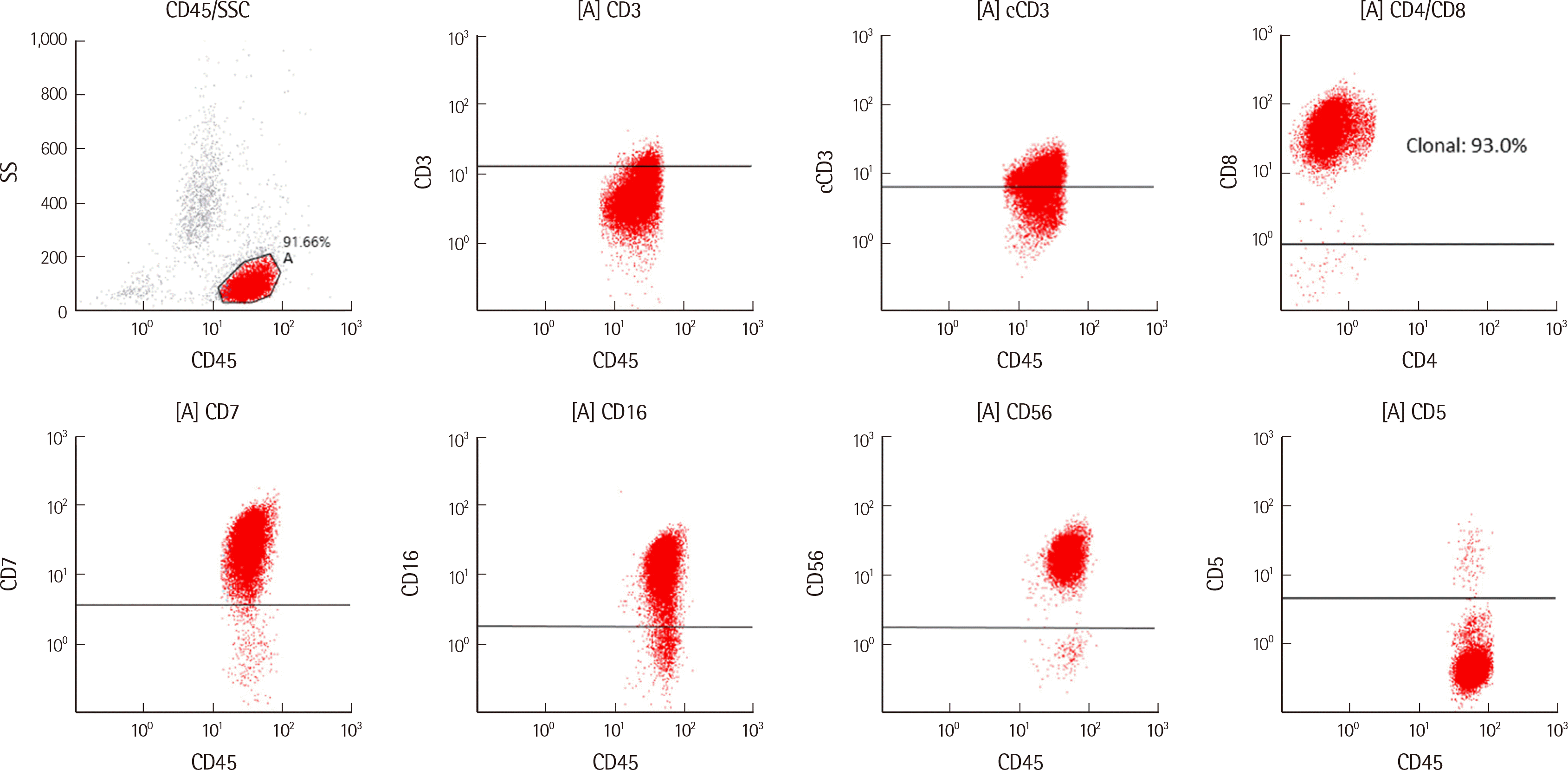
A 62-year-old man with a history of asthma was scheduled to undergo excision for a mass on his back. He visited the outpatient clinic in the plastic surgery department at Yongin Severance Hospital in November 2021 for a preoperative evaluation. His complete blood counts were as follows: white blood cell count, 206.94×10⁹/L (abnormal cells: 84%, segmented neutrophils: 5%, lymphocytes: 9%, monocytes: 2%, nucleated RBC: 1/100 white blood cells); hemoglobin level, 126 g/L; and platelet count, 68×109/L (Fig. 1A). Based on his results, he was referred to the hematology department and hospitalized for a BM biopsy and further evaluations. He complained of a weight loss of 5 kg over the last 6 months; however, there were no signs of coagulopathy, hemophagocytic syndrome, and multi-organ failure. His serum lactate dehydrogenase level had increased to 381 IU/L; nevertheless, the findings of other blood test results were not remarkable. EBV polymerase chain reaction (PCR) of whole blood was negative. Computed tomography of the abdomen and pelvis revealed splenomegaly (length: 24 cm). Computed tomography scans did not show any abnormal lymph node enlargement or neoplasms in the chest and neck. A BM aspiration smear showed 56.0% abnormal cells with atypical nuclei, distinct nucleoli, clumped chromatin, and basophilic cytoplasm containing azurophilic granules (Fig. 1B). BM biopsy showed approximately 50% interstitially scattered CD56+ and granzyme B focal+ atypical NK cell proliferation (Fig. 1C, D). Flow cytometry (Beckman Coulter, Miami, FL, USA) using a BM specimen showed an abnormal population (85.3% of total nucleated cells) with the following phenotype: CD3-, diminished cytoplasmic CD3 expression (cytoplasmic CD3 dim+), CD5-, CD7+, CD8+, CD16+, CD38+, and CD56+ (Fig. 2).
Next-generation sequencing and karyotype analysis were performed using the BM aspirate. A custom next-generation sequencing panel targeting 497 genes associated with hematologic neoplasms was performed using the NextSeq 550Dx platform (Illumina, San Diego, CA, USA), and a matched skin biopsy sample from the patient was used to differentiate somatic and germline variants. A somatic variant of DNMT3A p.Arg882His (variant allele frequency: 46.8%) was detected. Mutations of DNMT3A have been reported in hematologic malignancies such as myelodysplastic syndrome, acute myeloid leukemia, and angioimmunoblastic T-cell lymphoma [4]. An RNA fusion panel (FusionPlex Pan-Heme Panel, ArcherDX, Boulder, Colorado, USA) detected a gene fusion between exon 5 of RAB29 and exon 2 of NUCKS1 at chromosome 1q32.1 (Fig. 1F). The fusion reads of RAB29-NUCKS1 were 907 (25.69% of total reads). The G-banding karyotype was 46,XY,t(5;7)(p15.3;q11.2)[10]/46,XY[10] (Fig. 1E), which was not identified in the RNA fusion panel test. The t(5;7)(p15.3;q11.2) karyotype has been reported in a patient with monoclonal gammopathy [5]. However, the association or clinical implications in specific blood cancer of the t(5;7)(p15.3;q11.2) type have not been revealed. Cryptic RAB29-NUCKS1 fusion was confirmed using reverse-transcription PCR and Sanger sequencing (Fig. 1G) with forward primer (5´-TCAGCTGTGGGATATTGCAG-3´) and reverse primer (5´-GTTGTTGGCGCACATTTTTA-3´).

DISCUSSION

There were three possible diagnoses associated with NK cells: ANKL, extranodal NK/T-cell lymphoma, nasal type, and chronic lymphoproliferative disorder of NK cells. The neoplastic cells of ANKL typically show the following phenotype: CD2(+), surface CD3(-), CD3-epsilon(+), CD5(-), and CD56(+) and positivity for cytotoxic molecules. CD16 is frequently positive (75%) in ANKL but usually negative in extranodal NK/T-cell lymphoma [6]. In our patient, flow cytometry of the BM specimen showed CD3(-), CD5(-), CD16(+), and CD56(+). There was no definite involvement of the upper aerodigestive tract (the nasal cavity, nasopharynx, paranasal sinuses, and palate). Furthermore, given the clinical presentation and laboratory findings, chronic lymphoproliferative disorder of NK cells, which is clinically indolent and usually associated with diminished CD56 expression, could be ruled out. Therefore, he was diagnosed with ANKL and transferred to another hospital for a second opinion. Then, he began receiving chemotherapy consisting of vincristine, cyclophosphamide, and prednisolone.
Compared with classical cytogenetics, a target RNA panel allows accurate and more comprehensive identification of rare gene fusions [7]. However, it is important to exclude analytical errors and the possibility of chimeric RNAs in normal tissues to interpret the RNA fusion panel results [8]. We confirmed RAB29-NUCKS1 fusion using reverse-transcription PCR and Sanger sequencing to exclude false positivity. In addition, several studies have reported that RNA fusion such as BCR-ABL1 exists in normal individuals [9, 10]. However, in normal individuals, BCR-ABL1 fusion usually exists at a very low level [9]. However, the RAB29-NUCKS1 fusion detected in the case of our patient was detected with a high read count at the time of ANKL diagnosis. Since RAB29-NUCKS1 fusion was not previously observed in normal individuals or normal tissues, the possibility that this fusion gene is pathological cannot be completely excluded.
There are no recurrent cytogenetic abnormalities including leukemic fusion in ANKL. RAB29 and NUCKS1 have been reported to be associated with Parkinson’s disease [11]. RAB29 encodes Ras-related protein Rab-7L1, which belongs to the small GTPase superfamily. Rab proteins play important roles in the regulation of exocytic and endocytic pathways [12]. RAB29 maintains the integrity of the trans-Golgi network and is associated with the morphogenesis and retrograde trafficking of mannose-6-phosphate receptor [13]. Cation-independent mannose-6-phosphate receptor plays a role in binding and internalizing leukemia inhibitory factor, which stimulates the proliferation of some hematopoietic cells and has a direct inhibitory action on NK cells [14, 15]. NUCKS1 encodes nuclear ubiquitous casein and cyclin-dependent kinase substrate 1, a chromatin-associated protein involved in DNA damage response [16]. It has been reported to be associated with various types of diseases and is regarded as a potential biomarker [17]. NUCKS1 showed low expression in adult T-cell leukemia/lymphoma and pediatric acute lymphoblastic leukemia in an in vitro study [17]. A NUCKS1-PM20D1 fusion was previously identified in a glioblastoma sample [18]. How NUCKS1 contributes to the development of several cancers and other diseases including hematologic malignancies is currently unclear. However, based on the findings of our patient and the characteristics of RAB29 and NUCKS1, it is possible that the RAB29-NUCKS1 rearrangement contributes to the pathogenesis of ANKL in a way that is currently unknown.
In conclusion, to our knowledge, this is the first report of a case of ANKL with a RAB29-NUCKS1 fusion. Further studies will be needed to elucidate the molecular pathogenesis of ANKL related to a RAB29-NUCKS1 rearrangement. If the RAB29-NUCKS1 fusion is found to be involved in the molecular pathogenesis of ANKL, it may be a potential novel biomarker for the diagnosis and treatment of ANKL.

Acknowledgements

This work was supported by the National Research Foundation of Korea (NRF-2019R1C1C1010916).

Notes

Conflicts of Interest

None declared.

REFERENCES

1. Ruskova A, Thula R, Chan G. 2004; Aggressive natural killer-cell leukemia: report of five cases and review of the literature. Leuk Lymphoma. 45:2427–38. DOI: 10.1080/10428190400004513. PMID: 15621755.
2. Park S, Ko YH. 2014; Epstein-Barr virus-associated T/natural killer-cell lymphoproliferative disorders. J Dermatol. 41:29–39. DOI: 10.1111/1346-8138.12322. PMID: 24438142.
3. Ryder J, Wang X, Bao L, Gross SA, Hua F, Irons RD. 2007; Aggressive natural killer cell leukemia: report of a Chinese series and review of the literature. Int J Hematol. 85:18–25. DOI: 10.1532/IJH97.A10612. PMID: 17261497.
4. Yang L, Rau R, Goodell MA. 2015; DNMT3A in haematological malignancies. Nat Rev Cancer. 15:152–65. DOI: 10.1038/nrc3895. PMID: 25693834. PMCID: PMC5814392.
5. Hutchens C, Ketterling RP, Van Dyke DL. 2012; When are apparently non-clonal abnormalities in bone marrow chromosome studies actually clonal? Cancer Genet. 205:405–9. DOI: 10.1016/j.cancergen.2012.04.003. PMID: 22868001.
6. Suzuki R, Suzumiya J, Nakamura S, Aoki S, Notoya A, Ozaki S, et al. 2004; Aggressive natural killer-cell leukemia revisited: large granular lymphocyte leukemia of cytotoxic NK cells. Leukemia. 18:763–70. DOI: 10.1038/sj.leu.2403262. PMID: 14961041.
7. Heyer EE, Deveson IW, Wooi D, Selinger CI, Lyons RJ, Hayes VM, et al. 2019; Diagnosis of fusion genes using targeted RNA sequencing. Nat Commun. 10:1388. DOI: 10.1038/s41467-019-09374-9. PMID: 30918253. PMCID: PMC6437215.
8. Singh S, Qin F, Kumar S, Elfman J, Lin E, Pham LP, et al. 2020; The landscape of chimeric RNAs in non-diseased tissues and cells. Nucleic Acids Res. 48:1764–78. DOI: 10.1093/nar/gkz1223. PMID: 31965184. PMCID: PMC7038929.
9. Biernaux C, Loos M, Sels A, Huez G, Stryckmans P. 1995; Detection of major bcr-abl gene expression at a very low level in blood cells of some healthy individuals. Blood. 86:3118–22. DOI: 10.1182/blood.V86.8.3118.3118. PMID: 7579406.
10. Bose S, Deininger M, Gora-Tybor J, Goldman JM, Melo JV. 1998; The presence of typical and atypical BCR-ABL fusion genes in leukocytes of normal individuals: biologic significance and implications for the assessment of minimal residual disease. Blood. 92:3362–7. DOI: 10.1182/blood.V92.9.3362. PMID: 9787174.
11. Oh S, Shao J, Mitra J, Xiong F, D'Antonio M, Wang R, et al. 2021; Enhancer release and retargeting activates disease-susceptibility genes. Nature. 595:735–40. DOI: 10.1038/s41586-021-03577-1. PMID: 34040254.
12. Hutagalung AH, Novick PJ. 2011; Role of Rab GTPases in membrane traffic and cell physiology. Physiol Rev. 91:119–49. DOI: 10.1152/physrev.00059.2009. PMID: 21248164. PMCID: PMC3710122.
13. Wang S, Ma Z, Xu X, Wang Z, Sun L, Zhou Y, et al. 2014; A role of Rab29 in the integrity of the trans-Golgi network and retrograde trafficking of mannose-6-phosphate receptor. PLoS One. 9:e96242. DOI: 10.1371/journal.pone.0096242. PMID: 24788816. PMCID: PMC4008501.
14. Gary-Bobo M, Nirdé P, Jeanjean A, Morère A, Garcia M. 2007; Mannose 6-pho-sphate receptor targeting and its applications in human diseases. Curr Med Chem. 14:2945–53. DOI: 10.2174/092986707782794005. PMID: 18220730. PMCID: PMC2793280.
15. Abdoli Z, Assarehzadegan MA, Pipelzadeh MH, Iranparast S, Dashti Gerdabi N, Parsanahad M, et al. 2021; Leukemia inhibitory factor suppresses NKG2D mRNA expression and presentation on human natural killer cells. Iran J Allergy Asthma Immunol. 20:98–105. DOI: 10.18502/ijaai.v20i1.5416. PMID: 33639636.
16. Parplys AC, Zhao W, Sharma N, Groesser T, Liang F, Maranon DG, et al. 2015; NUCKS1 is a novel RAD51AP1 paralog important for homologous recombination and genome stability. Nucleic Acids Res. 43:9817–34. DOI: 10.1093/nar/gkv859. PMID: 26323318. PMCID: PMC4787752.
17. Huang P, Cai Y, Zhao B, Cui L. 2018; Roles of NUCKS1 in diseases: Susceptibility, potential biomarker, and regulatory mechanisms. Biomed Res Int. 2018:7969068. DOI: 10.1155/2018/7969068. PMID: 29619377. PMCID: PMC5830027.
18. Frattini V, Trifonov V, Chan JM, Castano A, Lia M, Abate F, et al. 2013; The integrated landscape of driver genomic alterations in glioblastoma. Nat Genet. 45:1141–9. DOI: 10.1038/ng.2734. PMID: 23917401. PMCID: PMC3799953.
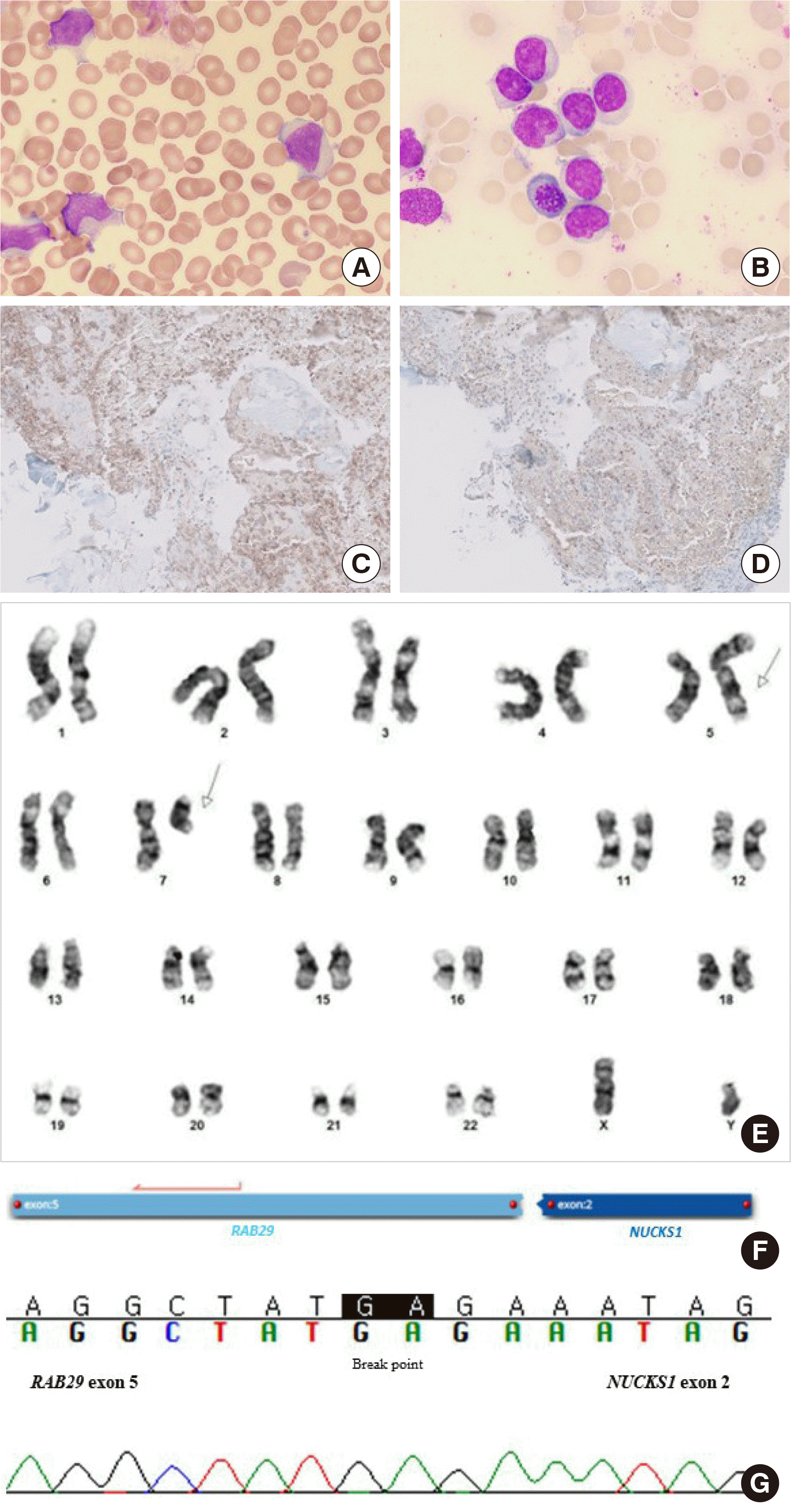
Fig. 1
Peripheral blood smear and bone marrow findings and genetic test results of the patient. (A) Abnormal cells resembling large granular lymphocytes (Wright-Giemsa stain, ×1,000). (B) Abnormal cells with irregular nuclear folding, clumped chromatin, distinct nucleoli, and lightly basophilic cytoplasm containing azurophilic granules in the bone marrow aspiration smear (Wright-Giemsa stain, ×1,000). Proliferation of neoplastic cells showing positive staining for (C) CD56 (×200) and (D) granzyme B (×200) in the bone marrow biopsy section. (E) G-banded karyogram showing 46,XY,t(5;7)(p15.3;q11.2). (F) RAB29-NUCKS1 gene rearrangement detected in the RNA fusion panel test. (G) Identification of the gene fusion breakpoint between exon 5 of the RAB29 gene (reference transcript: NM_003929.2) and exon 2 of the NUCKS1 gene (NM_022731.4) by using Sanger sequencing.
lmo-13-1-22-f1.tif
Fig. 2
Flow cytometry findings of the patient. Abnormal lymphoid cells highlighted in red exhibited the CD3-, cCD3 dim+, CD5-, CD7+, CD8+, CD16+, and CD56+ phenotypes at diagnosis.
Abbreviations: cCD3, cytoplasmic CD3; SSC, side scatter.
lmo-13-1-22-f2.tif
TOOLS
Similar articles