TO THE EDITOR: Coronavirus disease (COVID-19) is caused by SARS-CoV-2, a novel, highly infectious, single stranded RNA virus. An inappropriate immune response characterised by the excess production of pro-inflammatory cytokines (‘cytokine storm’) is common in severe cases of COVID-19 [1]. Such severe disease is frequently complicated by coagulopathy, often progressing to DIC and multi-organ failure [2]. The ‘cytokine storm’ accompanying severe COVID-19 as well as increased serum ferritin levels may be important sources of endogenous oxidative stress [3, 4] Such excess oxidative stress may lead to tissue damage in the lungs and elsewhere by generation of reactive oxygen species (ROS) [5].
The potential role of red blood cells (RBCs) in the pathophysiology of COVID-19, especially their possible contribution to hypoxia and to the thrombotic complications remains uncertain. Both anemia and increase in RBC distribution width have now been associated with increased mortality in hospitalized patients with SARS-CoV-2 infection [6, 7]. In addition, increased oxidation of structural proteins and impairment of membrane lipid homeostasis is reported in RBCs of COVID-19 positive patients, which may alter RBC deformability, potentially contributing to the thromboembolic complications seen in severe forms of COVID-19 infection [8].
To further investigate the possible role of RBC dysfunction in COVID-19, we measured ROS in RBCs of patients infected with COVID-19 at our hospital and the effect of the anti-oxidant N-acetyl cysteine (NAC). To look for evidence of increased adherence of RBCs to endothelial cells (ECs) and platelets in COVID-19 which might contribute to thrombosis, we measured RBC surface expression of adhesion molecules CD44, CD242 (ICAM-4) and CD47, as these red blood cell surface proteins have been implicated in interactions with blood vessels and/or platelets [9]. We also measured surface expression of CD147, as a surrogate marker of the lactate transporter monocarboxylate transporter 1 (MCT1) [10].
MATERIALS AND METHODS
Following approval from the hospital ethics committee, patients who tested positive for COVID-19 by reverse transcription polymerase chain reaction and age matched healthy controls were enrolled in the study after written informed consent. Blood samples were collected from COVID-19 patients within 48 hours of initial diagnosis. For immunophenotyping of RBC surface antigen expression, peripheral blood samples were incubated with fluorescent monoclonal antibodies to CD235a, CD44, CD242, CD47 and CD147 (BD Biosciences, San Jose, CA, USA) for 30 minutes and twice washed with cell wash before analysis by fluorescence activated cell analyzer (FACSCanto II) (BD Biosciences, San Jose, CA, USA). To measure RBC intracellular ROS, RBCs were incubated with 2’-7’-dichlorofluorescein diacetate (DCF) (Sigma-Aldrich, Arklow, Ireland), dissolved in Cellwash at 0.04 mM, at 37°C for 15 minutes [11]. To measure the effect of oxidant stress and the potential inhibition by anti-oxidant therapy prior to measuring DCF, RBCs were incubated at room temperature for 60 minutes with 2 mM hydrogen peroxide (H2O2) with/without initial incubation with 20 mM NAC at 37°C for 60 minutes. RBCs were analysed by FACSCanto II, with fluorescence measured in the FL1 channel. Forward scatters (FSS) versus side scatters (SSC) were set using the log scale and the mean fluorescence intensity (MFI) of a minimum of 50,000 events was recorded for each parameter. For COVID-19 positive patients, results of complete blood count (CBC), C-reactive protein (CRP), D-dimer, serum ferritin and serum lactate were also recorded. Results were expressed as mean±SD. Two-tailed Student t-test was used to compare the means of the COVID-19 and control groups. Two-tailed Pearson’s correlation was used to look for significant correlations between 2 variables. A P-value<0.05 was considered to be statistically significant.
RESULTS
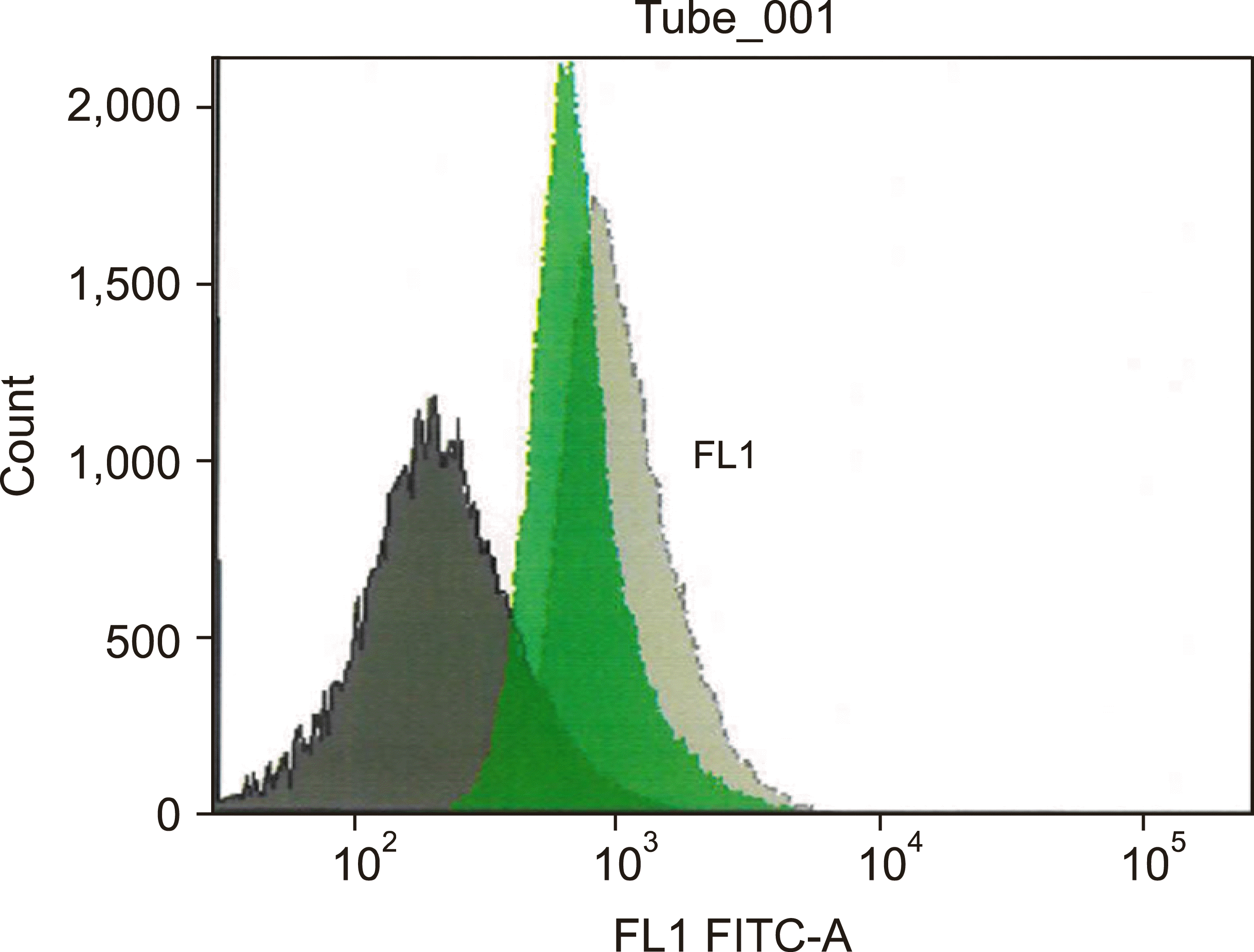
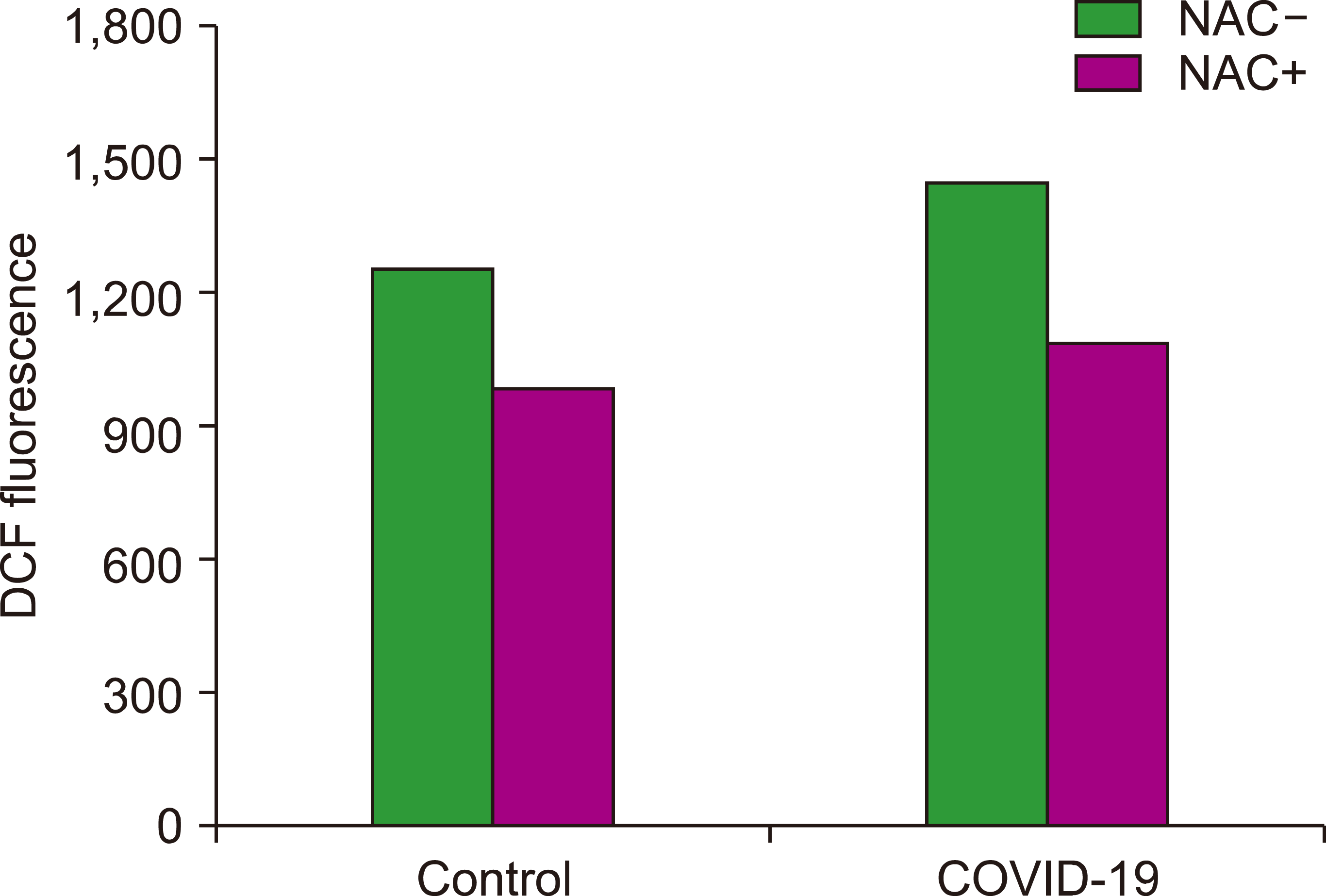
RBC ROS were measured in an initial consecutive cohort of COVID-19 patients [N=22, median age (range) 58 (30–91)] and in 10 age matched healthy controls. One patient died from COVID-19 induced respiratory failure, whilst only 3 others required care in the intensive care unit. There was no statistical difference in mean basal RBC DCF MFI levels between COVID-19 patients and healthy controls. However, mean increase in RBC DCF MFI following H2O2 incubation was significantly higher in the COVID-19 group (1105.7±336.3) compared to the control group (843.4±256.7) (P=0.042). Fig. 1 shows flow cytometric analysis of the ROS of H2O2 stimulated and unstimulated RBCs from a representative COVID-19 subject. The increase in RBC DCF MFI in the COVID-19 group correlated with CRP (R=0.514, P=0.014) but not with D-dimer, serum ferritin or any CBC parameters. Pre-incubation of RBC with NAC prior to H2O2 exposure caused a mean reduction in DCF MFI of 26.7% in the COVID-19 group (P=0.00008) (Fig. 2).
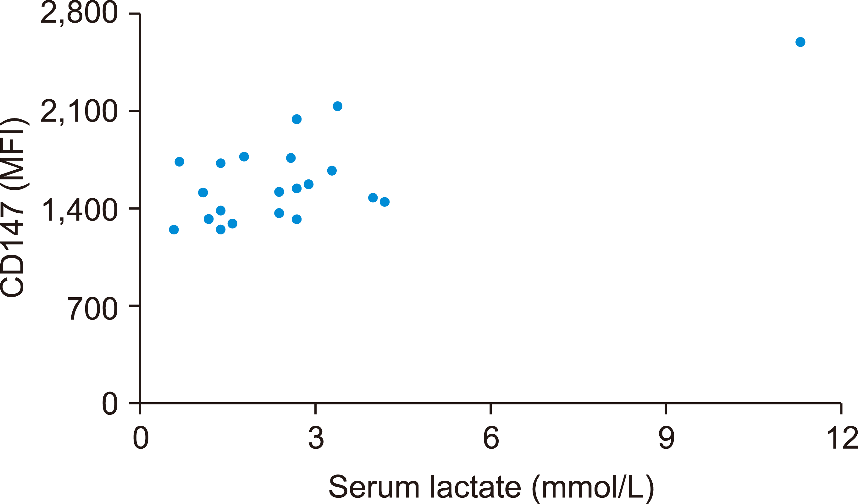
RBC expression of CD44, CD242, CD47 and CD147 was then measured in a second consecutive cohort of COVID-19 patients [N=32, median age (range) 76 (39–91)], and in 22 age matched healthy controls. There were no statistically significant differences in mean expression levels of CD44, CD242 and CD47 between the 2 groups. However, mean RBC CD147 MFI expression was higher in the COVID-19 group (1319.64±374.76) compared to controls (1061.59± 253.33) (P=0.018) (Fig. 2). There was no significant correlation between RBC CD147 MFI and D-dimer, CRP, serum ferritin or any CBC parameters in the COVID-19 group. 21 of the 32 COVID-19 patients had serum lactate levels measured on the same day as measurement of RBC CD147 MFI, with moderate positive correlation between CD147 MFI and serum lactate (R=0.65, P=0.0014) (Fig. 3). Six of these 21 patients died from COVID-19 related complications and had higher mean serum lactate (4.3±3.24 mMol/L) (P=0.028) and mean CD147 levels (1612.33±383.11 MFI) (P=0.023) than the surviving 15 patients (mean serum lactate 2±0.95 mMol/L; mean CD147 levels 1264.8±218.41 MFI).
DISCUSSION
Although basal ROS levels were not significantly different between our initial COVID-19 patient cohort and controls, we found that increase in ROS when stimulated by H2O2 was higher in the former. As there was only one death and 3 ITU admissions amongst the 22 COVID-19 patients, a greater difference in ROS levels compared to controls might have been detected if a larger number of severe cases of COVID-19 had been included. Although CRP is just one indirect biomarker of the ‘cytokine storm’ and it’s importance as a biomarker may differ according to the phase of the disease, an elevated CRP does predict worse survival in early COVID-19 among hospitalised geriatric patients [12]. As such, the positive correlation between increase in RBC ROS following H2O2 stimulation and CRP would support the supposition that ROS in RBCs reflects the severity of the ‘cytokine storm’ in COVID-19. Excessive oxidative stress is associated with accelerated RBC loss as RBCs enter suicidal death or eryptosis [13] and this may partly explain the anemia seen in severe cases of COVID-19. Pre-incubation of COVID-19 patient RBCs with NAC partially reversed the ROS inducing effects of H2O2, in keeping with previous reports that NAC acts as an anti-oxidant in normal RBCs and in RBCs with various disorders [11]. Despite paucity of clinical data to support efficacy of anti-oxidant therapy in COVID-19, our data provides some evidence of the effectiveness of anti-oxidant therapy and further studies into the pharmacological inhibition of oxidative stress in COVID-19 using single agents or combinations should be considered [13, 14].
There was no difference in mean expression levels of CD44, CD242 and CD47 between COVID-19 patients and controls and, therefore, no evidence of increased adherence of RBCs to platelets and ECs. We did detect increased expression of CD147 on RBCs of COVID-19 patients compared to controls. CD147 is a broadly distributed cell surface glycoprotein with a wide variety of functions, including association with and facilitation of cell surface expression of lactate transporter MCT1, the only MCT expressed by human RBCs [15]. Given the correlation between CD147 and serum lactate levels in our COVID-19 patients as well as the higher mean levels of CD147 and of serum lactate in those who died from COVID-19 related complications, we hypothesize that increased expression of CD147/MCT1 on RBCs facilitates transport of lactate from plasma into RBCs to protect against lactic acid induced organ damage which occurs in severe cases of COVID-19 [16]. As our results are preliminary, further research could test this hypothesis by measuring, in COVID-19 patients and controls, erythrocyte membrane expression of MCT1 protein by Western blot [10], as well as lactate distribution between plasma and RBCs [17]. The limitations of this study also include small sample size and weak statistical strength.


 PDF
PDF Citation
 Print
Print


 XML Download