

 PDF
PDF Citation
 Print
Print


INTRODUCTION
Multiple myeloma (MM) is a malignant disease of plasma cells that causes the overproduction of monoclonal light and heavy chains [1]. High-dose chemotherapy followed by autologous stem cell transplantation (ASCT) is the preferred standard therapy for eligible MM patients. The International Staging System (ISS) was created with beta-2 microglobulin and serum albumin values. In addition to the ISS, the Revised ISS (R-ISS) was created by using additional factors such as serum lactate dehydrogenase (LDH) and deletion of 17p, t(4;14), t(14;16) as detected by interphase fluorescent in situ hybridization [2-4].
The suppressor of cytokine signaling-1 (SOCS-1) gene is a short sequence located on chromosome 16 [5]. The coding sequence consists of two exons regulated by a promoter region characterized by a large CpG island spanning the gene from its promoter to the end of the second exon [5]. SOCS-1 functions to induce an appropriate immune response and is an essential physiological regulator of interferon signaling [5, 6]. SOCS-1 directly interacts with Janus kinases (JAKs), the main intracellular mediators of immune cytokine action, and inhibits their tyrosine-kinase activities [6]. The JAK signal transducer of activation (STAT) pathway is a cellular signaling pathway that is stimulated by interleukin-6 (IL-6), an important growth factor for myeloma cell survival [7]. SOCS-1 is methylated and down-regulated in patients with MM [7-10], leading to over-activation of the JAK-STAT pathway [7]. However, its role in the progression of SOCS-1 and MM epigenetics has not been established.
DNA methylation, an epigenetic regulatory mechanism frequently studied in MM and many tumors, involves adding a methyl group to the carbon 5 position of cytosine [11]. This reaction is catalyzed by DNA methyltransferases and occurs when cytosine is part of the 5’-CG-3’ sequence known as CpG or CG dinucleotide [12-14]. CpG islands are small regions of DNA usually located at the 5’ end of a gene. They range in size from 0.5 to 5 kb and are generally protected from methylation leading to down-regulation of their expression [12-14]. Global DNA and specific gene methylation constitute an important area of study.
Besides comparing SOCS-1 gene methylation status between patients with MM and the healthy control group, this study also aimed to demonstrate the effect of SOCS-1 rs33989964 gene distribution in patients with MM and the effect of the methylation status of SOCS-1 on progression-free survival (PFS) and overall survival (OS).
Go to : 
MATERIALS AND METHODS
Patient selection
This study included 120 MM patients in the Gaziantep University Hematology Clinic between January 2018 and January 2020. The control group involving 80 healthy individuals had no cancer and consisted of an unrelated individuals permanently residing in Turkey. In addition to demographic data, such as age and gender, the patients’ initial Durie-Salmon stages, ISS scores, Eastern Cooperative Oncology Group scores, laboratory data (hemoglobin, leukocytes, platelets, C-reactive protein, LDH, b2-microglobulin, albumin), first-line treatments, PFS and OS data, mortality rates, and mean follow-up duration were recorded.
All patients were found eligible for ASCT at the initial evaluation. ASCT was performed for all patients after four courses of VCD (bortezomib, cyclophosphamide, and dexamethasone) with at least a partial remission. LD (lenalidomide, dexamethasone) was used as maintenance therapy.
DNA isolation
Leukocytes were isolated from the blood samples taken into 2 mL EDTA tubes from the patients and control group individuals in the study. DNA isolation was performed from the leukocytes with the Quick-DNA Miniprep Plus Kit (Zymo Research) commercial kit according to the manufacturer’s instructions. DNA samples were stored at -20°C.
SOCS-1 -1478 CA/Del (rs33989964) polymorphism genotyping
SOCS-1 rs3398996 polymorphism was analyzed with the PCR-restriction fragment length polymorphism method. The primer sequence used for the SOCS-1 rs33989964 polymorphism is 5'-TGTCGTCCAGCTGCACCTC-3' (forward), 5'-ACCACAGGCTTCAGAGGAAC-3' (reverse). The size of the PCR product was determined as 250 bp. The DdeI enzyme was used as restriction endonuclease. The cut products were kept at 37 Celsius for one night, run on a 2.5% agarose gel, and visualized under UV light. Fragment lengths were determined as 250 bp, 145 bp, and 105 bp for the CA/Del genotype; 145 bp and 105 bp for the CA/CA genotype; and 250 bp and 105 bp for the Del/Del genotype [15].
Bisulfite modification
DNA bisulfite conversion was first performed for SOCS-1 gene methylation analysis using the EZ-96 DNA Methylation-Gold Kit (Zymo Research) protocol.
SYBR green-based Quantitative methylation-specific polymerase chain reaction (PCR) (qMSP)
Bisulfite-converted DNA samples were analyzed using a real-time quantitative methylation-specific PCR method to measure the methylation status of the SOCS-1 gene. The primer sequences for the SOCS-1 gene are 5-TTCGCG TGTATTTTTAGGTCGGTC-3 (forward) and 5-CGACACA ACTCCTACAACGACCG3 (reverse) [16]. The primer sequences for the control gene b-actin are 5'-TGGTGATG GAGGAGGTTTAGTAAGT-3' (forward) and 5'-AACCAAT AAAACCTACTCCTCCCTTAA-3' (reverse) [17]. Percent methylated reference (PMR) value of SOCS-1 in each sample was calculated using the 2-ΔΔCq method. ΔΔCq was calculated by subtracting the fully methylated DNA (Cq target gene-Cq ACTB control) from the sample DNA (Cq target gene-Cq ACTBcontrol) [18].
The CA/CA, CA/Del, and Del/Del genotypes and the CA and Del alleles of the SOCS-1 gene rs33989964 polymorphism were statistically compared between patients and healthy controls. Additionally, the statistically significant effects of these genotypes on PFS and OS were examined. In addition, the methylation status of the patients was compared to the healthy control group, and their effects on disease parameters were evaluated.
Ethical committee approval was received (Istanbul University, Faculty of Medicine, approval date and number: 29/05/2020-86529), and the patients and control subjects gave written informed consent before the beginning of the study. The present study was conducted following the principles of the Declaration of Helsinki.
Statistical analysis
The SPSS 21 package program was used for the statistical analysis of all data. The statistical significance of the differences between the patient and control groups was estimated by logistic regression analysis. Exp odds ratios (ORs) were calculated using a logistic regression model controlled for gender and age and reported at 95% confidence intervals. Differences in SOCS-1 rs33989964 polymorphism allele and genotype frequencies between the control and patient groups were compared using the c2 test, and when needed, the Fisher’s exact test was used. The Hardy–Weinberg equation was used to calculate estimated and observed genotype frequencies. The PFS was determined as the time from treatment initiation until disease progression, while OS was determined as the duration of patient survival from the time of treatment initiation. The PFS and OS results of patients diagnosed with MM were also analyzed according to the median PMR value of the healthy control group since there is no proven range or value in the literature. The survival probabilities were estimated by the Kaplan–Meier method, and differences were compared using the log-rank test. Cox stepwise regression analysis was employed to confirm the significance of risk factors. In multivariate analysis, we used eliminated variables stepwise (backward) with a significance of less than 10%. Values of P<0.05 were considered to indicate statistical significance.
Go to :
RESULTS
Of the 120 patients in our study, 56 (47%) were female and 53 (53%) were male. The median age of the patients was 55 (range, 32–70). There was no difference between the control and the patient group regarding gender distribution (P=0.064), and the 5-year PFS resulted in 52% and the 5-year OS in 80.5%. The mortality was 15% with 18 patients during follow-up. The median follow-up time was 38.3 months (range, 4.1–105.3 mo) (Table 1).
Table 1
Clinical features and treatment regimens of patients with MM.
![]()
In the statistical analysis of SOCS-1 rs33989964 polymorphism genotype distribution between patients with MM and healthy controls, the CA/CA genotype was statistically significantly higher in healthy controls (P=0.001). On the other hand, the Del/Del genotype was significantly higher in patients with MM (P=0.034). The Del allele was higher in MM patients, and the CA allele was higher in healthy controls (P=0.001). The PMR value of the SOCS-1 gene was found to be significantly higher in the healthy controls (median, 43.48; range, 2.76–247.75; P=0.001) (Table 2).
Table 2
Comparison of frequencies of SOCS-1 gene rs33989964 variants between patients with multiple myeloma and healthy controls.
![]()
In the statistical analysis of the factors affecting PFS and OS, the SOCS-1 rs33989964 genotype distribution did not affect PFS and OS (for PFS, P=0.229; for OS, P=0.763). No significant effect on PFS and OS was detected even when patients were grouped as CA/CA or CA/Del+Del/Del genotypes (for PFS, P=0.140; for OS, P=0.462).
In the statistical analysis performed according to the median PMR value of 43.48 of the healthy controls, it was observed that patients with a PMR value of ≥43.48 had a significantly shorter PFS (for <43.48% 56, for ≥43.48% 43; P=0.027) than those with a PMR value of <43.48 (Fig. 1, Table 3). There was no significant difference between these two groups regarding OS (P=0.484) (Table 3).
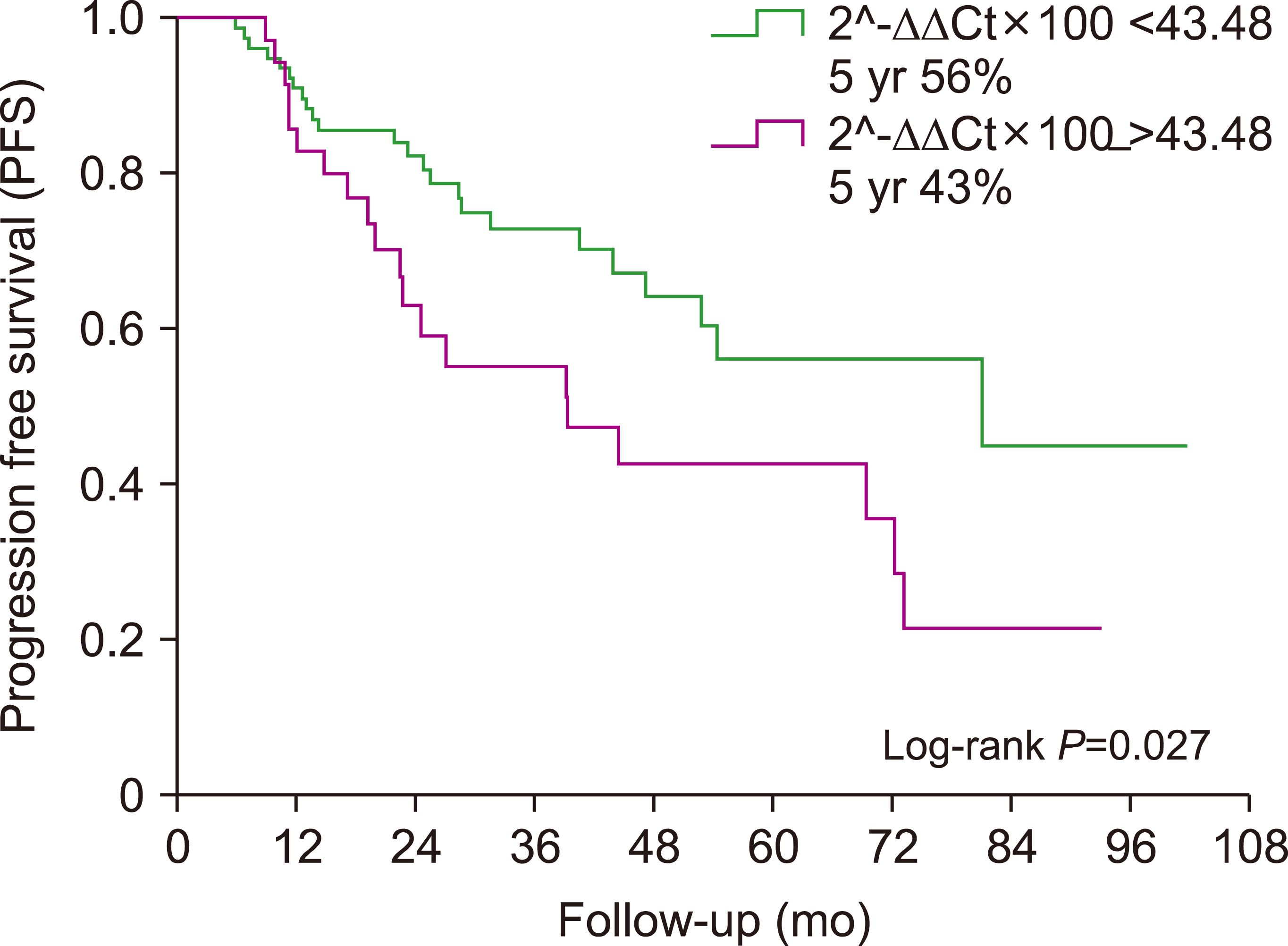 | Fig. 1Progression-free survival (PFS) analysis between patients’ subgroups in terms of SOCS-1 gene methylation status.
|
Table 3
Comparison of PFS and OS with prognostic factors of patients with MM.
![]()
In the statistical analysis performed by dividing the patients into two subgroups as “low” and “high” PMR values (<43.48 and ≥43.48), the only significant result was obtained in terms of the presence of progression. Patients with a PMR value of ≥43.48 were 3.125 times more likely to develop progression than those with a PMR value of <43.48 (95% CI, 1.390–7.025; P=0.006) (Table 4).
Go to :
DISCUSSION
This study includes significant and new literature contributions regarding MM epigenetics, SOCS-1 rs33989964 gene polymorphism, and the methylation status of the SOCS-1 gene. The CA/CA genotype of SOCS-1 gene was found to be significantly higher in healthy controls, while the Del/Del genotype was significantly higher in patients with MM. The PMR value of the SOCS-1 gene was significantly higher in healthy controls. Patients with a PMR value of ≥43.48 had a significantly shorter PFS, and patients with a PMR value of ≥43.48 were 3.125 times more likely to develop progression than those with a PMR value of <43.48.
SOCS-1 gene polymorphisms and gene expression have been revealed in more than one study. The SOCS-1 rs33989964 polymorphism represents a dinucleotide CA Insertion/Deletion (CA/Del) associated with respiratory immune-inflammatory diseases with high expression levels [19]. A functional SOCS-1 promoter polymorphism rs33989964 has increased SOCS-1 transcription in a Japanese population [6]. The fact that the SOCS-1 rs33989964 Del/Del genotype was more common in patients with MM also reveals the role of its low expression in the pathogenesis of MM. An increase in the CA allele was found protective against the disease, while an increase in the Del allele was a significant risk factor for developing the disease. Low SOCS-1 expression was found to increase MM susceptibility.
Global DNA and specific gene methylations are a field of study that includes literature data on MM epigenetics [20, 21]. In a study from our clinic [22], hypermethylation was significantly higher in patients with MM than in healthy controls. Global hypermethylation in post-treatment measurements was significantly increased compared with the pre-treatment state. Regarding adenomatous polyposis coli-2 promoter gene-specific hypermethylation, no significant differences were detected between pre-and post-treatment values [22].
There are different studies on hypermethylation in SOCS-1 and MM patients. In the study of Chim et al. [23], there were no significant results regarding SOCS-1 hypermethylation. On the other hand, the study of Galm et al. [8] showed that aberrant SOCS-1 methylation was found to be increased in MM cell lines. The exposure of these cell lines with the demethylating agent 5-aza-2’-deoxycytidine up-regulated SOCS-1 expression opened an important discussion point regarding myeloma treatment. In this study, SOCS-1 was hypermethylated in 62.9% of MM patient samples. It was concluded that SOCS-1 is frequently inactivated by hypermethylation in MM patients [8]. In the study of Depil et al. [9], SOCS-1 hypermethylation was observed up to 75%, while there was no significant difference in OS between the hypermethylated and unmethylated subgroups. In the study of Reddy et al. [10], SOCS-1 was shown to be hypermethylated in patients with MM and monoclonal gammopathy of undetermined significance (MGUS), where the number of patients with at least one gene hypermethylation was 93% in patients with MM and 60% in patients with MGUS.
In a study in which oral azacitidine (AZA) was combined with lenalidomide and dexamethasone [24], the efficacy of lenalidomide in cases of previously unsuccessful relapsed/refractory MM was examined. The ORR was 37.5% and the clinical benefit rate was 50%. The median OS was 10.3 months and the median PFS was 2.6 months [24]. In another study [25], 40 patients with a diagnosis of relapsed/refractory MM were included, and the effectiveness of subcutaneous AZA was evaluated. A clinical benefit response has been obtained in 30% of patients included in the study. AZA was well tolerated up to the target of 50 mg/m2 subcutaneously twice a week in combination with LD [25]. In another important study [26], AZA inhibited the elaboration of both IL-6 receptor-alpha and IL-6, resulting in the reduced expression of phospho-STAT3 and Bcl-xl. Its antagonist effects and efficacy against IL-6, the basis of MM pathogenesis, are considered important for the future. In this present study, we had the opportunity to examine the effects of SOCS-1 methylation status on survival in patients treated with the same regimen and who underwent ASCT. It was revealed that patients with a PMR value of ≥43.48 were 3.125 times more likely to develop progression than those with a PMR value of <43.48. In our study, no patient used hypomethylating agents. The use of hypomethylating agents and their effects on survival have opened a meaningful discussion topic. While the literature data reveals the effects of hypomethylating agents in relapsed/refractory myeloma cases, the patients included in our study received first-line therapy. These results contribute to another point regarding the discussion on early or first-line use of hypomethylating agents or their addition to the first-line treatment regimen.
There were significant limitations of our study. The first was the small patient population, which limited the statistical analysis. The second most important limitation was that it consisted of single-center data. Another limitation was that simultaneous SOCS-1 expressions of patients and healthy controls could not be measured.
In conclusion, while CA/CA genotype was statistically significantly higher in healthy controls, Del/Del genotype was significantly higher in patients with MM. The Del allele was higher in patients with MM, and CA genotype was higher in the healthy control group. The PMR value of the SOCS-1 gene was significantly higher in the healthy control group. Patients with a PMR value of ≥43.48 were 3.125 times more likely to develop progression than those with a PMR value of <43.48. The effects of SOCS-1 polymorphisms on the pathogenesis of MM and SOCS-1 methylation will shed light on the pathophysiology and treatment of MM.
Go to :
 XML Download