Journal List > Blood Res > v.57(S1) > 1516080603

Jang, Jo, Yoo, Kim, Lee, Jang, Chung, and Cho: Prognostic impact of total body irradiation dose in pediatric acute lymphoblastic leukemia patients treated with allogeneic hematopoietic stem cell transplantation in second complete remission

Abstract

Background

Allogeneic HSCT may improve survival in pediatric ALL patients who relapse. In this study, we analyzed the outcome and prognostic factors of 62 ALL patients (35 male, 56.5%) who received allogeneic HSCT in second complete remission (CR) at our institution between April 1st 2009 and December 31st 2019.

Methods

The median time from diagnosis to relapse was 35.1 months (range, 6.0‒113.6 mo). Fifty-three patients (85.5%) experienced bone marrow relapse only. The number of patients who received transplant according to each donor type was as follows HLA matched family donor 17 (27.4%), matched unrelated donor (UD) 22 (35.5%), mismatched donor 23 (37.1%). All patients received HSCT with a myeloablative conditioning, 58 patients (93.5%) with the incorporation of TBI [31 patients 12 Gray (Gy), 24 patients 13.2 Gy, 3 patients 8 Gy].

Results

The 5-year event-free survival (EFS), and overall survival of the study group was 41.3±6.3% (26/62), and 42.3±6.6% (27/62), respectively. The cumulative incidence of relapse and transplant-related mortality was 57.1±6.4% and 1.6±1.6%, respectively. Infant ALL, shorter time from diagnosis to relapse, and TBI dose of 12 Gy, rather than 13.2 Gy, resulted in significantly worse EFS. In multivariate analysis, infant ALL and TBI dose of 12 Gy during conditioning predicted significantly lower EFS.

Conclusion

In our study group, treatment with a higher dose of TBI during conditioning resulted in better EFS for ALL patients who underwent HSCT in second CR. Further study is needed to determine potential long-term complications associated with a higher TBI dose.

INTRODUCTION

Acute lymphoblastic leukemia (ALL) is the most common hematologic malignancy in the pediatric population [1]. Outcome has improved, and the overall survival (OS) has surpassed 90% in more recent studies [2]. However, more than 20% of children with ALL relapse, and survival following relapse remains poor, with relapsed ALL remaining one of the leading causes of childhood cancer death [3]. The OS of children with relapsed ALL was 56% according to the United Kingdom R2 trial [4]. Another study by the Children’s Oncology Group reported OS ranging from 11% to 85% depending on prognostic factors such as time from diagnosis to relapse and site of relapse [5]. The result of a multicenter trial by Acute Lymphoblastic Leukemia Relapse Berlin-Frankfurt-Munster 90 was 36% in terms of 10-year OS [6].
Patients with relapsed ALL may undergo allogeneic hematopoietic stem cell transplantation (HSCT) in second complete remission (CR). Outcome of allogeneic HSCT in second CR has improved with the advent of high-resolution human leukocyte antigen (HLA) typing, and the adoption of various conditioning regimens such as implementing total body irradiation (TBI) with cyclophosphamide (Cy) [7, 8]. A retrospective comparison of outcomes between TBI with Cy and busulfan with Cy as conditioning regimens concluded that the use of TBI resulted in a higher leukemia-free survival rate for children with ALL [8]. Fractionated administration of TBI up to a cumulative dose of 12 Gray (Gy) is commonly used in the preparative regimen prior to HSCT [9].
Whether an increase of the TBI dose results in better transplant outcomes remains unclear. A registry study based on a large number of pediatric ALL patients who received HSCT in second or subsequent CR or relapse showed that neither increasing the TBI dose beyond 12 Gy nor the addition of further chemotherapy during conditioning resulted in lower relapse rate [10]. In contrast, a recent study based on adult patients with hematologic malignancies found that higher doses of TBI led to a significant decrease in relapse rate; however, this was offset by greater transplant-related mortality (TRM) so that these patients did not have a survival advantage [11].
We previously published the outcomes of allogeneic HSCT for pediatric ALL in second CR done during a period of 17 years at our institution. The 5-year disease-free survival (DFS) and OS were 45.2% and 48.3%, respectively [12]. Donor type was the only significant factor for DFS in multivariate study, with patients who received a matched sibling donor (MSD) HSCT having better outcome than those who received an unrelated donor (UD) HSCT. In contrast, other studies have emphasized improved outcome of ALL patients who received UD HSCT in second CR, resulting in comparable survival rates between MSD and UD HSCT recipients [13-15]. Shorter duration of first CR resulted in a significantly higher incidence of post-HSCT relapse in these studies [14, 15].
In this study, we undertook a follow-up analysis to determine outcome and risk factors for patients with ALL who underwent allogeneic HSCT in second CR during a period of ten years at our institution.

MATERIALS AND METHODS

Patient characteristics at diagnosis

The study received approval from the institutional review board (KC21RASI0328). We retrospectively reviewed 62 patients (35 male, 56.5%) who received a first allogeneic HSCT in second CR between April 1st 2009 and December 31st 2019 at the Department of Pediatrics, Seoul St. Mary’s Hospital, The Catholic University of Korea (Table 1). Patients who had previously received allogeneic HSCT in first CR, and so received a second transplant in second CR were excluded, as were patients who received transplant in non-remission status.
The median age at diagnosis was 6.1 years (range, 0.2–16.4 yr). Fifty-eight patients (93.5%) were diagnosed with precursor B cell ALL. Of the 50 patients with available risk group classification at diagnosis, 38 (76%) were designated as high risk or very high risk according to our institution’s criteria [16].
The median time from diagnosis to relapse was 35.1 months (range, 6.0–113.6 mo). The number of patients who experienced early (<18 months from diagnosis), intermediate (18–36 months), and late relapse (>36 months from diagnosis) was 16 (25.8%), 16 (25.8%) and 30 patients (48.4%), respectively (Table 1). Fifty-three patients (85.5%) experienced bone marrow (BM) relapse only, while 5 patients (8.1%) showed isolated extramedullary (EM) relapse [central nervous system (CNS) relapse 3, testes 1, other EM 1]. Four patients (6.5%) showed combined BM and EM relapse (BM with CNS 3, BM with testes 1).

Transplantation

Median time from relapse to HSCT was 4.7 months (1.8–7.8 mo). Matched UD transplantation was the most common type of HSCT (Table 2). The majority of patients received a peripheral blood stem cell transplantation (PBSCT, 79.0%). The key HSCT procedures, including antimicrobial prophylaxis, were done as reported previously [17].
Total body irradiation (TBI), cyclophosphamide (Cy) ±rabbit antithymocyte globulin (ATG, Thymoglobulin, Sanofi, Paris, France) was the most commonly utilized conditioning regimen, followed by TBI, Cy, cytarabine (Ara) ±ATG. Since 2013, we increased the fractionated TBI dose from 12 Gy to 13.2 Gy. Patients who received 12 Gy and 13.2 Gy TBI received the total dose in fractions of 2 Gy and 1.65 Gy, respectively. Four of 17 patients (23.5%) who received a matched family donor HSCT received the higher TBI dose of 13.2 Gy, while 11 of 22 patients (50.0%) who received a matched UD HSCT received 13.2 Gy TBI (P=0.112).
Prior to 2014, ATG was given at a dose of 2.5 mg/kg/day for 3–4 days, the total cumulative dose depending on the HSCT donor type. Since 2014, the ATG dose was halved to 1.25 mg/kg/day [18]. Of the 39 patients who received ATG as part of the conditioning regimen, 16 (41.0%) received the lower dose of 1.25 mg/kg/day.
Median infused cell doses were as follows: total nucleated cell 12.28×10⁸/kg (range, 0.34–49.28×10⁸/kg), mononuclear cell 8.73×10⁸/kg (0.15–34.00×10⁸/kg), CD34+ 5.56×10⁶/kg (0.03–26.14×10⁶/kg), CD3+ 38.96×10⁷/kg (0.44–181.15×10⁷/kg).

Study endpoint

Important study objectives included calculation of 5-year event-free survival (EFS) and OS of the study group, as well as risk factors for EFS. We also evaluated the incidence of relapse and TRM in CR.
The following variables were analyzed for impact on EFS: patient gender, age at diagnosis, genetic abnormalities, time from diagnosis to relapse, HSCT donor type, cell source, donor-recipient gender, ABO compatibility, HLA compatibility, and TBI dose. EFS was determined from the time of HSCT to last follow-up in CR or first event, which included relapse or death in CR. OS was determined from time of HSCT to last follow-up or death.
The incidences of acute graft-versus-host disease (GVHD) (both grades 2–4 and grades 3–4), and chronic GVHD (overall, as well as moderate/severe) were analyzed. Acute and chronic GVHD were graded according to established criteria [19, 20].

Statistical analyses

The EFS and OS of the study group were calculated with the Kaplan-Meier method. Univariate study of risk factors for EFS was also done with the Kaplan-Meier method, with comparisons done with the log rank test. Multivariate analysis of risk factors for EFS was done with the Cox proportional hazard regression model. Variables found significant in univariate study (P<0.05) were entered simultaneously in multivariate analysis. Incidence of relapse and death in CR post-HSCT, and acute and chronic GVHD were calculated with the cumulative incidence function. Competing risks were death in CR for relapse, ALL relapse for TRM, relapse or TRM without acute GVHD within 100 days post-HSCT for acute GVHD, and relapse or TRM without chronic GVHD within 1 year post-HSCT for chronic GVHD. Comparison of categorical variables was done with Fisher’s exact test. Patient follow-up was done up till April 30th, 2022.

RESULTS

Outcome of the study group

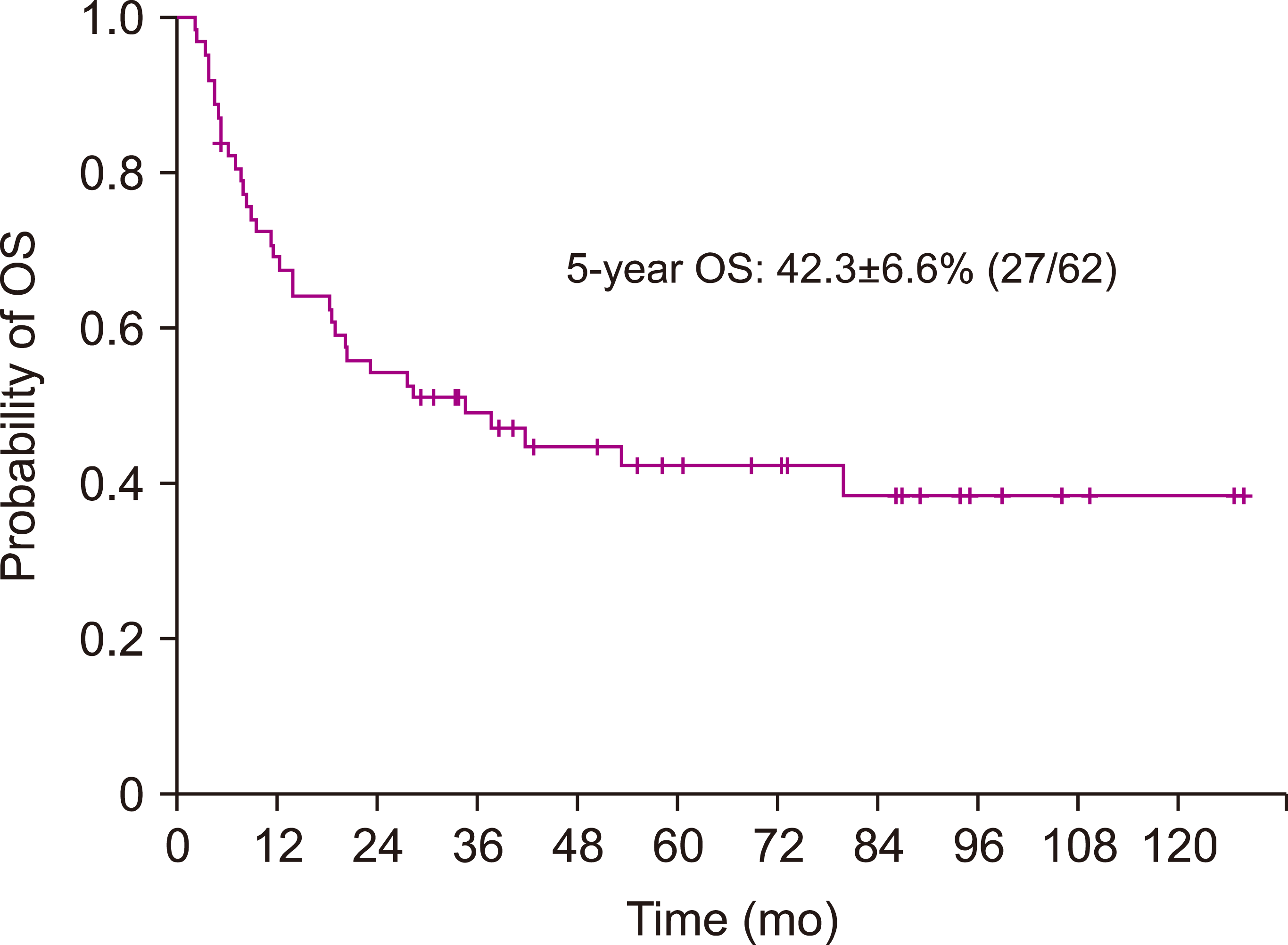
The 5-year EFS and OS of the study group was 41.3±6.3% (26/62) and 42.3±6.6% (27/62) (Fig. 1), respectively. The 5-year cumulative incidence of relapse was 57.1±6.4% (35/62). TRM was 1.6±1.6% (1/62), comprising 1 patient who died of grade 4 acute skin and gastrointestinal GVHD. Of the 35 patients who relapsed post-HSCT, only one patient, who achieved 3rd CR and underwent a second transplant survives.

Prognostic factors

Infant ALL, shorter time from diagnosis to relapse, and TBI dose of 12 Gy, rather than 13.2 Gy, resulted in significantly worse EFS (Fig. 2, Table 3). In multivariate analysis, infant ALL and TBI dose of 12 Gy during conditioning predicted significantly worse EFS (Table 4). TBI dose was a significant factor for EFS both in patients who received an HLA matched family donor HSCT, and in those who received an alternative donor (AD) HSCT, including matched UD transplant, or mismatched family donor/UD transplant: 5-year EFS of 75.0±21.7% (3/4) for 13.2 Gy vs. 7.7±7.4% (1/13) for 12 Gy in matched family donor transplant recipients (P=0.034), and 70.0±10.2 (14/20) for 13.2 Gy vs. 24.4±10.5 (5/18) for 12 Gy in AD transplant recipients (P=0.004).
As the two main modifications to the conditioning regimen during the study period were the increase in TBI dose from 12 Gy to 13.2 Gy, and the decrease in ATG dose from 2.5 mg/kg/day to 1.25 mg/kg/day, we further analyzed the impact of ATG dose in the AD transplant recipients, the majority of whom (38/45) received ATG as part of conditioning. Patients who received a lower cumulative dose of ATG had significantly better EFS than those who received a higher cumulative dose: 5-year EFS of 75.0±10.8% (12/16) for 1.25 mg/kg/day recipients vs. 22.7±8.9% (5/22) for 2.5 mg/kg/day recipients (P=0.002). Since both TBI dose and ATG dose had significant impact on outcome in univariate study, we undertook multivariate study to conclude upon the key factor for outcome; however, neither TBI dose nor ATG dose had a significant impact on EFS of AD transplant recipients in multivariate study.
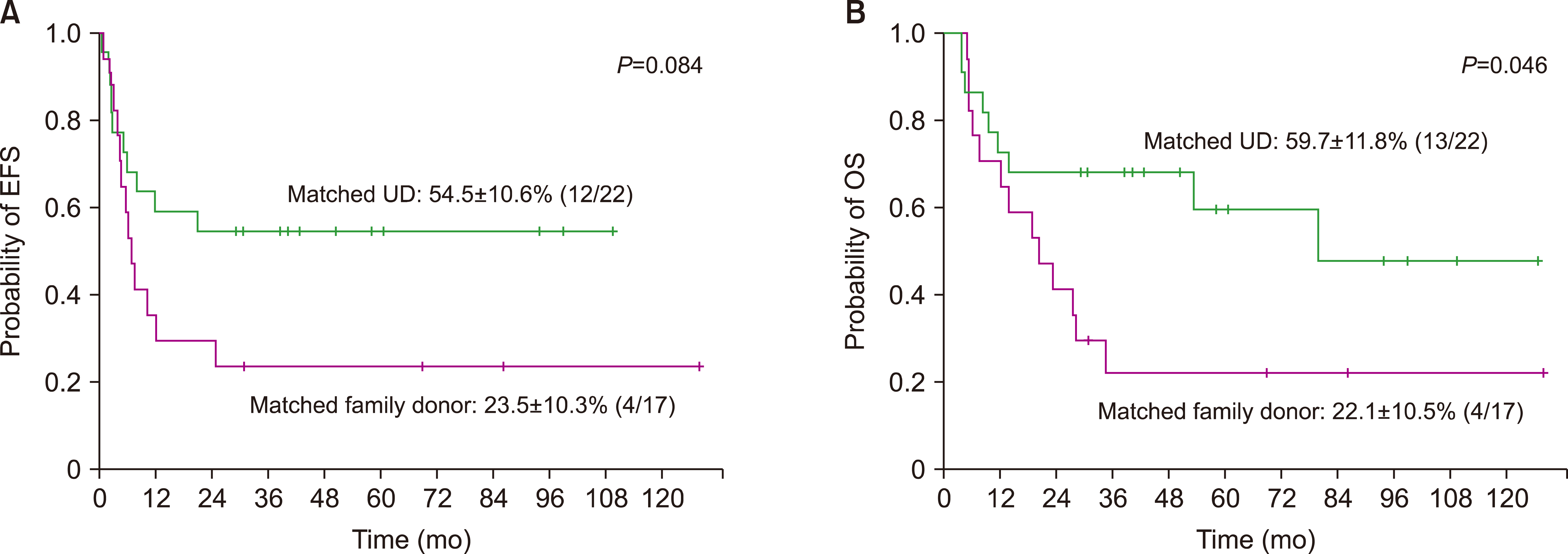
When comparing the EFS of matched family donor and matched UD transplant recipients, we found a better outcome in those who received a matched UD transplant: 5-year EFS of 54.5±10.6% (12/22) for matched UD transplant recipients vs. 23.5±10.3% (4/17) for matched family donor transplant recipients (P=0.084) (Fig. 3A). The OS was also higher in patients who received a matched UD transplant: 5-year OS of 59.7±11.8% (13/22) for matched UD transplant recipients vs. 22.1±10.5% (4/17) for matched family donor transplant recipients (P=0.046) (Fig. 3B).

Incidence of acute and chronic GVHD

The 100-day cumulative incidence of acute GVHD was as follows: 54.8±6.4% (standard error) (34 of 62 patients) for grades 2-4 acute GVHD, and 4.8±2.7% for grades 3-4 acute GVHD (3/62). Nine patients (14.5%) were diagnosed with chronic GVHD at a median of 5.0 months from transplant (range, 2.5–9.1 mo). The 1-year cumulative incidence of overall chronic GVHD and moderate/severe chronic GVHD was 14.7±4.6% (9/62) and 4.9±2.8% (3/62), respectively.

DISCUSSION

We report the outcomes of allogeneic HSCT for childhood ALL in second CR at a single institution during a period of 10 years. The estimated 5-year EFS and OS were 41.3±6.3% and 42.3±6.6%, respectively, comparable to survival rates from a previous study [12].
In our past study of ALL patients who received allogeneic HSCT in second CR, we reported that the donor type was the only major factor influencing transplant outcome, with patients who received a sibling donor transplant showing higher disease-free survival compared with UD transplant recipients [12]. However, in this follow-up study, we found a higher EFS for patients who received a matched UD transplant compared with those who received a matched family donor transplant, although the difference was not statistically significant. In our study group a lower percentage of matched family HSCT recipients received the higher TBI dose of 13.2 Gy than matched UD HSCT recipients, which may have contributed to the lower EFS for matched family donor transplant recipients. Overall, this result is consistent with comparable outcomes currently observed for ALL patients who receive either a sibling donor transplant or a UD transplant [14, 15, 21].
Age at diagnosis was a significant prognostic factor in our ALL patients who received HSCT in second CR, due mostly to the extremely poor outcome of infant ALL patients. Our previous study showed that infant ALL was a significant adverse prognostic factor for long-term EFS in a large cohort of newly diagnosed precursor B cell (Pre-B) ALL patients [22]. This study further emphasizes the low survival rate of infant ALL patients subsequent to relapse. In consideration of the poor outcome of high risk infant ALL patients after relapse, our current institutional strategy is to consolidate these patients with allogeneic HSCT in first CR, although undertaking such therapy may result in major long-term complications in the high risk infant ALL population. Regarding patient age, a previous study has also shown that older age at diagnosis may also be a risk factor for inferior survival after relapse [23]. Furthermore, our study confirmed the key role of time from diagnosis to relapse as a major prognostic factor in relapsed ALL patients, as found in previous studies [5, 13].
Pediatric ALL patients undergoing HSCT may receive either a TBI or chemotherapy-based conditioning regimen. A recent study undertaken mostly in ALL patients in first CR randomized patients to either a TBI-based or chemotherapy-based conditioning; the study found higher OS and lower relapse rate for patients who received TBI [24]. In our study of ALL patients we found that patients who received the higher fractionated dose of 13.2 Gy TBI had significantly better EFS than those who received 12 Gy TBI, with the TBI dose remaining a key prognostic factor in multivariate analysis, along with infant ALL. One adult study based on patients with various hematologic malignancies compared standard TBI dose of 12 Gy to higher doses up to 14 Gy. Although patients who received a higher dose of TBI had a lower rate of relapse, the rate of TRM was significantly greater, resulting in similar OS [11]. In our study group, the very low rate of TRM (1.6%) likely contributed to higher TBI dose recipients having a significantly better EFS.
The major modifications to the conditioning regimen during the study period included both an increase in the TBI dose, as well as a decrease in the cumulative ATG dose given to patients who received AD HSCT. We previously reported that this decrease in ATG dose resulted in lower relapse incidence and higher OS, with similar incidence of both acute and chronic GVHD in pediatric acute leukemia patients who received peripheral blood stem cell transplantation [18]. In our current study, ATG was primarily given to AD transplant recipients, and in this group of patients also, treatment with a lower ATG dose resulted in significantly better EFS. Hence, for patients who received AD HSCT in our study group, the ATG dose modification may have further augmented any improvement in outcome resulting from TBI dose increase. However, we emphasize that patients who received a higher TBI dose had better outcome in both the matched family donor and AD transplant subgroups.
The long-term toxicities of TBI may severely compromise patient outcome post-transplant. Late effects of TBI include cataract formation, and multiple endocrine abnormalities, such as gonadal insufficiency, thyroid hormone dysfunction, and growth impairment [9]. Above all, patients who receive a high TBI dose are at greater risk for secondary malignancies post-transplant. Whether the patients in our study who received a higher TBI dose are found to have a greater incidence of these complications requires further follow-up.
As the clear majority of events post-transplant in our study group were disease relapse, efforts to improve transplant outcome should focus on minimizing the disease burden prior to transplant. Immunotherapy and targeted therapy agents for Pre-B ALL, such as blinatumomab and chimeric antigen receptor T cells, may allow for an optimum decrease in ALL cells before consolidation with HSCT, resulting in improved transplant outcomes. In this regard, sensitive methods of detecting minimal residual disease (MRD) before HSCT such as next-generation sequencing based MRD detection may aid in predicting patient prognosis and allow for tailoring of the conditioning intensity according to pre-transplant MRD status [25].
In summary, patients who received the higher TBI dose of 13.2 Gy had better EFS in our study group. Long-term follow-up is necessary to monitor for TBI-related toxicities. Implementation of novel diagnostics and therapeutics prior to HSCT may allow for accurate prediction of patient survival post-transplant, and improvement in overall transplant outcome, while limiting the long-term toxicities related to therapy.

ACKNOWLEDGMENTS

We express sincerest gratitude to the nursing staff associated with the Division of Hematology/Oncology, Department of Pediatrics, The Catholic University of Korea, and, above all, to the patients and their families.

Notes

Authors’ Disclosures of Potential Conflicts of Interest

No potential conflicts of interest relevant to this article were reported.

REFERENCES

1. Ward E, DeSantis C, Robbins A, Kohler B, Jemal A. 2014; Childhood and adolescent cancer statistics, 2014. CA Cancer J Clin. 64:83–103. DOI: 10.3322/caac.21219. PMID: 24488779.
2. Malczewska M, Kośmider K, Bednarz K, Ostapińska K, Lejman M, Zawitkowska J. 2022; Recent advances in treatment options for childhood acute lymphoblastic leukemia. Cancers (Basel). 14:2021. DOI: 10.3390/cancers14082021. PMID: 35454927. PMCID: PMC9032060. PMID: 5e19730c94784a6c9c9fbb47a4904bfe.
3. Leung AWK, Vincent L, Chiang AKS, et al. 2012; Prognosis and outcome of relapsed acute lymphoblastic leukemia: a Hong Kong Pediatric Hematology and Oncology Study Group report. Pediatr Blood Cancer. 59:454–60. DOI: 10.1002/pbc.24162. PMID: 22610685.
4. Roy A, Cargill A, Love S, et al. 2005; Outcome after first relapse in childhood acute lymphoblastic leukaemia - lessons from the United Kingdom R2 trial. Br J Haematol. 130:67–75. DOI: 10.1111/j.1365-2141.2005.05572.x. PMID: 15982346.
5. Nguyen K, Devidas M, Cheng SC, et al. 2008; Factors influencing survival after relapse from acute lymphoblastic leukemia: a Children's Oncology Group study. Leukemia. 22:2142–50. DOI: 10.1038/leu.2008.251. PMID: 18818707. PMCID: PMC2872117.
6. Tallen G, Ratei R, Mann G, et al. 2010; Long-term outcome in children with relapsed acute lymphoblastic leukemia after time-point and site-of-relapse stratification and intensified short-course multidrug chemotherapy: results of trial ALL-REZ BFM 90. J Clin Oncol. 28:2339–47. DOI: 10.1200/JCO.2009.25.1983. PMID: 20385996.
7. Algeri M, Merli P, Locatelli F, Pagliara D. 2021; The role of allogeneic hematopoietic stem cell transplantation in pediatric leukemia. J Clin Med. 10:3790. DOI: 10.3390/jcm10173790. PMID: 34501237. PMCID: PMC8432223. PMID: f9f53fe197454e1eb6ad545c983760aa.
8. Davies SM, Ramsay NK, Klein JP, et al. 2000; Comparison of preparative regimens in transplants for children with acute lymphoblastic leukemia. J Clin Oncol. 18:340–7. DOI: 10.1200/JCO.2000.18.2.340. PMID: 10637248.
9. Hill-Kayser CE, Plastaras JP, Tochner Z, Glatstein E. 2011; TBI during BM and SCT: review of the past, discussion of the present and consideration of future directions. Bone Marrow Transplant. 46:475–84. DOI: 10.1038/bmt.2010.280. PMID: 21113184.
10. Tracey J, Zhang MJ, Thiel E, Sobocinski KA, Eapen M. 2013; Transplantation conditioning regimens and outcomes after allogeneic hematopoietic cell transplantation in children and adolescents with acute lymphoblastic leukemia. Biol Blood Marrow Transplant. 19:255–9. DOI: 10.1016/j.bbmt.2012.09.019. PMID: 23041605. PMCID: PMC3553255.
11. Sabloff M, Chhabra S, Wang T, et al. 2019; Comparison of high doses of total body irradiation in myeloablative conditioning before hematopoietic cell transplantation. Biol Blood Marrow Transplant. 25:2398–407. DOI: 10.1016/j.bbmt.2019.08.012. PMID: 31473319. PMCID: PMC7304318.
12. Lee EJ, Han JY, Lee JW, et al. 2012; Outcome of allogeneic hematopoietic stem cell transplantation for childhood acute lymphoblastic leukemia in second complete remission: a single institution study. Korean J Pediatr. 55:100–6. DOI: 10.3345/kjp.2012.55.3.100. PMID: 22474465. PMCID: PMC3315619.
13. Locatelli F, Zecca M, Messina C, et al. 2002; Improvement over time in outcome for children with acute lymphoblastic leukemia in second remission given hematopoietic stem cell transplantation from unrelated donors. Leukemia. 16:2228–37. DOI: 10.1038/sj.leu.2402690. PMID: 12399966.
14. Smith AR, Baker KS, Defor TE, Verneris MR, Wagner JE, Macmillan ML. 2009; Hematopoietic cell transplantation for children with acute lymphoblastic leukemia in second complete remission: similar outcomes in recipients of unrelated marrow and umbilical cord blood versus marrow from HLA matched sibling donors. Biol Blood Marrow Transplant. 15:1086–93. DOI: 10.1016/j.bbmt.2009.05.005. PMID: 19660721. PMCID: PMC5225985.
15. Kennedy-Nasser AA, Bollard CM, Myers GD, et al. 2008; Comparable outcome of alternative donor and matched sibling donor hematopoietic stem cell transplant for children with acute lymphoblastic leukemia in first or second remission using alemtuzumab in a myeloablative conditioning regimen. Biol Blood Marrow Transplant. 14:1245–52. DOI: 10.1016/j.bbmt.2008.08.010. PMID: 18940679.
16. Lee JW, Kim SK, Jang PS, et al. 2016; Treatment of children with acute lymphoblastic leukemia with risk group based intensification and omission of cranial irradiation: a Korean study of 295 patients. Pediatr Blood Cancer. 63:1966–73. DOI: 10.1002/pbc.26136. PMID: 27463364.
17. Chung NG, Lee JW, Jang PS, Jeong DC, Cho B, Kim HK. 2013; Reduced dose cyclophosphamide, fludarabine and antithymocyte globulin for sibling and unrelated transplant of children with severe and very severe aplastic anemia. Pediatr Transplant. 17:387–93. DOI: 10.1111/petr.12073. PMID: 23551397.
18. Kang HM, Kim SK, Lee JW, Chung NG, Cho B. 2021; Efficacy of low dose antithymocyte globulin on overall survival, relapse rate, and infectious complications following allogeneic peripheral blood stem cell transplantation for leukemia in children. Bone Marrow Transplant. 56:890–9. DOI: 10.1038/s41409-020-01121-9. PMID: 33199818.
19. Przepiorka D, Weisdorf D, Martin P, et al. 1995; 1994 Consensus conference on acute GVHD grading. Bone Marrow Transplant. 15:825–8. PMID: 7581076.
20. Jagasia MH, Greinix HT, Arora M, et al. 2015; National Institutes of Health consensus development project on criteria for clinical trials in chronic graft-versus-host disease: I. The 2014 Diagnosis and Staging Working Group Report. Biol Blood Marrow Transplant. 21:389–401. e1. DOI: 10.1016/j.bbmt.2014.12.001. PMID: 25529383. PMCID: PMC4329079.
21. Peters C, Schrappe M, von Stackelberg A, et al. 2015; Stem-cell transplantation in children with acute lymphoblastic leukemia: a prospective international multicenter trial comparing sibling donors with matched unrelated donors-The ALL-SCT-BFM-2003 trial. J Clin Oncol. 33:1265–74. DOI: 10.1200/JCO.2014.58.9747. PMID: 25753432.
22. Lee JW, Kim S, Jang PS, Chung NG, Cho B. 2021; Differing outcomes of patients with high hyperdiploidy and ETV6-RUNX1 rearrangement in Korean pediatric precursor B cell acute lymphoblastic leukemia. Cancer Res Treat. 53:567–75. DOI: 10.4143/crt.2020.507. PMID: 33070555. PMCID: PMC8053883.
23. Oskarsson T, Söderhäll S, Arvidson J, et al. 2016; Relapsed childhood acute lymphoblastic leukemia in the Nordic countries: prognostic factors, treatment and outcome. Haematologica. 101:68–76. DOI: 10.3324/haematol.2015.131680. PMID: 26494838. PMCID: PMC4697893.
24. Peters C, Dalle JH, Locatelli F, et al. 2021; Total body irradiation or chemotherapy conditioning in childhood ALL: a multinational, randomized, noninferiority phase III study. J Clin Oncol. 39:295–307. DOI: 10.1200/JCO.20.02529. PMID: 33332189. PMCID: PMC8078415.
25. Friend BD, Bailey-Olson M, Melton A, et al. 2020; The impact of total body irradiation-based regimens on outcomes in children and young adults with acute lymphoblastic leukemia undergoing allogeneic hematopoietic stem cell transplantation. Pediatr Blood Cancer. 67:e28079. DOI: 10.1002/pbc.28079. PMID: 31724815.
Fig. 1
5-year OS of the patient group.
br-57-4-256-f1.tif
Fig. 2
5-year EFS according to (A) age at diagnosis (B) time from diagnosis to relapse and (C) TBI dose during HSCT conditioning.
br-57-4-256-f2.tif
Fig. 3
5-year (A) EFS and (B) OS according to donor type (matched family donor vs. matched UD).
br-57-4-256-f3.tif
Table 1
Patient characteristics at diagnosis and relapse.
N=62 (%)
Median age at diagnosis, years (range) 6.1 (0.2–16.4)
Gender
Male 35 (56.5)
Female 27 (43.5)
Immunophenotype
Pre-B 58 (93.5)
T cell 3 (4.8)
Mixed phenotype (Pre-B/T) 1 (1.6)
Genetics at diagnosisa)
ETV6-RUNX1 13 (21)
High hyperdiploidy 6 (9.7)
E2A-PBX1 4 (6.5)
MLL (+) 4 (6.5)
Hypodiploidy 1 (1.6)
Others 18 (29)
Normal karyotype 5 (8.1)
Overall risk group at diagnosisa)
Low 6 (9.7)
Standard 6 (9.7)
High 21 (33.9)
Very high 17 (27.4)
Median age at relapse, years (range) 9.8 (1.1–19.8)
Time from diagnosis to relapse (mo)
Early (<18) 16 (25.8)
Intermediate (18–36) 16 (25.8)
Late (>36) 30 (48.4)
Type of relapse
BM only 53 (85.5)
Isolated EM 5 (8.1)
BM+EM 4 (6.5)

a)Initial diagnosis and treatment at a different institution and subsequent transfer after relapse resulted in missing data for 11 and 12 patients for ‘Genetics’ and ‘Overall risk group at diagnosis’ respectively.

Abbreviations: BM, bone marrow; EM, extramedullary; MLL (+), MLL rearrangement (+); Pre-B, precursor B cell.

Table 2
Hematopoietic stem cell transplantation characteristics.
N=62 (%)
Donor typea)
Matched familial 17 (27.4)
Matched unrelated 22 (35.5)
Mismatched familial 6 (9.7)
Mismatched unrelated 17 (27.4)
Cell source
BM 7 (11.3)
PBSC 49 (79.0)
CB 6 (9.7)
Donor to recipient gender
Female to male 24 (38.7)
Others 38 (61.3)
ABO compatibility
Match 23 (37.1)
Mismatch 39 (62.9)
Conditioning regimen
TBI-Cy±ATG 28 (45.2)
TBI-Ara-Cy±ATG 22 (35.5)
TBI-Flu-Ara 5 (8.1)
TBI-Bu-Flu±ATG 3 (4.8)
Bu-Flu-ATG 3 (4.8)
Bu-Cy 1 (1.6)
Fractionated TBI doseb)
13.2 Gy 24 (41.4)
12 Gy 31 (53.4)
8 Gy 3 (5.2)
ATG dose, 2.5 mg/kg/dayc)
Cumulative 7.5 mg/kg 20 (51.3)
Cumulative 10 mg/kg 3 (7.7)
ATG dose, 1.25 mg/kg/dayc)
Cumulative 3.75 mg/kg 15 (38.5)
Cumulative 5 mg/kg 1 (2.6)
GVHD prophylaxis
CS-MTX 55 (88.7)
Tac-MMF 4 (6.5)
CS-PTCy 3 (4.8)

a)HLA compatibility based on matching for HLA-A, B, C, DRB1 alleles. b)Based on 58 patients who received TBI as part of conditioning. c)Based on 39 patients who received rabbit ATG as part of conditioning.

Abbreviations: Ara, cytarabine; ATG, rabbit anti-thymocyte globulin; BM, bone marrow; Bu, busulfan; CB, cord blood; CS, cyclosporine; Cy, cyclophosphamide; Flu, fludarabine; GVHD, graft-versus-host disease; Gy, Gray; MTX, methotrexate; MMF, mycophenolate mofetil; PBSC, peripheral blood stem cells; PTCy, post-transplantation cyclophosphamide; Tac, tacrolimus; TBI, total body irradiation.

Table 3
Analysis of risk factors for EFS (N=62).
Patients (events) 5-year EFS (±SE, %) P
Gender 0.538
Male 35 (22) 37.1±8.2
Female 27 (14) 46.9±9.8
Age at diagnosis (yr) <0.001
<1 4 (4) 0
1–9.9 40 (21) 46.5±8.0
≥10 18 (11) 38.9±11.5
Genetics 0.212
ETV6-RUNX1 13 (9) 30.8±12.8
High hyperdiploidy 6 (1) 83.3±15.2
E2A-PBX1 4 (3) 25.0±21.7
MLL (+) 4 (3) 25.0±21.7
Hypodiploidy 1(0) 100
Others 18 (13) 27.8±10.6
Normal karyotype 5 (3) 40.0±21.9
Time from diagnosis to relapse 0.005
<18 months 16 (13) 18.8±9.8
18–36 months 16 (10) 34.4±12.3
>36 months 30 (13) 56.7±9.0
Donor typea) 0.332
Matched familial 17 (13) 23.5±10.3
Matched unrelated 22 (10) 54.5±10.6
Mismatched familial 6 (4) 33.3±19.2
Mismatched unrelated 17 (9) 44.6±12.4
Cell source 0.158
Bone marrow 7 (6) 14.3±13.2
PBSC 49 (27) 44.9±7.1
Cord blood 6 (3) 41.7±22.2
Donor-recipient gender 0.548
Female to male 24 (15) 37.5±9.9
Others 38 (21) 43.7±8.2
ABO compatibility 0.941
Match 23 (13) 43.5±10.3
Mismatch 39 (23) 39.9±7.9
TBI doseb) <0.001
12 Gy 31 (25) 17.0±6.9
13.2 Gy 24 (7) 70.8±9.3

a)HLA compatibility based on matching for HLA-A, B, C, DRB1 alleles. b)Based on 55 patients who received either 12 Gy or 13.2 Gy TBI as part of conditioning.

Abbreviations: Gy, Gray; PBSC, peripheral blood stem cells; TBI, total body irradiation.

Table 4
Multivariate study of risk factors for EFS.
Hazard ratio 95% CI P
Age at diagnosis (yr)
1–9.9 1
≥10 1.30 0.57–3.00 0.535
<1 20.46 2.59–161.56 0.004
Time from diagnosis to relapse
>36 months 1
18–36 months 1.41 0.56–3.55 0.463
<18 months 2.07 0.83–5.19 0.120
TBI dose
13.2 Gy 1
12 Gy 4.38 1.84–10.43 0.001

Abbreviations: Gy, Gray; TBI, total body irradiation.

TOOLS
Similar articles