Journal List > J Korean Neurosurg Soc > v.66(1) > 1516080586

Cho, Ryu, Jang, Park, Ha, Kuh, Chin, Kim, Cho, and Kim: Clinical Effect of Transverse Process Hook with K-Means Clustering-Based Stratification of Computed Tomography Hounsfield Unit at Upper Instrumented Vertebra Level in Adult Spinal Deformity Patients

Abstract

Objective

This study aimed to investigate the efficacy of transverse process (TP) hook system at the upper instrumented vertebra (UIV) for preventing screw pullout in adult spinal deformity surgery using the pedicle Hounsfield unit (HU) stratification based on K-means clustering.

Methods

We retrospectively reviewed 74 patients who underwent deformity correction surgery between 2011 and 2020 and were followed up for >12 months. Pre- and post-operative data were used to determine the incidence of screw pullout, UIV TP hook implementation, vertebral body HU, pedicle HU, and patient outcomes. Data was then statistically analyzed for assessment of efficacy and risk prediction using stratified HU at UIV level alongside the effect of the TP hook system.

Results

The screw pullout rate was 36.4% (27/74). Perioperative radiographic parameters were not significantly different between the pullout and non-pullout groups. The vertebral body HU and pedicle HU were significantly lower in the pullout group. K-means clustering stratified the vertebral body HU ≥205.3, <137.2, and pedicle HU ≥243.43, <156.03. The pullout rate significantly decreases in patients receiving the hook system when the pedicle HU was from ≥156.03 to < 243.43 (p<0.05), but the difference was not statistically significant in the vertebra HU stratified groups and when pedicle HU was ≥243.43 or <156.03. The postoperative clinical outcomes improved significantly with the implementation of the hook system.

Conclusion

The UIV hook provides better clinical outcomes and can be considered a preventative strategy for screw-pullout in the certain pedicle HU range.

INTRODUCTION

Long segment pedicle screw fixation is the standard technique to correct global spine malalignments and spinal stabilization among patients with adult spine deformity (ASD). Long segment spinal fixation is associated with various postoperative complications such as instrument malfunction, pseudoarthrosis, deep wound infection, and junction failure [6,18]. Pedicle screw loosening is a common instrument malfunction, occurring in 10–60% of long-term follow-ups. This complication can cause nonunion, screw pullout, screw breakage, and junctional kyphosis [27]. The known risk factors of screw pullout include age, number of fusion levels, overcorrection of sagittal alignment, and osteoporosis [11,21,22].
For preventing screw pullout, the transverse process (TP) hook system has been applied in ASD surgery [1,4,7,23]. Although those reports, there is still controversy about the efficacy of TP hook for UIV screw pullout prevention with clinical and basic pieces of evidence [10,16].
Bone mineral density (BMD) directly affects the stability of fusion instrumentation and can be measured by dual energy X-ray absorptiometry (DXA), which is considered the gold standard for measuring BMD to evaluate osteoporosis. However, BMD measurement using DXA has several limitations including overestimation of BMD in patients with vertebral compression fractures, hardware, osteoarthritis, osteophytes, and vascular calcifications [8]. In addition, patients with ASD have a heterogeneous bone density distribution due to a coronal and sagittal imbalance.
The Hounsfield unit (HU) value, measured on computed tomography (CT) scans, is another simple and rapid technique that can assess heterogeneous bone quality [28] and predict osteoporosis [3,17], fusion rate [14], and instability of implants [2,19,29].
Although some studies have associated the vertebral body or pedicle HU value to screw loosening, to the author’s knowledge, this is the first study to examine the clinical effect of the TP hook according to CT HU value at UIV level as a defender of screw pullout in deformity correction surgery for patients with ASD.
This study aimed to investigate whether the use of the TP hook system can be effective as a screw pullout prevention strategy in adult spinal deformity surgery using K-means stratification of CT HU at the UIV level.

MATERIALS AND METHODS

Patients

This retrospective study, approved by the Institutional Review Board of Gangnam Severance Hospital (IRB No. 3-2019-0056), included patients who underwent pedicle screw fixation for deformity correction from a single institution from November 2011 to December 2020, either as a primary surgery or revision surgery. Seventy-four patients were included in the study and divided into two groups i.e., the pedicle screw pullout group (n=27) and the pedicle screw non-pullout group (n=47).
The inclusion criteria for this study were as follows : 1) long posterior spinal screw fixation involving four or more levels; 2) age >50 years at the time of surgery; 3) preoperative spine CT scans obtained within 1 month before surgery; 4) follow-up conducted for at least 12 months with postoperative standing radiographs to evaluate coronal/sagittal plane alignment and spinopelvic parameters; 5) indications for pedicle screw fixation, including adult spinal scoliosis (degenerative or idiopathic), iatrogenic spinal deformity, and primary degenerative sagittal imbalance; and 6) instrumentation at the UIV with pedicle screws or TP hooks. Patients with a history of spinal tumors, ankylosing spondylitis, and metabolic bone disease were excluded from the study. Baseline patient data was collected, including age at surgery, sex, height, weight, body mass index (BMI), and BMD.

Estimation of bone density

Using the technique described by Schreiber et al. [17], bone density was determined using HU values from preoperative CT scans (Siemens, Erlangen, Germany) within 1 month prior to surgery.
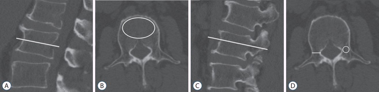
The HU value for the average region of interest (ROI) was determined using the picture archiving and communication system (PACS) software (GE Healthcare, Barrington, IL, USA). The vertebral body HU value was measured in the axial plane of the mid-vertebral body. The elliptical ROI included the maximum possible cancellous bone region and to avoid cortical edges and heterogeneous regions, such as osseous abnormalities and the posterior venous plexus. The pedicle HU value was measured on the axial plane of the mid-pedicle, which commonly lies within the screw trajectory. The elliptical ROI was drawn in the same way as the vertebral body HU, except for the cortical bone. The pedicle HU was determined as the average HU value on both sides. Additionally, the diameter of the pedicle was measured at the narrowest segment on the axial plane of the mid-pedicle. We measured the HU values of the vertebral body and pedicles on preoperative CT at the level of screw pullout during the follow-up (Fig. 1).

Radiologic assessment and definition of screw pullout

Standing whole-spine radiography was performed before and 1 week after surgery to assess global spinal alignment and spinopelvic parameters. The pelvic tilt (PT), pelvic incidence (PI), sacral slope (SS), C7-S1 sagittal vertical alignment (C7SVA), lumbar lordosis (LL), and thoracic kyphosis (TK) were measured.
Screw pullout was defined as a change in pullout length ≥5 mm in the sagittal view on the follow-up spine radiography.

Clinical outcomes

Clinical outcome parameters collected during the preoperative hospital stay, and 12–24 months post-operatively, were used to quantify pain and functional improvement. These parameters included Visual analog scales (VAS) for back and leg pain, Oswestry disability index (ODI) scores, and the 36-Item Short Form Survey (SF-36).

Statistical analysis

Statistical analyses were performed using IBM SPSS Statistics for Windows version 22 (IBM Corporation, Armonk, NewYork, USA). Standard analyses were performed using the paired t-test and the independent Student’s t-test for continuous variables. The chi-square test was used for categorical variables. The vertebral body HU and pedicle HU were divided into three groups using the clustering method with two centroids. For survival analyses of screw pullout, Kaplan-Meier survival curves for each group were drawn and compared using the log-rank test. Statistical significance was determined using a p-value <0.05.

RESULTS

Baseline characteristics

The screw pullout rate was 36.4% (27/74) in the patients undergoing pedicle screw fixation. No statistically significant differences were identified in age, sex, height, weight, BMI, BMD, follow-up periods, fusion level or UIV level between the two groups. The mean fusion levels in the pullout and non-pullout groups were 7.6±2.7 and 8.6±2.1, respectively (p=0.093). The incidence of TP hook instrumentation at UIV was 14.8% (4/27) in the pullout group and 44.7% (21/47) in the non-pullout group, and there were significant differences between the two groups (p=0.009) (Table 1).
Perioperative radiographic parameters showed no significant differences in PT, PI, SS, LL, TK, and C7SVA between the two groups.

HU values of vertebral body and pedicle with stratification using K-means clustering

The HU values of the UIV vertebral body and pedicle in the screw pullout group were significantly lower than the non-pullout group (vertebral body HU, p<0.001; pedicle HU, p<0.001). Although the mean pedicle diameter was higher in the screw pullout group than the non-pullout group, the difference was not statistically significant (p=0.352) (Table 1).
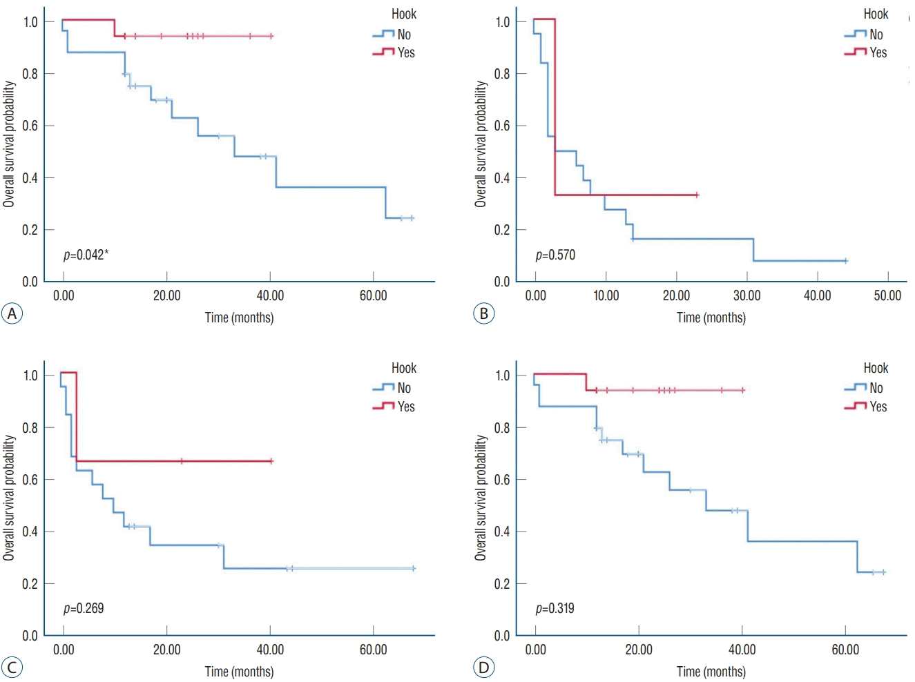
Using K-means clustering, they were divided into three groups with two centroids. The vertebral body HU were group 1 ≥205.3, 205.3> group 2 ≥137.2, group 3 <137.2, and pedicle HU were group 1 ≥243.43, 243.43> group 2 ≥156.03, group 3 <156.03.

Survival analyses on screw pullout

The Kaplan-Meier curves for each analysis are shown in Fig. 2. The application of the TP hook statistically appeared to reduce the occurrence of screw pullout in group 2 from 243.43> pedicle HU to ≥156.03 (p=0.042), and there was no statistical significance was observed when pedicle HU was ≥243.43 or <156.03. Vertebra body HU groups did not show the differences in screw-pullout occurrence whether the application of the TP hook or not.

Pain and functional outcomes

Clinical outcome parameters included the VAS for back and leg pain, ODI scores, and SF-36 scores. No statistically significant differences were observed in the preoperative outcomes assessment between the pullout and non-pullout groups. However, postoperative VAS, ODI, SF-PF, and SF-RP scores were significantly higher in the non-pullout group than patients with screw pullout (Table 2). Similarly, no significant preoperative differences were found with or without hook implementation; however, the postoperative VAS, ODI, SF-PF, and SF-RP showed significant improvement when a hook was used (Table 3).

DISCUSSION

In this study, the screw pullout rate was 36.4% (27/74), which was similar to the rate reported in another recent study (34.2%) [22]. Unlike previous studies, age, number of fusion levels, overcorrection of sagittal alignment, and osteoporosis were not significantly different between the two groups in this study. BMD suggested a trend towards lower values in the pullout group; however, the difference was not statistically significant.
Traditional DXA-based bone density measurements tend to be overestimated in patients with ASD; furthermore, such measurements do not reflect local heterogeneous changes due to the unequal distribution of load caused by malalignment. The HU value is a simple and rapid technique to assess heterogeneous bone quality [28] and is used to predict osteoporosis [3,15,17], pseudoarthrosis [14], cage subsidence [13], and screw loosening [2,19,29].
In this study, vertebral body HU, pedicle HU, and hook application were significantly lower in the screw pullout group. This is consistent with a previous study, which showed that vertebral body HU alone is insufficient for accurate evaluation of screw loosening risk, and that including pedicle HU would facilitate greater accuracy in risk assessment [25].
Furthermore, we shed light on the clinical efficacy of TP hook system as a screw pullout prevention strategy in adult spinal deformity surgery using K-means clustering of pedicle HU at the UIV, suggesting that the pedicle segment is a more significant contributor to TP hook semi-rigid potential than the vertebra body segment.
In biochemical study, the bone density of the intrapedicular segment has a significantly stronger correlation with pedicle screw pull-out strength than other segments [24], and the finite element method study also reported more von Mises stress at the pedicle than vertebra body [20,26].
Although the use of TP hook as a UIV implant was associated with a lower incidence of proximal junctional kyphosis and better functional outcomes than using pedicle screws [5,9,12], some researches raise questions about its clinical effectiveness [10,16]. Although the importance of pedicle HU has been reported in the spine surgical field, consideration of the UIV bone environment of patients with spinal deformity corrective surgery was insufficient. In particular, since there were no observative studies of the effect of the TP hook in specific bone characteristics and environments, it seems that the discussion on the clinical effect of the TP hook is still ongoing. In our study, the effect of TP hook was mainly observed in the mid-range pedicle HU group. First of all, in the group with a sufficiently high UIV pedicle HU, group 1, the application of the TP hook would not show a significant difference in screw pullout because the pedicle screw alone could withstand sufficient shear force. Furthermore, in the case of group 3, even though the TP hook was applied, as shown in Fig. 2B, the shear force itself was too small to protect the screw pullout with the TP hook. On the other hand, in the mid-range pedicle HU, the effect of dynamic stabilization of TP hook is considered to be statistically significant with proper distribution of shear force from UIV pedicle screw. For direct understanding, representative case for UIV screw pullout without TP hook and non-pullout with TP hook in mid-range pedicle HU group (156.03≤ UIV pedicle HU <243.43) was described in Supplementary Fig. 1.
This study has some limitations. Since this a retrospective study from a single institution, it is difficult to generalize the findings. A prospective study is required to validate our results. The control group was established by considering various factors affecting screw pullout, such as age, BMI, radiographic parameters, and fusion level. We confirmed that there was no significant difference of those factors between the two groups. To overcome the results of BMD, HU of VB, HU of pedicle, and pedicle diameter of the subject in the no-pullout group showed more favorable results than the pullout group in entire data, we further analyzed subgroup analysis using k-means clustering algorithm, and compared the efficacy of TP hook under no statistically significant different condition in mid-range pedicle HU group (Supplementary Table 1). However, we did not consider the length or diameter of the screw and mainly assessed UIV screw pullouts. Additional studies are required to assess the factors related to lowest instrumented vertebra (LIV) screw pullouts, features of screws, and the role of hooks. In the case of lumbosacral fixation, it is also necessary to consider a technique for measuring HU values at the LIV. The sample sizes for hook assessment were insufficient because the hook system has been implemented only recently for deformity correction surgery for patients with ASD. Because we focused on the comparison of the screw-pull out or not, we didn’t show the results about pre and postoperative comparative analysis. Further studies are needed to validate the ability of hooks to prevent screw pullout in patients with ASD, and the use of pedicle HU as a predictor of proximal and distal junctional failure.

CONCLUSION

The UIV hook can be considered a preventive strategy against screw pullout after corrective surgery for adult spinal deformity with certain pedicle HU window. Furthermore, The UIV hook application can contribute to good postoperative clinical outcomes with specific categories of SF-36.

Notes

Conflicts of interest

No potential conflict of interest relevant to this article was reported.

Informed consent

This type of study does not require informed consent

Author contributions

Conceptualization : YH, KHK, SR; Data curation : JC, HJJ, JYP, SUK, YH, DKC, KSK, YEC, KHK, SR; Formal analysis : JC, SR; Methodology : HJJ, JYP, SUK, YH, DKC, KSK, YEC, KHK; Project administration : SR; Visualization : JC, SR; Writing - original draft : JC, SR; Writing - review & editing : JC, SR

Data sharing

None

Preprint

None

ACKNOWLEDGMENTS

This research was supported by Korea Health Industry Development Institute (KHIDI) funded by the Ministry of Health & Welfare, Republic of Korea (grant number : HI22C007800).

Supplementary materials

The online-only data supplement is available with this article at https://doi.org/10.3340/jkns.2022.0174.
Supplementary Fig. 1.
Representative case for UIV screw pullout without TP hook and non-pullout with TP hook in mid-range pedicle HU group (156.03≤ UIV pedicle HU <243.43). A : The immediate post-operative X-ray of a patient without TP hook, the patient is 70-years-old female who underwent spinal deformity correction surgery. In pre-operative evaluation, UIV HU pedicle value was 182.3, BMI was 30.6 and BMD was -2.5. B : After 1 year of follow-up, UIV screw was pulled out. C : The immediate post-operative X-ray of a patient with TP hook, the patient is 72-years-old female who underwent spinal deformity correction surgery. In pre-operative evaluation, UIV HU pedicle value was 176.2, BMI was 22.6 and BMD was -2.6. D : After 1 year of follow-up, UIV screw was not pulled out. UIV : upper instrumented vertebra, TP : transverse process, HU : Hounsfield unit, BMI : body mass index, BMD : bone mineral density.
jkns-2022-0174-suppl1.pdf
Supplementary Table 1.
Patient characteristics in mid-range pedicle HU group
jkns-2022-0174-suppl2.pdf

References

1. Bess RS, Lenke LG, Bridwell KH, Cheh G, Mandel S, Sides B. Comparison of thoracic pedicle screw to hook instrumentation for the treatment of adult spinal deformity. Spine (Phila Pa 1976). 32:555–561. 2007.
2. Bredow J, Boese CK, Werner CM, Siewe J, Löhrer L, Zarghooni K, et al. Predictive validity of preoperative CT scans and the risk of pedicle screw loosening in spinal surgery. Arch Orthop Trauma Surg. 136:1063–1067. 2016.
3. Buckens CF, Dijkhuis G, de Keizer B, Verhaar HJ, de Jong PA. Opportunistic screening for osteoporosis on routine computed tomography? An external validation study. Eur Radiol. 25:2074–2079. 2015.
4. Cazzulino A, Gandhi R, Woodard T, Ackshota N, Janjua MB, Arlet V, et al. Soft Landing technique as a possible prevention strategy for proximal junctional failure following adult spinal deformity surgery. J Spine Surg. 7:26–36. 2021.
5. Hassanzadeh H, Gupta S, Jain A, El Dafrawy MH, Skolasky RL, Kebaish KM. Type of anchor at the proximal fusion level has a significant effect on the incidence of proximal junctional kyphosis and outcome in adults after long posterior spinal fusion. Spine Deform. 1:299–305. 2013.
6. Hicks JM, Singla A, Shen FH, Arlet V. Complications of pedicle screw fixation in scoliosis surgery: a systematic review. Spine (Phila Pa 1976). 35:E465–E470. 2010.
7. Hongo M, Gay RE, Zhao KD, Ilharreborde B, Huddleston PM, Berglund LJ, et al. Junction kinematics between proximal mobile and distal fused lumbar segments: biomechanical analysis of pedicle and hook constructs. Spine J. 9:846–853. 2009.
8. Jain RK, Vokes T. Dual-energy X-ray absorptiometry. J Clin Densitom. 20:291–303. 2017.
9. Line BG, Bess S, Lafage R, Lafage V, Schwab F, Ames C, et al. Effective prevention of proximal junctional failure in adult spinal deformity surgery requires a combination of surgical implant prophylaxis and avoidance of sagittal alignment overcorrection. Spine (Phila Pa 1976). 45:258–267. 2020.
10. Mac-Thiong JM, Levasseur A, Parent S, Petit Y. The influence of proximal anchors on the risk of proximal junctional fracture in the osteoporotic spine: biomechanical comparison between pedicle screws and transverse process hooks. J Spinal Disord Tech. 27:E49–E54. 2014.
11. Maruo K, Ha Y, Inoue S, Samuel S, Okada E, Hu SS, et al. Predictive factors for proximal junctional kyphosis in long fusions to the sacrum in adult spinal deformity. Spine (Phila Pa 1976). 38:E1469–E1476. 2013.
12. Matsumura A, Namikawa T, Kato M, Oyama S, Hori Y, Yabu A, et al. Effect of different types of upper instrumented vertebrae instruments on proximal junctional kyphosis following adult spinal deformity surgery: pedicle screw versus transverse process hook. Asian Spine J. 12:622–631. 2018.
13. Mi J, Li K, Zhao X, Zhao CQ, Li H, Zhao J. Vertebral body hounsfield units are associated with cage subsidence after transforaminal lumbar interbody fusion with unilateral pedicle screw fixation. Clin Spine Surg. 30:E1130–E1136. 2017.
14. Nguyen HS, Shabani S, Patel M, Maiman D. Posterolateral lumbar fusion: relationship between computed tomography hounsfield units and symptomatic pseudoarthrosis. Surg Neurol Int. 6(Suppl 24):S611–S614. 2015.
15. Pickhardt PJ, Pooler BD, Lauder T, del Rio AM, Bruce RJ, Binkley N. Opportunistic screening for osteoporosis using abdominal computed tomography scans obtained for other indications. Ann Intern Med. 158:588–595. 2013.
16. Rosinski A, Odeh K, Ungurean V Jr, Leasure J, Kondrashov D. Nonpedicular fixation techniques for the treatment of spinal deformity: a systematic review. JBJS Rev. 8:e0150. 2020.
17. Schreiber JJ, Anderson PA, Rosas HG, Buchholz AL, Au AG. Hounsfield units for assessing bone mineral density and strength: a tool for osteoporosis management. J Bone Joint Surg Am. 93:1057–1063. 2011.
18. Schwab F, Lafage V, Patel A, Farcy JP. Sagittal plane considerations and the pelvis in the adult patient. Spine (Phila Pa 1976). 34:1828–1833. 2009.
19. Schwaiger BJ, Gersing AS, Baum T, Noël PB, Zimmer C, Bauer JS. Bone mineral density values derived from routine lumbar spine multidetector row CT predict osteoporotic vertebral fractures and screw loosening. AJNR Am J Neuroradiol. 35:1628–1633. 2014.
20. Sensale M, Vendeuvre T, Schilling C, Grupp T, Rochette M, Dall’Ara E. Patient-specific finite element models of posterior pedicle screw fixation: effect of screw’s size and geometry. Front Bioeng Biotechnol. 9:643154. 2021.
21. Shin HK, Koo HW, Kim KH, Yoon SW, Sohn MJ, Lee BJ. The usefulness of trabecular CT attenuation measurement at L4 level to predict screw loosening after degenerative lumbar fusion surgery: consider number of fused levels and postoperative sagittal balance. Spine (Phila Pa 1976). 47:745–753. 2022.
22. Sumiya S, Fukushima K, Kurosa Y, Hirai T, Inose H, Yoshii T, et al. Comparative analysis of clinical factors associated with pedicle screw pull-out during or immediately after surgery between intraoperative conebeam computed tomography and postoperative computed tomography. BMC Musculoskelet Disord. 22:55. 2021.
23. Thawrani DP, Glos DL, Coombs MT, Bylski-Austrow DI, Sturm PF. Transverse process hooks at upper instrumented vertebra provide more gradual motion transition than pedicle screws. Spine (Phila Pa 1976). 39:E826–E832. 2014.
24. Wichmann JL, Booz C, Wesarg S, Bauer RW, Kerl JM, Fischer S, et al. Quantitative dual-energy CT for phantomless evaluation of cancellous bone mineral density of the vertebral pedicle: correlation with pedicle screw pull-out strength. Eur Radiol. 25:1714–1720. 2015.
25. Xu F, Zou D, Li W, Sun Z, Jiang S, Zhou S, et al. Hounsfield units of the vertebral body and pedicle as predictors of pedicle screw loosening after degenerative lumbar spine surgery. Neurosurg Focus. 49:E10. 2020.
26. Xu M, Yang J, Lieberman IH, Haddas R. Finite element method-based study of pedicle screw-bone connection in pullout test and physiological spinal loads. Med Eng Phys. 67:11–21. 2019.
27. Yuan L, Zhang X, Zeng Y, Chen Z, Li W. Incidence, risk, and outcome of pedicle screw loosening in degenerative lumbar scoliosis patients undergoing long-segment fusion. Global Spine J. 2021; [Epub ahead of print].
28. Zaidi Q, Danisa OA, Cheng W. Measurement techniques and utility of hounsfield unit values for assessment of bone quality prior to spinal instrumentation: a review of current literature. Spine (Phila Pa 1976). 44:E239–E244. 2019.
29. Zou D, Muheremu A, Sun Z, Zhong W, Jiang S, Li W. Computed tomography Hounsfield unit-based prediction of pedicle screw loosening after surgery for degenerative lumbar spine disease. J Neurosurg Spine. 32:716–721. 2020.

Fig. 1.
Estimation of bone density using Hounsfield unit (HU) values with preoperative computed tomography scan. A : The vertebral body HU value was measured on the axial plane of the mid-vertebral body. B : The elliptical region of interest (ROI) was drawn to include maximum possible amount of cancellous bone, while avoiding cortical edges and heterogeneous regions (white circle). C : The pedicle HU value was measured on the axial plane of the mid-pedicle where screw trajectory is commonly directed (white line). D : The elliptical ROI for the pedicle resembled that of vertebral body HU, except for the cortical bone (white circle). The diameter of the pedicle was measured at the narrowest segment of the pedicle (white line).
jkns-2022-0174f1.tif
Fig. 2.
The Kaplan-Meier curves for screw pullout-free survival : (A) 243.43 >UIV pedicle HU ≥156.03, (B) UIV pedicle HU <156.03, (C) 205.3> UIV vertebral body HU ≥137.2, and (D) UIV vertebral body HU <137.2 whether transverse process hook application or not. *p<0.05. UIV : upper instrumented vertebra, HU : Hounsfield unit.
jkns-2022-0174f2.tif
Table 1.
Patient characteristics
Screw pullout
p-value
Yes (n=27) No (n=47)
Male/female ratio 6 : 21 3 : 44 0.084
Age (years) 68.8±6.1 69.4±5.4 0.649
Height (cm) 155.5±8.3 154.0±5.9 0.435
Weight (kg) 61.5±9.6 59.8±9.4 0.441
BMI (kg/m2) 25.1±2.8 25.2±3.5 0.982
BMD -2.1±1.1 -1.9±1.0 0.484
Follow-up periods (months) 35.1±19.6 30.4±23.4 0.387
Fusion levels 7.6±2.7 8.6±2.1 0.093
UIV level 11.5±2.8 10.9±2.0 0.375
UIV TP hook* 4 (14.8) 21 (44.7) 0.009
UIV Vertebral body HU* 135.0±33.8 183.4±46.9 <0.001
UIV pedicle HU* 147.1±43.1 218.8±48.7 <0.001
UIV pedicle diameter (mm) 7.7±3.1 6.9±1.6 0.352
Pre-operative radiographic parameters
 PT (°) 35.1±12.5 33.4±11.4 0.565
 PI (°) 54.5±11.1 53.6±11.4 0.736
 SS (°) 19.5±12.9 20.2±14.2 0.827
 LL (°) 9.9±16.6 11.2±22.5 0.807
 TK (°) -17.8±15.1 -18.2±17.8 0.703
 C7SVA (mm) 118.4±70.8 95.2±60.6 0.142
Post-operative radiographic parameters
 PT (°) 24.8±9.5 27.4±18.2 0.490
 PI (°) 49.4±12.9 52.1±10.1 0.344
 SS (°) 24.7±11.7 26.8±11.1 0.431
 LL (°) 35.7±14.2 34.8±17.1 0.809
 TK (°) -9.5±14.9 -8.3±11.3 0.700
 C7SVA (mm) 21.6±51.2 39.5±47.2 0.133

Values are presented as mean±standard deviation or number (%) unless otherwise indicated.

* p<0.05.

BMI : body mass index, BMD : bone mineral density, UIV : upper instrumented vertebra, TP : transverse process, HU : Hounsfield unit, PT : pelvic tilt, PI : pelvic incidence, SS : sacral slope, LL : lumbar lordosis, TK : thoracic kyphosis, C7SVA : C7-S1 sagittal vertical alignment

Table 2.
Comparison of clinical outcomes with VAS, ODI, SF-36 before and after surgery according to incidence of screw pullout
Pre-operation
Post-operation
Pullout (n=27) Non-pullout (n=47) p-value Pullout (n=27) Non-pullout (n=47) p-value
VAS* 7.8±1.6 7.3±1.4 0.212 4.6±1.9 3.3±1.4 0.018
ODI (%)* 66.7±16.5 64.6±11.8 0.605 53.8±13.9 42.7±13.2 0.014
PF* 9.3±13.5 17.2±16.8 0.121 17.6±16.2 44.1±17.9 <0.001
SF 25.0±18.3 34.8±28.7 0.238 38.4±24.3 34.8±22.9 0.652
RP* 6.3±9.4 16.7±22.9 0.099 13.1±16.2 36.8±22.7 0.001
PCS 18.1±10.3 26.2±16.6 0.072 30.3±16.1 34.9±17.4 0.424
MCS 29.8±12.3 40.5±22.9 0.102 43.5±17.3 40.2±23.9 0.659

Values are presented as mean±standard deviation.

* p<0.05.

VAS : Visual analog scales, ODI : Oswestry disability index, SF-36 : the 36-Item Short Form Survey, PF : physical functioning, SF : social functioning, RP : role limitations caused by physical problems, PCS : physical component summary, MCS : mental component summary

Table 3.
Comparison of clinical outcomes with VAS, ODI, SF-36 before and after surgery according to incidence of hook implementation
Pre-operation
Post-operation
With hook (n=25) Without hook (n=49) p-value With hook (n=25) Without hook (n=49) p-value
VAS* 7.4±1.9 7.5±1.2 0.212 3.2±1.4 4.1±1.7 0.039
ODI (%)* 67.5±12.7 63.3±14.8 0.605 41.7±13.9 53.4±12.5 0.006
PF* 15.6±17.2 13.6±15.2 0.121 41.2±19.9 25.3±20.4 0.012
SF 28.8±24.8 34.4±27.2 0.238 35.6±25.1 36.8±21.4 0.884
RP* 14.7±24.2 11.3±13.7 0.099 34.6±26.2 19.1±16.5 0.028
PCS 24.6±18.4 21.4±10.5 0.072 32.3±19.3 34.2±13.9 0.734
MCS 35.4±22.8 38.4±17.6 0.102 39.1±22.9 44.3±19.9 0.474

Values are presented as mean±standard deviation.

* p<0.05.

VAS : Visual analog scales, ODI : Oswestry disability index, SF-36 : the 36-Item Short Form Survey, PF : physical functioning, SF : social functioning, RP : role limitations caused by physical problems, PCS : physical component summary, MCS : mental component summary

TOOLS
Similar articles