INTRODUCTION
MATERIALS AND METHODS
Patient population
Radiographic parameters
Statistical analysis
RESULTS
Table 1.
Values are presented as mean±standard deviation or number unless otherwise indicated. ALIF : anterior lumbar interbody fusion, OLIF : oblique lumbar interbody fusion, TLIF : transforaminal lumbar interbody fusion; BMD : bone mineral density, ASA PS : American Society of Anesthesiologists physical status
Table 2.
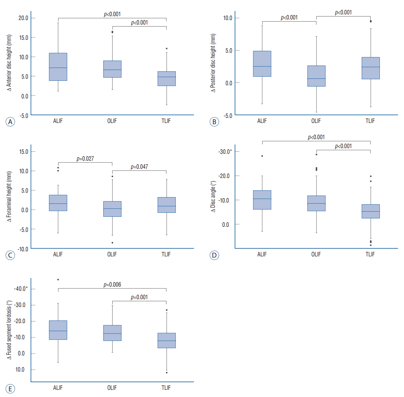
 | Fig. 1.Changes in local radiographic parameters after multilevel lumbar interbody fusion according to the approaches. A : changes in anterior disc height. B : changes in posterior disc height. C : changes in foraminal height. D : changes in disc angle. E : changes in fused segment lordosis. *Negative values indicate lordosis. ALIF : anterior lumbar interbody fusion, OLIF : oblique lumbar interbody fusion, TLIF : transforaminal lumbar interbody fusion. |
Table 3.
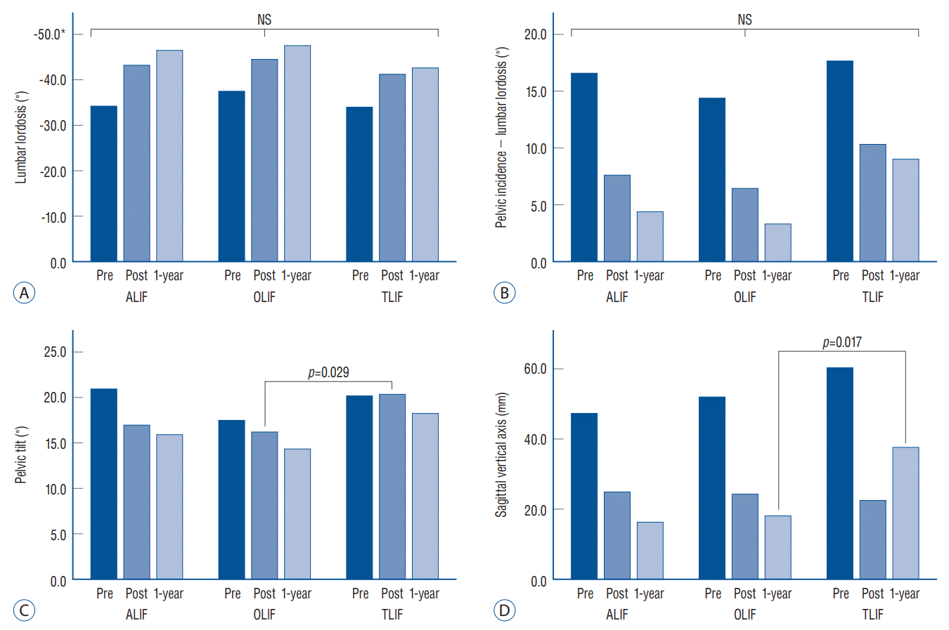
 | Fig. 2.Changes in global sagittal alignment after multilevel lumbar interbody fusion according to the approaches. A : changes in lumbar lordosis. B : changes in pelvic incidence – lumbar lordosis. C : changes in pelvic tilt. D : changes in sagittal vertical axis. *Negative values indicate lordosis. NS : not significant, ALIF : anterior lumbar interbody fusion, OLIF : oblique lumbar interbody fusion, TLIF : transforaminal lumbar interbody fusion. |
 | Fig. 3.A 74-year-old woman underwent anterior lumbar interbody fusion (ALIF) at L4-S1. A and B : Preoperative and postoperative radiographs revealed an increased disc angle following ALIF. D and D : Preoperative and 1-year postoperative standing radiographs, respectively. Improvements in fused segment lordosis (FSL) (from -32.3° to -44.0°) and pelvic incidence (PI) (from 25.2° to 16.2°) were observed after surgery. However, the change in lumbar lordosis (LL) was trivial (from -45.1° to -45.3°), possibly due to a decrease in intraspinal compensation. PT : pelvic tilt, SVA : sagittal vertical axis. |
 | Fig. 4.A 66-year-old woman underwent oblique lumbar interbody fusion (OLIF) at L3-5. A and B : Preoperative and postoperative radiographs revealed an increased disc angle following OLIF. C and D : Preoperative and 1-year postoperative standing radiographs, respectively. Improvements in fused segment lordosis (FSL) (from -12.7° to -38.6°) and pelvic incidence (PI) (from 32.4° to 13.1°) were observed following surgery. Lumbar lordosis (LL) was markedly increased following surgery (from -25.1° to -46.4°). PT : pelvic tilt, SVA : sagittal vertical axis. |
 | Fig. 5.A 70-year-old man underwent transforaminal lumbar interbody fusion (TLIF) at L3-5. A and B : Preoperative and postoperative radiographs revealed an increased disc angle following TLIF. C and D : Preoperative and 1-year postoperative standing radiographs, respectively. Fused segment lordosis (FSL) (from -4.0° to -25.4°) was improved following surgery. Spinopelvic parameters, including lumbar lordosis (LL) (from -25.5° to -47.4°), pelvic incidence (PI) (from 21.3° to 18.9°), and sagittal vertical axis (SVA) (from 140.1 mm to -12.5 mm) were also improved following surgery. PT : pelvic tilt. |


 PDF
PDF Citation
 Print
Print


 XML Download