

 PDF
PDF Citation
 Print
Print


Introduction
Sudden onset of abdominal distension in children is a common cause of emergency department (ED) visits. The distension can be considered medical or surgical according to the intensity, duration of associated symptoms, and findings of physical examination. In mild cases, abdominal distension is often caused by transient gastroenteritis, overeating or constipation. Previously healthy children are treated with medications or enema. Prolonged pain or distension has various causes, such as Hirschsprung’s disease (HD), intussusception, volvulus, appendicitis, Meckel’s diverticulum, lymphatic malformation or malignant tumor1). Plain radiography, ultrasonography, and computed tomography (CT) can help diagnose the underlying causes. This article describes a recent case of intestinal obstruction caused by acute perforated appendicitis misdiagnosed as HD in a 31-month-old boy who had several enemas for an unknown cause of abdominal distension prior to the diagnosis. This study was approved by the institutional review board of Dong-A University Medical Center (IRB no. DAUHIRB-22-196).
Case
A previously healthy 31-month-old boy was referred to the ED with severe abdominal distension that had persisted for 10 days. He had been treated with glycerin enema at a local clinic 8 days before, and at an outside ED 5 days before the referral. Abdominal bloating started 10 days prior to the referral. A pediatrician at the local clinic prescribed oral medication with the enema, assuming that the boy had gastroenteritis. He gradually became lethargic and unable to eat well, and developed abdominal pain. At the visit to the outside ED for the pain, he received another enema. After 3 days (2 days before the referral), he developed fever with ongoing abdominal pain. On the same day of referral, non-enhanced CT was performed at the outside ED. The CT showed marked small bowel distension with the colon collapsed, prompting the referral for evaluation for HD and possible surgery.
At presentation, the boy was ill-looking, lethargic, and dehydrated. The abdominal pain made him lie in the right decubitus position with irritability. The initial vital signs were as follows: blood pressure, 119/80 mmHg; heart rate, 121 beats/minute; respiratory rate, 40 breaths/minute; and temperature, 37.4°C. His height was 84.3 cm (< 3 percentile) and weight was 14.3 kg (50-75 percentile). Physical examination showed the tense and distended abdomen. Abdominal tenderness was uncheckable due to his irritability. The gastric juice was drained via a nasogastric tube drainage with a rate of less than 30 mL/day. The anus was located within the normal sphincter. There was no explosive passage of feces on digital rectal examination. Although saline irrigation enema was performed, a small amount of feces were evacuated.
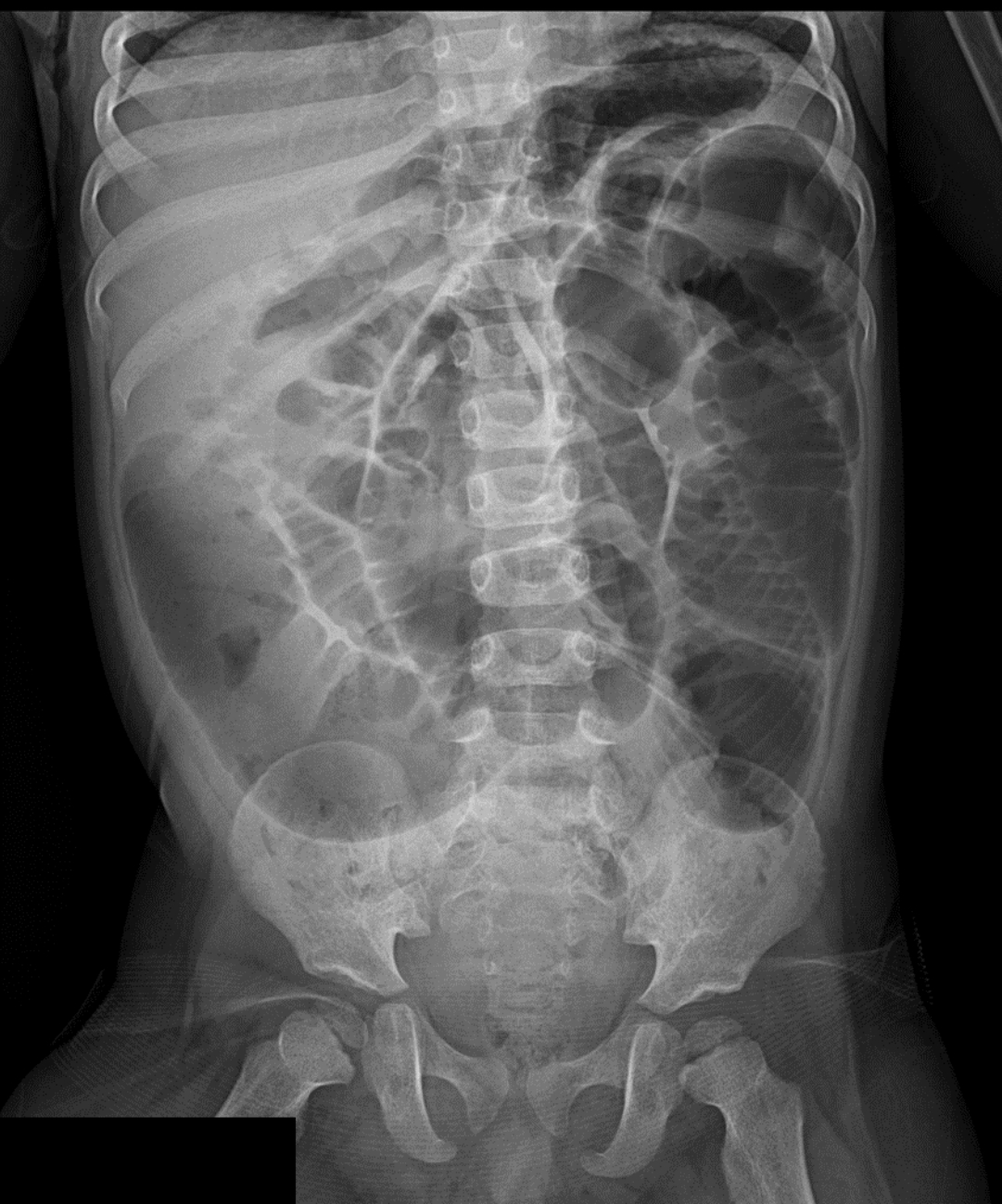
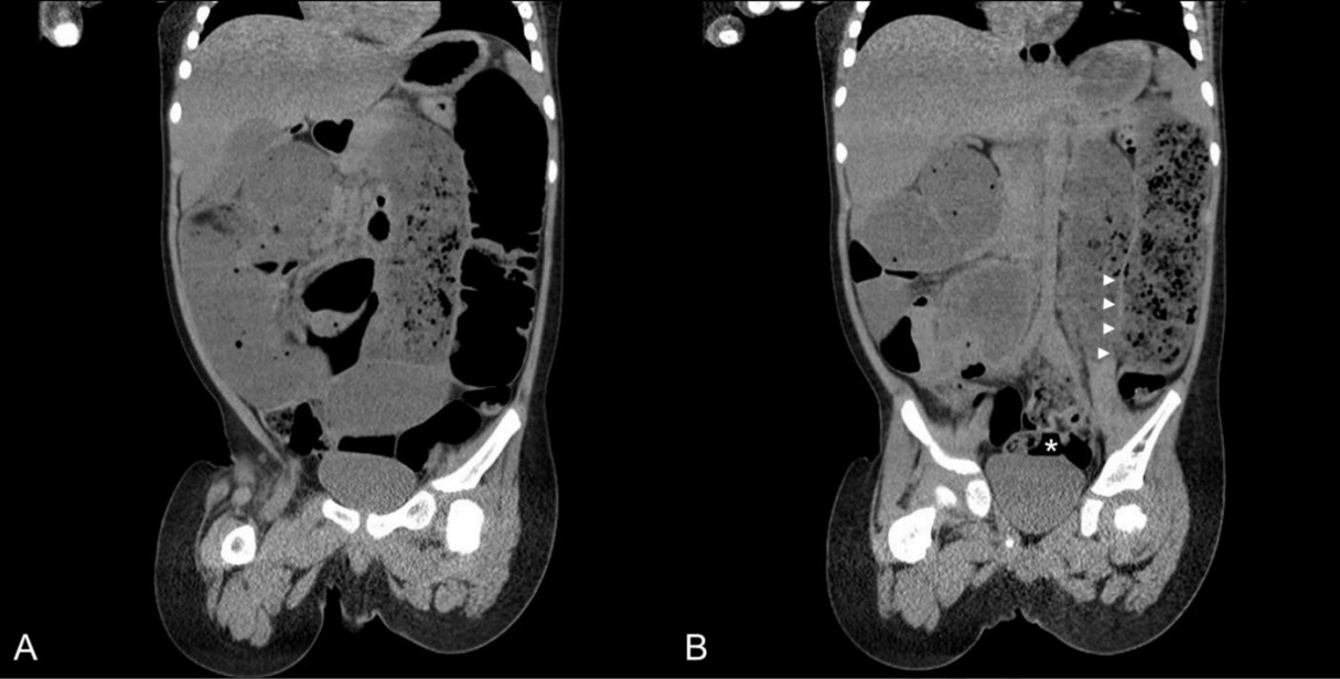
The initial laboratory findings on the day of hospitalization were as follows: hemoglobin, 11.9 g/dL; hematocrit, 33.5%; leukocytes, 20,040/mm3 (neutrophils, 60%); platelets, 464,000/mm3; C-reactive protein, 5.0 mg/dL; protein, 6.4 g/dL; albumin, 3.7 g/dL; total bilirubin, 0.7 mg/dL; direct bilirubin, 0.1 mg/dL; aspartate aminotransferase, 64 IU/L; alanine aminotransferase, 37 IU/L; blood urea nitrogen, 19 mg/dL; creatinine, 0.4 mg/dL; pH, 7.37; sodium, 130 mmol/L; potassium, 3.7 mmol/L; chloride, 92 mmol/L; and total carbon dioxide, 23.4 mmol/L. Stool study was not performed. A plain radiograph showed the dilated bowel loops (Fig. 1). On review of the CT performed at the outside ED, we noted the findings of intestinal obstruction (Fig. 2).
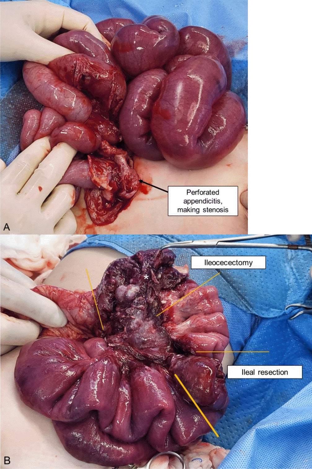
On day 4, exploratory laparotomy was performed to differentiate the potential causes of the obstruction, such as an inflammatory lesion or late-onset HD. When the abdominal cavity was opened, abundant pus was drained. The entire small bowel and colon were explored using blunt dissection. The dissection was technically difficult because of the severely inflamed small bowel loop adhered to the retroperitoneum. The terminal ileum, 10 cm above the ileocecal valve, firmly adhered to another loop of mesentery, and grossly showed a stenotic lesion following being squashed from the entire small bowel. After mobilization of the cecum, the perforated appendix was found adhered to the retroperitoneum and posterior mesentery (Fig. 3A). The entire colon had normal caliber with peristalsis. The author resected approximately 10 cm of the necrotic terminal ileum and anastomosed it side-to-side using surgical staplers (Fig. 3B). Ileocecectomy, including the necrotic appendix, was performed with functional end-to-end anastomosis using a surgical stapler. Pathologically, the resected terminal ileum showed a transmural hemorrhagic infarction, and the resected appendix severe inflammation with perforation.
On day 7 (postoperative day 3), the boy became able to take sips of water, and recovered well. On day 11, a wound seroma was detected and treated with a wound drain insertion and reinforced sutures. He was discharged without a complication on day 14.
Discussion
Intestinal obstruction can occur from various conditions, such as adhesions (congenital or acquired after surgery), hernias, tumors, strictures from an intestinal inflammation caused by Crohn’s disease or vasculitis, intussusception, bezoars, and foreign bodies2-5). Particularly in young children, it is important to ensure whether the obstruction originates from congenital gastrointestinal anomalies. Thus, HD may manifest as a late-onset intestinal obstruction presenting as abdominal distension or chronic constipation. The absence of intestinal ganglion cells leads to functional obstruction with incomplete relaxation of the affected bowel segment. Onset time is dependent on the length of the involved segment6). Therefore, the case patient was referred with a presumptive diagnosis of HD. In the author’s opinion, retrospectively, late-onset HD was less likely to be present in the boy without constipation. Most cases of the HD present with encopresis with less than 2 episodes of defecation per week and severe bowel dilation7). His plain radiography did not show findings suggestive of HD, such as transitional zone or reversed rectosigmoid ratio.
The author focused on the medical history of sudden onset of abdominal bloating, mild fever, leukocytosis, and findings of rectal examination. The non-enhanced CT was not helpful in the differential diagnosis. Meckel’s diverticulum and inflammatory lesions, such as appendicitis and enterocolitis, could not be excluded by imaging studies. Given the presumptive diagnosis of HD, the author planned the elective laparotomy to check the absence of ganglion cells on a frozen section, in case of indefinite cause of the obstruction.
The cause of the intestinal obstruction was eventually proven to be acute perforated appendicitis. As per a review by Makama et al.3), 45 case reports of small bowel obstruction due to appendicitis were published between 1950 and 2016. There were 4 categories: adynamic ileus, mechanical ileus, strangulation, and mesentery ischemia. The cited reports explained appendiceal morphology of mechanical ileus as follows: the inflamed tip directly adheres to the bowel wall or posterior peritoneum across the ileum or mesentery near the ileocolic artery, internal herniation between the gap of the appendiceal base and tip, and formation of a complex knot3). Mobility of the appendix can cause various types of obstruction. In this case, the author speculated that the inflamed appendix directly adhered to the small bowel mesentery-like fibrous band. This lesion might have acted as a stricture to the small bowel, and delayed treatment contributed to adjacent mesentery ischemia following thromboembolism and pressure necrosis.
Without imaging findings suggestive of acute appendicitis, it is difficult to pinpoint it as the primary cause of intestinal obstruction4-5,8-10). Additionally, the clinical suspicion of appendicitis in a 3-year-old child could be difficult given the incidence of appendicitis lower than 0.5 per 1,000 person-years in the young children11).
Sudden onset of intestinal obstruction without a previous surgery can be caused by inflammatory lesions, particularly acute appendicitis, even though preoperative diagnosis is difficult. This case may facilitate a timely diagnosis of intestinal obstruction in young children without a history of surgery.
 XML Download