Methods
This was a prospective, double-blind, randomized clinical trial. The institutional Research Ethics Committee of Cairo University El-Kasr Alainy Hospital approved this study (IRB no. MD-250-2020). The trial was registered on clinicaltrials.gov (Ref no. NCT 04417179) and was conducted from January 2021 to February 2022 in accordance with the Helsinki Declaration-2013. All patients who were screened and met the eligibility criteria were invited to participate in the trial, and all enrolled patients provided written informed consent. Consent was requested from patients upon arrival to the operating suite for surgery or on the ward if they were admitted the night before surgery.
The inclusion criteria were as follows: patients of any gender aged 18–60 years with American Society of Anesthesiologists physical status classifications II–III and a body mass index (BMI) of 40–50 kg/m2 who did not exhibit any of the following: contraindications to peripheral regional anesthesia blocks, existing infection at the block site, contraindication to regional anesthesia, history of opiate abuse, pre-existing chronic pain or cognitive dysfunction (which would impede accurate engagement with postoperative quality of recovery and analgesia assessment), refusal of the regional block, any neurological or psychological disorders, inability to cooperate, patients scheduled for concomitant laparoscopic cholecystectomy or paraumbilical hernia repair, those with a history of previous bariatric surgery or obstructive sleep apnea, patients with anatomic abnormalities at the site of injection, and those with skin lesions or a wound at the site of proposed needle insertion.
The individual indications for surgery were laparoscopic bariatric surgery, that is, sleeve gastrectomy and/or Roux-en-Y gastric bypass (RYGB) surgery.
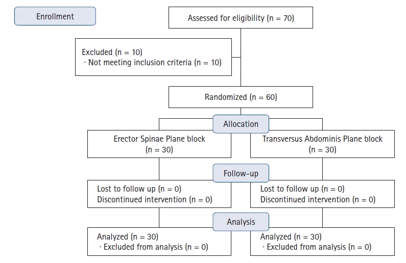
The patients were assigned to one of the trial groups using a computer-generated random number table. Patients with even numbers were allocated into the ESP block group and those with odd numbers were allocated into the TAP block group. The patient study code number and group allocation were typed on separate pages, folded, and concealed in sequentially numbered, sealed envelopes. Block randomization in groups of six individuals was applied to ensure an even number in each group as the study progressed. The groups were named “ESP” and “TAP”. An independent third party held the randomization key. Both patients and anesthetists involved in postoperative data collection were blinded to the group to which the patients were allocated.
Upon arrival in the operating room, perioperative monitoring, which included continuous electrocardiogram (GE-Datex Ohmeda 5-lead ECG cable, USA), pulse oximetry (GE-Datex Ohmeda finger SpO2 sensor), and non-invasive arterial blood pressure (GE-Datex Ohmeda NIBP cuff), were initiated. Baseline vital signs were recorded, including non-invasive measurements of systolic, mean, and diastolic arterial pressures, and HR and oxygen saturation. After intravenous (IV) access, the patient was premedicated with metoclopramide at 0.1–0.2 mg/kg. The patient was randomly assigned to one of two groups according to the intervention used: Group A (30 patients), which received the TAP block, and Group B (30 patients), which received the ESP block.
Both blocks were performed by the primary investigator and supervised by consultant anesthesiologists who had experience in regional anesthesia and were familiar with the ESP and TAP blocks. Additionally, prior to the administration of either block, 1 mg of IV midazolam and 5 ml of lidocaine 1% infiltration were administered on both sides at the site of the block needle injection.
Ultrasound-guided blocks were performed under full aseptic conditions according to randomization before the surgery. All patients received bupivacaine 0.25% at a total volume of 40 ml regardless of the block they received. All blocks were performed with a blunted tip, 20-gauge, short bevel needle (Pajunk Sonoplex, Germany) using the same ultrasound machine (high-frequency linear ultrasound transducer, Siemens Acuson x300 3–5 MHz ultrasound), which was placed in a sterile cover.
The patients randomized to the ESP group were first placed in the prone position. The ESP block was then performed using a high-frequency linear ultrasound transducer that was placed sagittally against the target vertebral level (T5 transverse process) in the prone position and moved approximately 3 cm laterally to the spinous process. The erector spinae muscle and transverse process were then identified, and a blunted tip, 20-gauge, short bevel needle (Pajunk Sonoplex, Germany) was advanced using the in-plane approach in a cephalad-to-caudal direction through the interfascial plane between the erector spinae and the underlying transverse process under strict aseptic precautions until the tip had been advanced deep into the erector spinae muscle, as evidenced by visible hydro-dissection below the muscle plane and a 5-ml injection of normal saline to confirm the correct needle position. The block was performed bilaterally by injecting 40 ml of 0.25% bupivacaine (20 ml on each side) into the fascial plane between the deep surface of the erector spinae muscle and transverse processes of the lumbar vertebrae laterally (at the most lateral part of the transverse process).
Patients randomized to the TAP group were placed in the supine position. The TAP block was then administered using a high-frequency linear ultrasound transducer. After skin preparation and isolation, the transducer was placed 2 cm subxiphoidian and moved along the subcostal edge to identify the rectus abdominis muscle and transversus abdominis. Once these structures were identified, a blunted-tip, 20-gauge, short bevel needle (Pajunk Sonoplex, Germany) was introduced using the in-plane approach 2–3 cm laterally to the transducer under direct ultrasound visualization, and 1–2 ml of saline was injected between the rectus abdominis and transversus abdominis muscles. After confirming the correct placement of the needle and negative aspiration probe, the rest of the anesthetic was injected along the subcostal line in the TAP (20 ml 0.25% bupivacaine), and dissection of the plane was observed. The block was performed bilaterally. A total of 20 ml of 0.25% bupivacaine was injected on each side after aspiration to avoid intravascular placement.
Thirty minutes after performing each block, all patients received general anesthesia induced with fentanyl (1.5 g/kg) based on lean body weight (maximum dose of 200 g), and propofol (2 mg/kg) was administered based on total body weight. Tracheal intubation was facilitated with 0.5 mg/kg of atracurium based on the ideal body weight. Anesthesia was maintained using isoflurane in oxygen and air. Additional doses of 0.1 mg/kg atracurium were administered every 30 min. A urinary catheter was placed to control diuresis. Surgical intervention was permitted 20 min after completion of the block procedure. Volume-controlled ventilation was adjusted to maintain normocapnia. Anesthesia was maintained using 1–1.5% isoflurane in a mixture of oxygen and air (50/50) and atracurium top-ups at a dose of 0.1 mg/kg every 30 min. All participants were then administered 1 g of IV paracetamol (maximum dose of 4 g/24 h) together with 4 mg of ondansetron 10 min prior to the end of surgery for postoperative nausea and vomiting prophylaxis.
After skin closure, the inhaled anesthesia was discontinued and muscle relaxation was reversed with IV atropine (0.02 mg/kg) and neostigmine (0.05 mg/kg) after the return of spontaneous breathing. Patients were transferred to the post-anesthesia care unit (PACU) for 60 min for complete recovery and monitoring.
If at any point hypotension occurred (defined as a decrease in mean arterial pressure > 25% from the baseline value or systolic arterial pressure of 100 mmHg), it was treated with 5 mg of IV bolus ephedrine, which was repeated every 3 min until the hypotension resolved. Bradycardia (defined as an HR of 40 beats/min) was treated with intravenous atropine (0.5 mg).
In the PACU, the visual analog scale (VAS) score was assessed 15 min after extubation by the attending anesthetist. When the score was ≥ 4/10, rescue analgesia in the form of nalbuphine 0.1 mg/kg (individual dose not to exceed 20 mg and a maximum dose of 50 mg/24 h) was administered. Another dose of nalbuphine 0.1 mg/kg was given in the PACU to patients with a VAS score > 4 30 min after the first dose.
After discharge from the PACU, the analgesia plan was intravenous paracetamol (1 g/8 h), nalbuphine (0.1 mg/kg/8 h) if the VAS score was ≥ 4, and ketorolac as a second rescue analgesia (0.5 mg/kg/6 h) as long as the pain score remained ≥ 4 (reassessment was done by the nurses in the ward 30 min after administration of the first rescue analgesia).
The primary outcome was the analgesic efficacy of the ESP block versus the TAP block, as assessed by the mean VAS score in the first 24 h postoperatively.
Secondary outcomes were as follows: the time required for a successful block (measured as the time from initiation of ultrasound scanning to completion of the block on both sides), incidence of complications, time to first rescue analgesia, time to first flatus or stool passage, and total opioid consumption.


 PDF
PDF Citation
 Print
Print


 XML Download