Introduction
Methods
1. Patients and preoperative assessment
Table 1.
M, male; F, female; TDAP, thoracodorsal artery perforator; SA, serratus anterior; PTV, posterior tibial vessels; MTB, Mycobacterium tuberculosis; HERZ, isoniazid, ethambutol, rifampicin, pyrazinamide; CKD, chronic kidney disease; HTN, hypertension; DM, diabetes mellitus; NTM, nontuberculous mycobacteria; M., Mycobacterium; CV, cephalic vein; TB, tuberculosis; ATV, anterior tibial vessels; SLE, systemic lupus erythematosus; LDmc, latissimus dorsi musculocutaneous; MSGA, medial superior genicular artery; MCFA, medial circumflex femoral artery; RA, rheumatoid arthritis; ESRD, end-stage renal disease; +, yes; –, no.
2. Surgical techniques
3. Antimycobacterial medication
Results
1. Demographics
2. Case reports
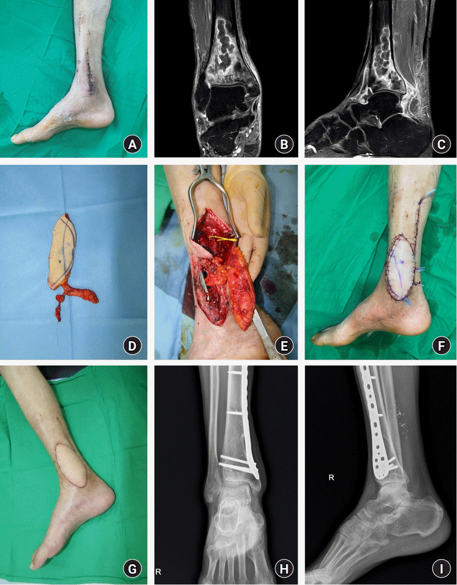
Case 10
 | Fig. 1.(A) A patient with tuberculous osteomyelitis on the right distal tibia. A preoperative photograph shows the previous operative wound with recurrent dehiscence. (B) Coronal view of preoperative T1-weighted magnetic resonance imaging (MRI). (C) Sagittal view of preoperative T1-weighted MRI. An intramedullary abscess measuring approximately 6×2×2 cm was identified. (D) A thoracodorsal artery perforator (TDAP) chimeric flap containing a 6×11-cm skin paddle and 1.5×5-cm serratus anterior (SA) fascial component was harvested. (E) The harvested TDAP chimeric flap is prepared for inset, covering the previously fixed plate containing an SA fascial component. (F) Immediate postoperative photograph of reconstruction using a TDAP chimeric flap. (G) One-year postoperative follow-up photograph shows no problems, including recurrence. (H) Anteroposterior view of the 1-year postoperative follow-up X-ray. (I) Lateral view of the 1-year postoperative follow-up X-ray, showing no remaining osteomyelitis, while the previously fixed plate remained in place. |
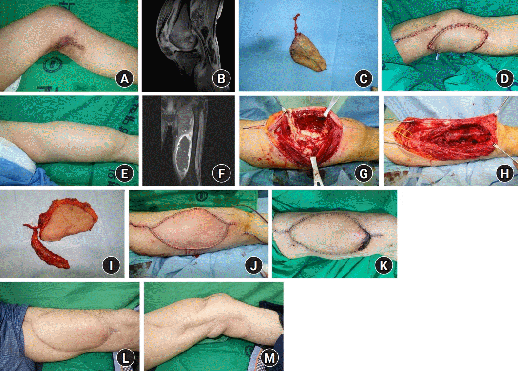
Case 12
 | Fig. 2.(A) A patient with tuberculous arthritis of the left knee joint. A preoperative photograph shows a defect measuring approximately 2×2 cm aligned with the healed wound from previously attempted closure. (B) Sagittal view of preoperative T1-weighted magnetic resonance imaging (MRI). An abscess with ill-defined septa was identified on the semimembranous bursa. (C) A 12×8-cm thoracodorsal artery perforator (TDAP) flap was harvested. (D) Immediate postoperative photograph of reconstruction using a TDAP flap. (E) Broad swelling was observed on the left thigh, adjacent to the prior arthritis region. (F) Coronal view of follow-up T1-weighted MRI shows an approximately 9×7×25-cm abscess arising from the suprapatellar bursa, superiorly extending along the vastus medialis and vastus intermedius muscle. (G) Intraoperative photograph shows whitish necrotic debris in the abscess. (H) The wound was cleared with application of negative-pressure wound therapy. (I) A TDAP chimeric flap containing a 25×12-cm skin paddle and a 10×3-cm serratus anterior muscular component was harvested. (J) Immediate postoperative photograph of reconstruction using a TDAP chimeric flap on the thigh. (K) Partial distal flap necrosis was found after 2 weeks but healed without additional surgery. (L, M) Fifteen-month postoperative follow-up photographs show no problems, including recurrence, in the thigh and popliteal region. |


 PDF
PDF Citation
 Print
Print


 XML Download