Abstract
Conservative treatment shows favorable results for minimally displaced fractures of the distal radius. A commonly reported complication associated with distal radius fractures is delayed tendon rupture. The extensor pollicis longus tendon is the tendon that has most commonly been reported to be injured in distal radius fractures. This report provides a new case of delayed rupture of the extensor carpi radialis longus and extensor carpi radialis brevis tendons after conservative treatment of a distal radius fracture, along with a review of the literature.
The distal radius fracture is one of the most common fractures in the forearm. Acceptable alignment fractures can be treated nonoperatively and commonly have a good clinical outcome [1]. However, it is necessary to inform patients about complications associated with conservative treatment, like delayed tendon injury or carpal tunnel syndrome [2].
Delayed tendon injury is a common complication of distal radius fractures. After conservative treatment, the tendon rupture most commonly involved is the extensor pollicis longus (EPL). If delayed rupture of the EPL occurs, a tendon transfer or tendon graft is usually required to restore function [3].
We found delayed rupture of extensor carpi radialis longus (ECRL) and extensor carpi radialis brevis (ECRB) associated with distal radius fracture. Delayed rupture of ECRB and ECRL due to unrecognized distal radius fracture has not been reported, so we report the clinical course and literature review.
This report was approved by the Institutional Review Board of SMG-SNU Boramae Medical Center (No. 20-2022-79). Written informed consent for publication of the clinical images was obtained from the patient.
A 71-year-old female patient presented to our institution with weakness during extension for 1 year and a palpable mass on the dorsum of the left wrist. She had a trauma history of the left wrist 16 months ago, but she did not seek medical attention. She experienced the right distal radius fracture and was treated by open reduction and internal fixation by volar locking plate fixation technique four years ago. On the physical examination, motor power for wrist extension was grade 4, and about 2.0×2.0 cm-sized mass was palpated on the dorsal aspect of the 2nd to 3rd metacarpal bone base area. Plain radiographs of the left wrist showed a slightly reduced volar tilt by 5° that suspected a previous fracture (Fig. 1). However, it was impossible to compare with the contralateral wrist because of previous surgery. The patient had not received any other treatment, such as steroid injections for her left wrist, and she was a housewife, not a heavy wrist worker. The musculoskeletal radiologist suspected a fibroma or giant cell tumor of the tendon sheath by confirming a 2.0×2.0×1.5-cm mass on the ECRB and ECRL on the ultrasound examination (Fig. 2). On ultrasonography, the continuity of ECRB and ECRL appeared intact.
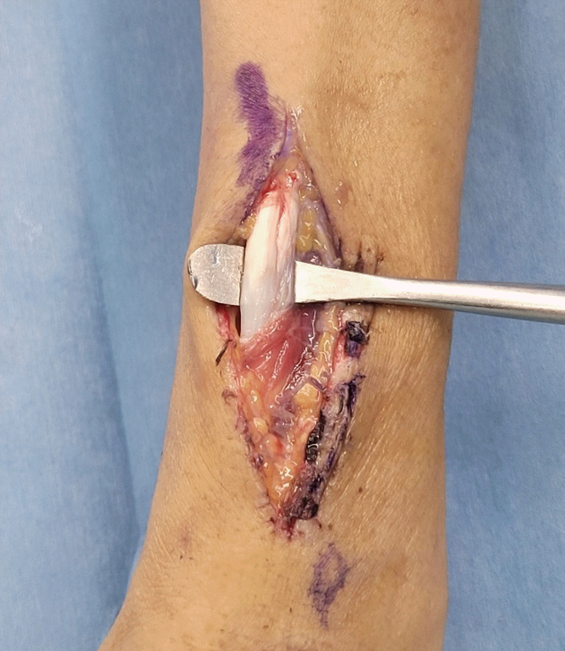
The first operation under local anesthesia was performed for excision with a biopsy of the mass. However, the mass was not soft tissue and was identified as a distal remnant of ruptured ECRL and ECRB tendon (Fig. 3). Additional surgical exploration was conducted in the 3-cm range near the Lister’s tubercle, but proximal stumps could not be observed. It was difficult to take more time to search the proximal stumps and reconstruction due to the tourniquet. The operator ended the operation for further evaluation and reconstruction planning. The magnetic resonance imaging (MRI) demonstrated a total rupture of ECRL and ECRB tendon and showed the proximal stumps were at 6 cm proximal from Lister’s tubercle. An osteophyte, sharply protruding to the second extensor compartment, was seen on implying a previous fracture (Fig. 4).
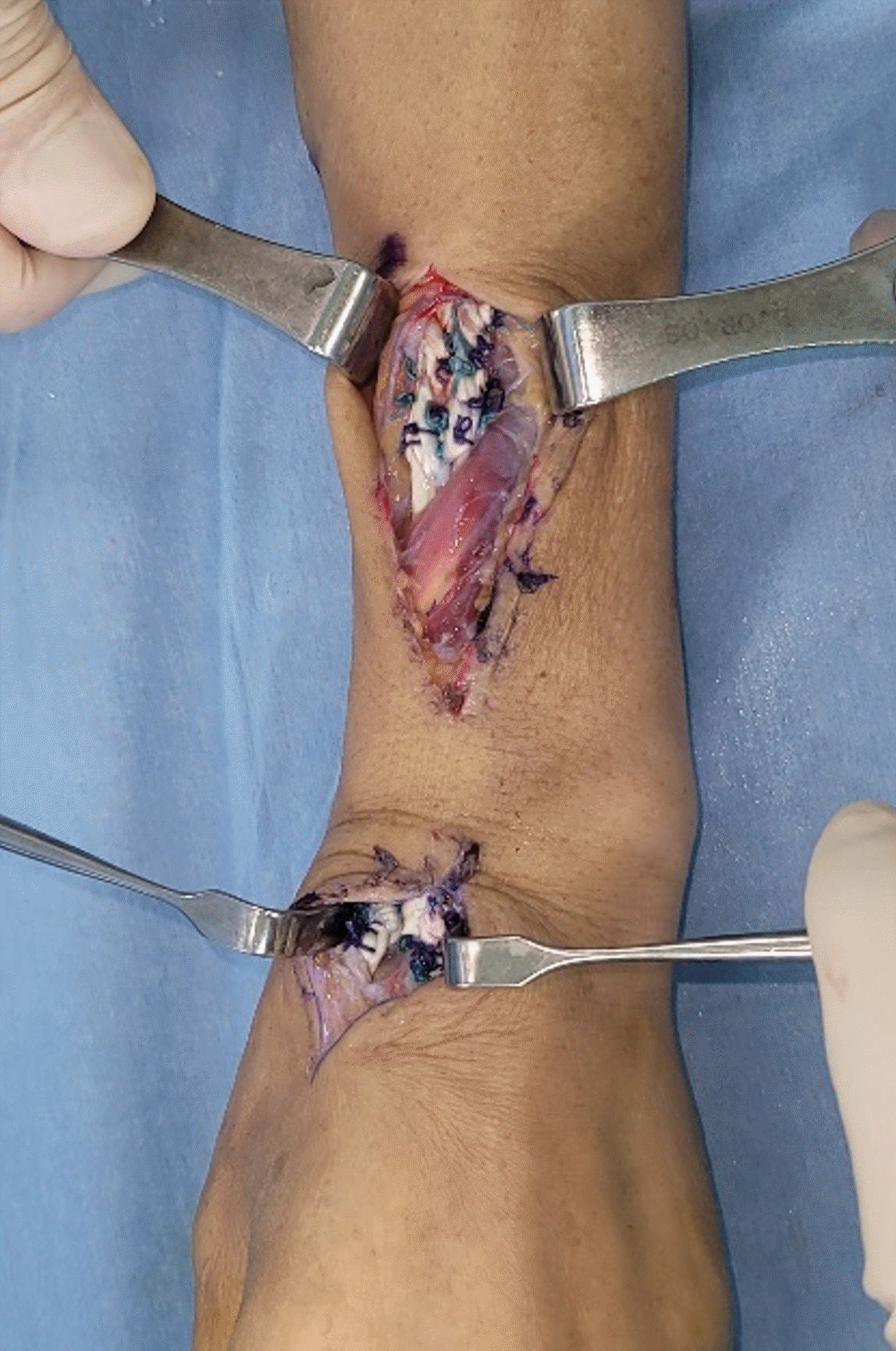
The second operation was performed 2 weeks after the first surgery. On surgical exploration, each proximal end of the ECRL and ECRB tendon was totally ruptured, but the tendon sheath was intact, which was misread intact tendon by sonography (Fig. 5). Each distal end of tendon rupture was debrided and the protruded osteophyte was removed. Two palmaris longus tendons were harvested from each forearm, and ECRB and ECRL were reconstructed (Fig. 6). Postoperatively, the hand was immobilized in a short arm splint with the wrist in 30° extension for 2 weeks. After the suture stitch-off, a removable splint was applied for 4 weeks with mild wrist joint exercise. Eight weeks after surgery, the patient regained full range of motion and strength of wrist extension as contralateral wrist (Fig. 7).
Tendon injury is one of the common major complications of distal radius fracture and EPL rupture is a common tendon injury associated with conservative treatment [2]. The incidence of EPL tendon rupture is about 3% to 5% of conservative treatment [4]. Extensor tendon ruptures other than the EPL tendon are very rare. Only two case reports described concomitant rupture of the EPL and extensor digitorum communis tendon [5,6]. The rupture usually occurs from 6 weeks to 3 months postoperatively without pain. Extensor rupture is believed to occur more frequently after minimally displaced and nondisplaced distal radius fractures [4]. The extensor retinaculum on the third dorsal compartment is intact in non- or minimal displaced distal radius fracture. Therefore hematoma, swelling, edema, and fracture callus contribute to narrowing of space available for the EPL tendon in the third dorsal compartment. Eventually, the EPL tendon gradually becomes devitalization and ruptures [7].
Flexor tendon ruptures in conservative treatment of distal radius fractures were different mechanisms from the EPL rupture such as malunited bones or sharp bony spurs [8]. Malunion-induced flexor tendon injury was caused by attrition of the ulnar head or radius volar cortex prominence. Flexor tendon injuries commonly occur in the flexor digitorum profundus and are most often caused by a protruded ulnar head [8].
This case demonstrates a rare type of delayed tendon rupture after conservative treatment of distal radius fracture in both the ECRL and ECRB tendon, not the EPL tendon. Considering the previous literature, the rupture of the ECRL and ECRB tendons is most likely due to osteophyte, unlike EPL. Hirasawa et al. [7] reported the ECRL and ECRB tendon has a more abundant vascularized sheath than the EPL tendon and may be less ruptured. There are reports about a closed rupture of the ECRL and ECRB tendon, but in case of multiple extensor tendon ruptures during high-energy injury like a motor vehicle accident [9], or of abrupt extension/flexion of the wrist like striking a boxing bag [10]. This case was a minimally displaced fracture, not heavy labor, and only a sharp osteophyte on the second compartment was identified. Therefore, the rupture of ECRL and ECRB is likely due to osteophyte.
This report means the treating physicians should be noted and explain to patients the possibility of rupture in extensor tendons other than the EPL tendon when treating nondisplaced distal radius fracture. In addition, after fracture union is achieved, computed tomography or MRI evaluation may be performed to confirm risky osteophytes to evaluate the possibility of tendon injury. Furthermore, if ECRB and ECRL injury occurred, a normal function could be regained through reconstruction. For early detection of sharp osteophyte, it seems that a plain radiograph comparison with the unaffected side could be helpful. However, in this case, there was a malunion deformity of the contralateral radius due to the previous surgical history so it was hard to initially find sharp osteophyte which result in the extensor tendon rupture by plain radiograph. The physicians should notice the normal range of radiologic features on the distal radius and be aware of the possibility of a rare type of extensor rupture.
References
1. Nellans KW, Kowalski E, Chung KC. The epidemiology of distal radius fractures. Hand Clin. 2012; 28:113–25.

2. McKay SD, MacDermid JC, Roth JH, Richards RS. Assessment of complications of distal radius fractures and development of a complication checklist. J Hand Surg Am. 2001; 26:916–22.

3. Schaller P, Baer W, Carl HD. Extensor indicis-transfer compared with palmaris longus transplantation in reconstruction of extensor pollicis longus tendon: a retrospective study. Scand J Plast Reconstr Surg Hand Surg. 2007; 41:33–5.

4. Roth KM, Blazar PE, Earp BE, Han R, Leung A. Incidence of extensor pollicis longus tendon rupture after nondisplaced distal radius fractures. J Hand Surg Am. 2012; 37:942–7.

5. Sadr B. Sequential rupture of extensor tendons after a Colles’ fracture. Plast Reconstr Surg. 1985; 76:487.

6. de Boer SW, van Kooten EO, Ritt MJ. Extensor pollicis longus tendon rupture with concomitant rupture of the extensor digitorum communis II tendon after distal radius fracture. J Hand Surg Eur Vol. 2010; 35:679–81.

7. Hirasawa Y, Katsumi Y, Akiyoshi T, Tamai K, Tokioka T. Clinical and microangiographic studies on rupture of the E.P.L. tendon after distal radial fractures. J Hand Surg Br. 1990; 15:51–7.

8. Komura S, Hirakawa A, Yamamoto K, et al. Delayed rupture of the flexor tendons as a complication of malunited distal radius fracture after nonoperative management: a report of two cases. Trauma Case Rep. 2019; 21:100198.

9. Vandeputte G, De Smet L. Avulsion of both extensor carpi radialis tendons: a case report. J Hand Surg Am. 1999; 24:1286–8.

10. Mundell T, Miladore N, Ruiter T. Extensor carpi radialis longus and brevis rupture in a boxer. Eplasty. 2014; 14:ic40.
Fig. 1.
Plain radiographs of the left wrist. (A) Posterior anterior view and (B) lateral view. A decreased volar tilt angle was seen on the lateral view.
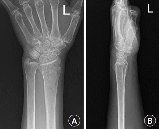
Fig. 2.
Transverse ultrasonography image shows a well-circumscribed, hypoechoic mass (arrow) on the dorsum of the second and third metacarpal bases.
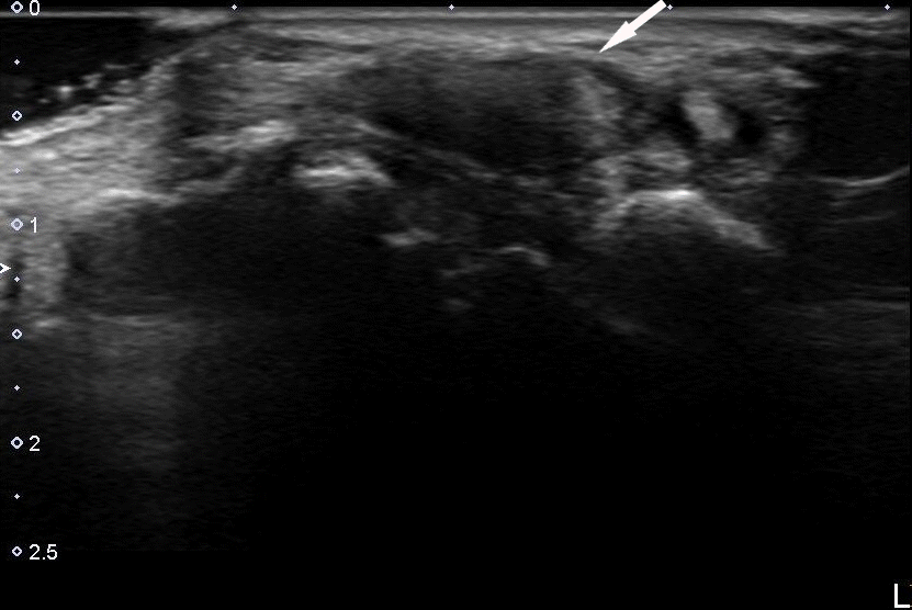
Fig. 3.
Intraoperative photograph of the first operation planned for mass excision. The mass was the remnant of the ruptured extensor carpi radialis longus and extensor carpi radialis brevis tendons.
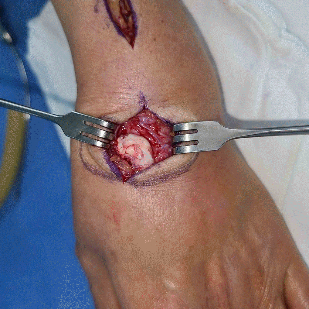
Fig. 4.
Magnetic resonance imaging obtained after the first operation. The images revealed a large osteophyte (arrows) sharply protruding into the second extensor compartment in (A) the coronary plane and (B) axial plane.
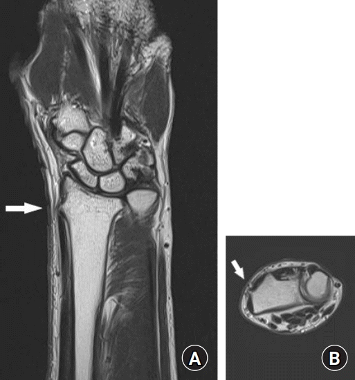
Fig. 5.
The proximal ends of the ruptured extensor carpi radialis longus and extensor carpi radialis brevis tendons were identified with intact sheaths.


 PDF
PDF Citation
 Print
Print



 XML Download