Abstract
Penetrating injuries due to a plant thorn in the hand may occur during outdoor activities. We experienced two patients with rupture of the extensor digitorum communis (EDC) tendon in extensor zone V caused by plant thorn injuries. Both patients, presenting with pain and swelling around the third metacarpophalangeal joint, had a history of penetrating injuries due to plant thorns. The injuries had been ineffectively treated with oral antibiotics. Plain radiographs showed soft tissue swelling without a foreign body and bony lesions. Enhanced magnetic resonance imaging confirmed a rupture of the EDC tendon at the third extensor zone V. Tendon ruptures were diagnosed 27 and 60 days after injury, respectively. A plant thorn injury in the hand can cause persistent synovitis around the tendon, and as a result, progress to tendon rupture. A magnetic resonance imaging examination may be helpful if symptoms do not improve for a prolonged period after plant thorn injury.
A penetrating injury due to a plant thorn in the hand may cause pain, swelling, stiffness, or decreased range of motion in the affected finger joint, and sometimes may present with chronic monoarthritis, infectious tenosynovitis, pyogenic arthritis, or osteomyelitis. Most of these injuries can be managed with conservative treatment including oral antibiotics administration. However, surgical treatment such as debridement, removal of the remnant foreign body, or synovectomy of the tendon sheath or affected finger joint may be required depending upon the progression [1-4].
The authors experienced two patients who underwent conservative treatment for prick injuries due to a plant thorn, resulting in the rupture of the extensor digitorum communis (EDC) tendon. The characteristics of these patients are discussed along with literature review.
This case report was approved by the Institutional Review Board of Konyang University Hospital (No. KYUH 2021-12-023-003). Written informed consent was obtained for the publication of this case report including accompanying images.
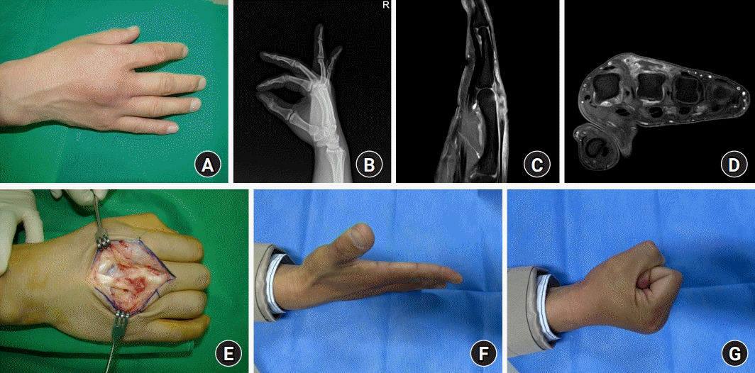
A 47-year-old male patient presented with pain and swelling of the right dorsal hand (Fig. 1A). He had a history of being stabbed by a plant thorn in the dorsal hand while working outdoors about 2 months earlier. He stated that he was injured by the thorn of Japanese Elm during environmental cleanup work in a military unit. He was treated with oral antibiotics at a private hospital with pain and swelling around the third metacarpophalangeal (MCP) joint, but the symptoms did not improve. The physical examination revealed tenderness and an approximate 10°extension lag in the third MCP joint. There was no local heating or fluid fluctuation around the third MCP joint. He had no comorbidities such as diabetes mellitus, gout, or rheumatoid arthritis. Plain radiographs of the right hand showed mild swelling of the dorsal hand, but no bony abnormality or residual foreign material (Fig. 1B). On enhanced magnetic resonance imaging (MRI), subcutaneous edema and rupture with retraction of the EDC tendon in the dorsal aspect of the third metacarpal head were confirmed (Fig. 1C, 1D). The results of laboratory tests at the time of presentation were within the normal ranges. Intraoperatively, turbid fluid collection, tenosynovitis, and about 90% rupture of the third EDC tendon in extensor zone V were noted (Fig. 1E). However, residual foreign body and perforation of joint capsule were not observed. The necrotic tissue was debrided, and the ruptured tendon was sutured directly. Intraoperative cultures including gram stain, acid-fast bacteria stain, bacterial culture, fungal culture, and the tuberculosis/non-tuberculosis polymerase chain reaction test had negative results. The histopathological examination confirmed chronic inflammation without foreign material. Postoperatively, antibiotic treatment using first-generation cephalosporin was performed for 2 weeks. And cast immobilization including the MCP joint was applied for 4 weeks. Three months after surgery, the patient showed a 10° extension lag and 90° flexion of the third MCP joint, without interfering with daily activities or signs of recurrent infection (Fig. 1F, 1G). Since no residual foreign body could be identified during the operation, a telephone interview was conducted to check whether the symptom has recurred. And there has been no recurrence of symptoms so far, more than 2 years after surgery.
A 54-year-old male complained of swelling of the left dorsal hand, and pain and limitations of motion in the left third finger (Fig. 2A). His job was to hunt harmful animals such as wild boars, and he stated that he had felt a penetrating feeling on his dorsal hand while walking around the mountains about 4 weeks ago. At that time, he said he did not find any visible thorns. Although he took oral antibiotics, the symptoms did not improve. He had no comorbidities. On physical examination, swelling and tenderness around the left third MCP joint were found and active motion of affected finger was limited. Plain radiographs showed swelling of the dorsal hand without other abnormal findings. MRI revealed a rupture of the EDC tendon at the dorsal aspect of the third metacarpal head (Fig. 2B). Also, because enhanced MRI showed diffuse synovial thickening and enhancement around affected joint, infectious arthritis was suspected (Fig. 2C, axial image). The results of laboratory tests were within the normal ranges. Intraoperatively, tenosynovitis with synovial hypertrophy, residual foreign material, and complete rupture of the third EDC tendon in the extensor zone V were noted (Fig. 2D). Synovitis and perforation of the third MCP joint capsule, and erosion of the articular cartilage were also confirmed (Fig. 2E). Since no findings suggestive of the infectious arthritis were observed, intraarticular debridement was not performed, only around perforation of joint capsule. After the necrotic tissues were debrided, the ruptured tendon was directly repaired (Fig. 2F). The intraoperative cultures had negative results. Histopathological analysis confirmed chronic inflammation with foreign material. Postoperatively, antibiotics were administrated for 2 weeks. Cast immobilization was applied for 4 weeks. Three months after surgery, the patient showed a 10° extension lag and 75° flexion of the third MCP joint, without interfering with daily activities or signs of recurrent infection.
A penetrating injury of the hand can easily occur during daily or professional activities and can be caused by various agents such as metal, nails, needles, glass, fish bones, wood splinters, or thorns. The risk of infection depends upon the type and properties of the foreign objects, as well as the timing and duration of symptom presentation, the location and depth of penetration, the environment of the injury, previous treatment, and the immune status of the patient [3,4]. A penetrating injury due to foreign material should be properly evaluated to determine whether or not surgical exploration is needed in the early stages. Because patients may not be aware of retained foreign material, physicians should make efforts to confirm the presence of foreign material and localize it [3]. Plain radiography, ultrasonography, and computed tomography can be used to detect retained foreign material, but MRI has the limitations of cost, inspection time, expense, and the characteristics of the images [4]. The composition of the foreign material must be confirmed because organic matters such as wood and vegetable are related to increased inflammation in soft tissue compared to metal or glass objects and as a result, has a higher risk of infection [3,4]. Foreign materials penetrating the skin can induce granuloma formation related to a foreign body reaction, and can mostly be treated with conservative management or removal of the foreign material [1]. However, this injury may present as various complications, which include persistent pain, foreign body sensation or reaction, local cellulitis, synovitis or tenosynovitis, monoarthritis, abscess, infectious tenosynovitis, septic arthritis, and osteomyelitis [5,6]. The treatment of patients with complications should be approached by dividing the presentation into superficial and deep involvement [7]. Also, if an infection is observed, active treatment such as antibiotics or surgical debridement should be performed [1,6,8].
Because of anatomical characteristics of the dorsal hand, even deep structures such as the extensor tendon sheath and joint capsule are vulnerable by the penetrating injury, resulting in deep infections like infectious tenosynovitis or infectious arthritis. In this study, deep infection was observed as a form of delayed rupture of the EDC tendon, which has not been previously reported in the English literatures. Flexor tendon rupture secondary to a catfish spine injury was reported to occur 1 to 2 days after the initial injury, but this rupture was likely caused by the residual sharp spine [9]. The presented cases were treated for 2 months and 4 weeks after the incident, respectively, and surgery was delayed because there was no foreign material on the initial radiography. It seems that sustained inflammation caused by a foreign body reaction continued and as a result, the tendon ruptured, and the histological examination confirmed chronic granulomatous inflammation, not a suppurative infection.
Doctors often encounter patients who are stabbed by the pointed part of a plant during outdoor activities. Although oral antibiotic administration is sometimes necessary, most can be treated conservatively or by only removing the foreign body. Since the symptoms are not severe in the early stage of injury, the diagnosis is likely to be delayed even in patients with complications [2]. In the cases of this study, the occurrence of tendon ruptures related to plant thorn injuries was not be considered in course of conservative treatment. Therefore, if the symptoms of a finger affected by a penetrating injury do not improve, the involvement of deep structures should be suspected. MRI examination may be helpful in determining the extent of the involved lesion and the condition of the tendons. Moreover, because organic matter induces more severe inflammatory response than inorganic matter, the penetrating injury due to this matter should be observed more carefully.
References
1. Rerucha CM, Ewing JT, Oppenlander KE, Cowan WC. Acute hand infections. Am Fam Physician. 2019; 99:228–36.
2. Vaishya R. A thorny problem: the diagnosis and treatment of acacia thorn injuries. Injury. 1990; 21:97–100.
3. Halaas GW. Management of foreign bodies in the skin. Am Fam Physician. 2007; 76:683–8.
4. Rupert J, Honeycutt JD, Odom MR. Foreign bodies in the skin: evaluation and management. Am Fam Physician. 2020; 101:740–7.
5. Chang MC, Huang YL, Liu Y, Lo WH. Infectious complications associated with toothpick injuries of the hand. J Hand Surg Am. 2003; 28:327–31.
7. Racz RS, Ramanujam CL, Zgonis T. Puncture wounds of the foot. Clin Podiatr Med Surg. 2010; 27:523–34.
8. Resnick CD, Fallat LM. Puncture wounds: therapeutic considerations and a new classification. J Foot Surg. 1990; 29:147–53.
9. Spitler C, Rehm J. A zone-II flexor tendon injury secondary to a catfish spine: a case report. JBJS Case Connect. 2017; 7:e61.