Journal List > Ann Surg Treat Res > v.103(5) > 1516080385

Byun, Lee, Kim, You, Jung, Jang, Lee, Kim, Cho, Kang, and on behalf of the Korean Liver Cancer Association: Prognostic value of preoperative protein-induced vitamin K absence or antagonist II after liver resection for hepatitis B-related hepatocellular carcinoma: a nationwide multicenter study

Abstract

Purpose

Although protein-induced vitamin K absence or antagonist II (PIVKA-II) has been used as a diagnostic tool for hepatocellular carcinoma (HCC), its prognostic value remains unclear.

Methods

This was a nationwide multicenter study using the database of the Korean Liver Cancer Association. Patients with hepatitis B-related HCC who underwent liver resection as the first treatment after initial diagnosis (2008–2014) were selected randomly. Propensity score matching (1:1) was performed for comparative analysis between those with low and high preoperative PIVKA-II. Univariable and multivariable Cox proportional-hazards regression were used to identify prognostic factors for HCC-specific survival.

Results

Among 6,770 patients, 956 patients were included in this study. After propensity score matching, the 2 groups (n = 245, each) were well balanced. The HCC-specific 5-year survival rate was 80.9% in the low PIVKA-II group and 78.7% in the high PIVKA-II group (P = 0.605). In univariable analysis, high PIVKA-II (>106.0 mAU/mL) was not a significant predictor for worse HCC-specific survival (hazard ratio [HR], 1.183; 95% confidence interval [CI], 0.76–1.85; P = 0.461). In multivariable analysis, hyponatremia of <135 mEq/L (HR, 4.855; 95% CI, 1.67–14.12; P = 0.004), preoperative ascites (HR, 4.072; 95% CI, 1.59–10.43; P = 0.003), microvascular invasion (HR, 3.112; 95% CI, 1.69–5.74; P < 0.001), and largest tumor size of ≥5.0 cm (HR, 2.665; 95% CI, 1.65–4.31; P < 0.001), but not preoperative high PIVKA-II, were independent predictors for worse HCC-specific survival.

Conclusion

Preoperative PIVKA-II is not an independent prognostic factor for HCC-specific survival after liver resection for hepatitis B-related HCC.

INTRODUCTION

Primary liver cancer is the sixth most commonly diagnosed cancer and the third leading cause of cancer death worldwide in 2020 [1]. Hepatocellular carcinoma (HCC) accounts for the majority (75%–85%) of primary liver cancer cases. Its incidence has been increasing on a global scale [12].
Main clinical prognostic factors in patients with HCC are related to tumor status (defined by number and size of nodules, presence of vascular invasion, and extrahepatic spread), liver function (defined by Child-Pugh class, bilirubin, albumin, clinically relevant portal hypertension, and ascites), and general tumor-related health status [23456]. Tissue and serum biomarkers predicting prognosis have been less explored in patients with HCC [2]. Regarding serum markers, increased α-FP is associated with poorer prognosis [2]. Elevated α-FP levels can predict the risk of tumor recurrence after resection [789], survival and risk of tumor recurrence after liver transplantation [1011], response to locoregional therapies [1213], and survival in advanced HCC [1415]. Protein-induced vitamin K absence or antagonist II (PIVKA-II) was first described by Liebman et al. [16] as a serum marker in patients with HCC in 1984. Since then, it has been used as a diagnostic tool for HCC with serum α-FP. Although a few studies have reported the prognostic effect of baseline PIVKA-II levels, the prognostic value of baseline PIVKA-II before treatment including liver resection in patients with HCC has been insufficiently elucidated [21718].
Therefore, the aim of the present study was to compare HCC-related survival according to baseline PIVKA-II level. The prognostic value of preoperative serum PIVKA-II in patients after liver resection for hepatitis B-related HCC was also evaluated.

METHODS

This study was performed in accordance with the ethical guidelines of the 1975 Declaration of Helsinki. The present study protocol was reviewed and approved by the Institutional Review Board of Eulji University College of Medicine (No. 2020-05-009).

Study design

This study was a nationwide multicenter registry-based comparative analysis of low (LP) vs. high (HP) preoperative PIVKA-II in patients with hepatitis B-related HCC who were initially diagnosed between January 1, 2008 and December 31, 2014 in Korea. All eligible patients with hepatitis B-related HCC were assigned to 1 of 2 groups (LP or HP) at a 1:1 ratio using propensity score (PS) matching.

Registry and data collection

The Korea Central Cancer Registry is a governmental organization with a statutory nationwide cancer registry. This population-based registry began in 1999. Registry completeness accounted for more than 95% of all cancers. The present study was based on data from the Korean Primary Liver Cancer Registry, a joint project between the Korean Liver Cancer Association and the Korea Central Cancer Registry. The Korean Primary Liver Cancer Registry is a retrospective randomly selected nationwide database from the Korea Central Cancer Registry using a systematic random sampling. It has a random sample consisting of approximately 15% of patients newly diagnosed with primary HCC in Korea. The present study was focused on those with newly diagnosed HCC between 2008 and 2014 from the Korean Primary Liver Cancer Registry.
The database included information such as age, sex, date of diagnosis, etiology (hepatitis B or C virus-related, alcoholic liver disease, etc.), Child-Pugh class, Model for End-Stage Liver Disease (MELD) score, performance status, laboratory results such as albumin or bilirubin, tumor markers (serum α-FP and PIVKA-II levels), portal vein invasion, tumor number, tumor size (defined as the diameter of the largest tumor), American Joint Committee on Cancer/International Union Against Cancer TNM stage, Barcelona Clinic Liver Cancer staging, data on first treatment modality and timing, and survival outcomes until December 31, 2016. Individuals with missing data for any of the above variables were excluded from the analysis. Individuals with Child-Pugh classifications B and C were excluded from the analysis. Only individuals with pathologically confirmed HCC were included in this study.

Propensity score matching

PS matching is increasingly used for reducing the effects of confounding in observational studies. A matching was performed for 956 primarily selected patients using the PS matching method. The PS for probability assigned to each group was estimated using logistic regression with baseline characteristics including age, sex, histologic tumor number, largest tumor size based on diagnostic imaging, preoperative serum α-FP level, and underlying liver disease (combined hepatitis C virus-related liver disease, or alcoholic liver disease). After PS estimation, we matched patients using a 1:1 nearest neighbor matching. The 2 matched groups were compared to examine covariate balance [1920] and to determine whether there were statistically significant differences in baseline covariates between groups.

Outcomes

Outcomes included long-term disease (HCC)-specific survival in matched groups and comparative prognostic factors using univariable and multivariable analyses. HCC-specific survival was measured from the date of liver resection of HCC until the date of HCC-related death or the last follow-up. HCC-specific death was based on data obtained from the registry. Variables such as age, sex, obesity (body mass index of ≥27 kg/m2), smoking, diabetes, hypertension, MELD score, hepatitis C virus-related liver disease, alcoholic liver disease, pathologic tumor number, pathologic largest tumor size, preoperative tumor markers (α-FP, PIVKA-II), ascites on preoperative imaging, preoperative laboratory test results (indocyanine green-R15, serum total bilirubin, serum albumin, serum ALT, PT/international normalized ratio, serum sodium, platelets, and serum creatinine levels), and pathologic results (microvascular invasion, bile duct invasion, and lymph node positivity) were analyzed to determine prognostic factors of HCC-specific survival.

Statistical analysis

Receiver-operating characteristic (ROC) curve analysis was used to identify the optimal cutoff for baseline α-FP and PIVKA-II that had the highest sensitivity and specificity in discriminating between HCC-related survivors and nonsurvivors. Survival rates were estimated using the Kaplan-Meier method. They were compared using the log-rank test for proportional hazard. PS matching [21] and other statistical analyses were performed with IBM SPSS Statistics ver. 28.0 (IBM Corp., Armonk, NY, USA). In the process of PS matching, Student t-tests or Wilcoxon rank sum test was used for continuous variables, while the chi-square test or Fisher exact test was used for categorical variables for analysis and comparison of baseline covariates. After PS matching, baseline characteristics and survival outcomes of matched groups were compared using paired t-test or Wilcoxon signed-rank test for continuous variables and McNemar test for categorical variables. All categorical data are expressed as numbers or frequencies with percentages. All continuous data are presented as mean ± standard deviation. Comparative prognostic factors were assessed by univariable and multivariable analyses of Cox proportional-hazards regression models. Multivariable analysis was performed using all variables that showed P-values less than 0.05 in univariable analyses. All reported P-values are 2-sided. Statistical significance was defined as a P-value of <0.05.

RESULTS

Patient selection and study population

Patient selection is shown in Fig 1. A total of 10,578 patients initially diagnosed with HCC between 2008 and 2014 in Korea were randomly selected by systematic random sampling from the Korea Central Cancer Registry. Among these, 6,770 patients had HCC related to hepatitis B. Excluding liver transplantations, 1,367 patients underwent liver resection as the first treatment after initial diagnosis of hepatitis B-related HCC. Excluding patients with Child-Pugh B/C and without preoperative PIVKA-II values, a total of 956 patients with preoperative PIVKA-II values were finally enrolled in this study.

Validation of the optimal cutoff value of preoperative α-FP and PIVKA-II

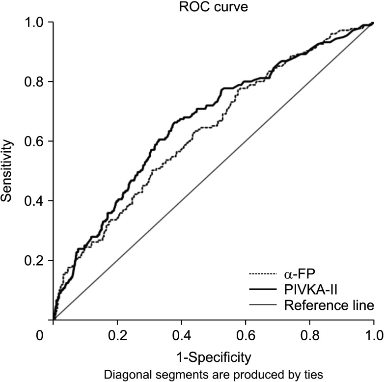
Fig. 2 shows ROC curve of α-FP and PIVKA-II in this study population. The cutoff value for baseline α-FP was 40.5 ng/mL, with a sensitivity of 61.7% and a specificity of 56.4% for HCC-specific survival after liver resection. The area under the ROC curve was 0.632 (P < 0.001). The cutoff value for baseline PIVKA-II was 106.0 mAU/mL, with a sensitivity of 64.6% and a specificity of 62.9% for HCC-specific survival after liver resection. The area under the ROC curve was 0.662 (P < 0.001).

Propensity score-matched analysis

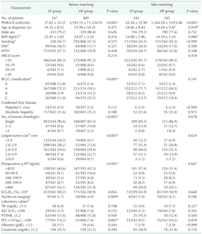
After PS matching, LP and HP groups each contained 245 patients (44.8% and 59.9%, respectively). Baseline characteristics of the 2 groups before and after PS matching are summarized in Table 1. Before PS matching, there were significant differences in several baseline variables between the 2 groups (Table 1). After PS matching, there were no statistically significant differences in baseline variables between the 2 groups, except baseline PIVKA-II (Table 1). Hence, these 2 groups were well-matched.

Overall survival and hepatocellular carcinoma-specific survival in matched groups

Overall survival time and HCC-specific survival time of patients in the 2 matched groups are shown in Fig. 3. In overall and HCC-specific survival rates, there were no significant differences between the LP group and the HP group (P = 0.460 and P = 0.605, respectively). The 1-, 2-, 3-, and 5-year overall survival rates were 95.6%, 92.2%, 89.5%, and 83.7% in the LP group and 95.6%, 90.7%, 86.4%, and 80.0% in the HP group, respectively (Fig. 3A). The 1-, 2-, 3-, and 5-year HCC-specific survival rates were 94.7%, 90.7%, 88.0%, and 80.9% in the LP group and 95.9%, 90.7%, 85.5%, and 78.7% in the HP group, respectively (Fig. 3B).

Analysis of prognostic factors

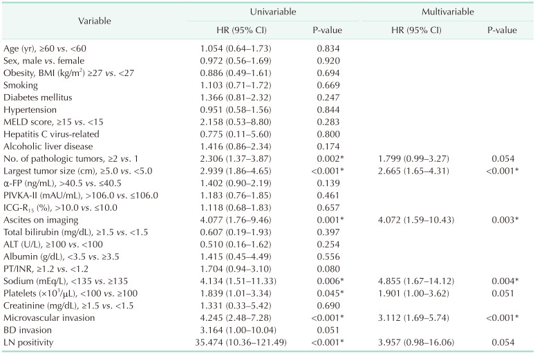
Prognostic factors were analyzed for variables obtained from the database using a Cox proportional-hazards model. Results of univariable and multivariable analyses are summarized in Table 2. In univariable analysis, lymph node positivity (hazard ratio [HR], 35.474; 95% confidence interval [CI], 10.36–121.49; P < 0.001), microvascular invasion (HR, 4.245; 95% CI, 2.48–7.28; P < 0.001), hyponatremia of <135 mEq/L (HR, 4.134; 95% CI, 1.51–11.33; P = 0.006), preoperative ascites (HR, 4.077; 95% CI, 1.76–9.46; P = 0.001), largest tumor size of ≥5.0 cm (HR, 2.939; 95% CI, 1.86–4.65; P < 0.001), multiple HCC (HR, 2.306; 95% CI, 1.37–3.87; P = 0.002), and thrombocytopenia (<100 × 103/µL) (HR, 1.839; 95% CI, 1.01–3.34; P = 0.045) were significant prognostic factors for worse HCC-specific survival (Table 2). Preoperative high α-FP (>40.5 ng/mL) (HR, 1.402; 95% CI, 0.90–2.19; P = 0.139), and preoperative high PIVKA-II (>106.0 mAU/mL) (HR, 1.183; 95% CI, 0.76–1.85; P = 0.461) were not significant prognostic factors for worse HCC-specific survival (Table 2).
For multivariable analysis, Cox regression analysis was performed in a backward manner. In multivariable analysis, hyponatremia of <135 mEq/L (HR, 4.855; 95% CI, 1.67–14.12; P = 0.004), preoperative ascites (HR, 4.072; 95% CI, 1.59–10.43; P = 0.003), microvascular invasion (HR, 3.112; 95% CI, 1.69–5.74; P < 0.001), and largest tumor size of ≥5.0 cm (HR, 2.665; 95% CI, 1.65–4.31; P < 0.001) were independent predictors for worse HCC-specific survival (Table 2).

DISCUSSION

Evaluating predictors of prognosis is of clinical importance for defining treatment strategies. As previously described, the main prognostic factors in patients with HCC are related to tumor status, liver function, and general tumor-related health [23456]. Currently, measuring biomarker levels both before and after HCC treatment is clinically valuable as a simple way to monitor treatment outcomes (usually in combination with radiological analysis) and to predict prognosis, recurrence, and survival [22]. With regard to the prognostic effect of baseline PIVKA-II levels before liver resection in patients with HCC, little is known.
To the best of our knowledge, this study has the largest number of patients among similar studies conducted so far regarding PIVKA-II. In overall and HCC-specific survival rates in the present study, there were no significant differences between the LP group and the HP group (P = 0.460 and P = 0.605, respectively). Univariable analysis showed that preoperative high PIVKA-II (>106.0 mAU/mL) was not a significant prognostic factor for worse HCC-specific survival in patients with hepatitis B-related HCC. After multivariable analysis in the present study, hyponatremia of <135 mEq/L, preoperative ascites on imaging, microvascular invasion, and largest tumor size of ≥5.0 cm, but not preoperative high PIVKA-II (>106.0 mAU/mL), were independent prognostic factors for worse HCC-specific survival in patients with hepatitis B-related HCC.
Imamura et al. [23] first revealed significantly associated relationships between preoperative PIVKA-II elevation and pathological parameters implicating more aggressive tumor characteristics (intrahepatic metastasis, vascular invasion, and tumor cell differentiation). After that, several studies have reported that elevated serum PIVKA-II level is related to larger tumor size, more frequent or extent of vascular invasion including portal vein thrombosis, more intrahepatic metastasis and extrahepatic disease extension, and recurrence after treatment, all of which can affect the prognosis of patients with HCC [242526]. To state the obvious, tumor invasiveness, metastasis, and recurrence can result in poor clinical outcomes for patients with HCC including operable cases [622]. Tumor microvascular invasion is a critical determinant of HCC recurrence and prognosis [427]. It is highly correlated with adverse biological markers including elevated serum PIVKA-II [6]. Therefore, it is reasonably anticipated that preoperative serum PIVKA-II could be related to prognosis in patients with HCC.
However, studies on the prognostic value of preoperative PIVKA-II as an independent factor for survival in patients with HCC are limited, especially in liver resection. Several studies have shown that preoperative PIVKA-II levels do not always reflect prognosis after curative liver resection [282930]. In the present study, preoperative high PIVKA-II was not a powerful marker for predicting HCC-specific survival after liver resection in hepatitis B-related HCC either. Not surprisingly, this means that other well-known main prognostic factors (related to tumor status, liver function, and general tumor-related health status) are more strongly related to survival in patients with HCC (especially after liver resection) than PIVKA-II or α-FP. Large tumor size and microvascular invasion as independent prognostic factors in the present study were included as already known main prognostic factors of tumor status. Other independent prognostic factors (hyponatremia, preoperative ascites) in this study were associated with main prognostic factors related to liver function and/or general tumor-related health status. In addition, the prognostic role of PIVKA-II may need to be evaluated in the context of considering combined α-FP and PIVKA-II as complementary markers. To clarify the prognostic value of PIVKA-II in HCC, further well-designed large-scale studies or randomized controlled trials are needed.
The present study had some limitations despite it being a well-matched comparative study using randomly selected data from a large nationwide registry with PS matching. There might be selection bias despite using systematic random sampling and PS matching. The study population included patients with only hepatitis B virus-related HCC in Korea. Therefore, caution is needed when extending or applying the findings of the present study to other general cohorts. Additionally, important variables like recurrence-free interval or disease-free survival were missing.
Notwithstanding, this study was performed with the largest number of patients to date, compared to previous other studies. Methodologically, this study had several strengths, including highly reliable data from a nationwide multicenter cohort, systematic random sampling, and a PS matching which minimized selection bias. The present study could serve as valuable background for future studies on the prognostic value of PIVKA-II in HCC patients, especially in those with hepatitis B-related liver disease.
In conclusion, preoperative PIVKA-II is not an independent prognostic factor for HCC-specific survival after liver resection for hepatitis B-related HCC.

ACKNOWLEDGEMENTS

The authors thank the Korea Central Cancer Registry and Korean Liver Cancer Association. This work was supported by the Research Supporting Program of the Korean Liver Cancer Association based on data from the Primary Liver Cancer Registry, a joint project supported by the Korean Liver Cancer Association and Korea Central Cancer Registry, Ministry of Health and Welfare, Republic of Korea.

Notes

This study was presented in part at HBP Surgery Week 2021 (Seoul, March 2021) and 2021 The Congress of the Korean Surgical Society (Daegu, May 2021).

Fund/Grant Support: None.

Conflict of Interest: No potential conflict of interest relevant to this article was reported.

Author Contribution:

  • Conceptualization, Formal analysis: DB, SGL, HK.

  • Investigation, Methodology: All authors.

  • Writing – Original Draft: DB, SGL, HK.

  • Writing – Review & Editing: All authors.

References

1. Sung H, Ferlay J, Siegel RL, Laversanne M, Soerjomataram I, Jemal A, et al. Global cancer statistics 2020: GLOBOCAN estimates of incidence and mortality worldwide for 36 cancers in 185 countries. CA Cancer J Clin. 2021; 71:209–249. PMID: 33538338.
crossref
2. European Association for the Study of the Liver. EASL Clinical practice guidelines: management of hepatocellular carcinoma. J Hepatol. 2018; 69:182–236. PMID: 29628281.
3. Tsilimigras DI, Moris D, Hyer JM, Bagante F, Sahara K, Moro A, et al. Hepatocellular carcinoma tumour burden score to stratify prognosis after resection. Br J Surg. 2020; 107:854–864. PMID: 32057105.
crossref
4. Lim KC, Chow PK, Allen JC, Chia GS, Lim M, Cheow PC, et al. Microvascular invasion is a better predictor of tumor recurrence and overall survival following surgical resection for hepatocellular carcinoma compared to the Milan criteria. Ann Surg. 2011; 254:108–113. PMID: 21527845.
crossref
5. Hong SK, Lee KW, Lee S, Hong SY, Suh S, Han ES, et al. Impact of tumor size on hepatectomy outcomes in hepatocellular carcinoma: a nationwide propensity score matching analysis. Ann Surg Treat Res. 2022; 102:193–204. PMID: 35475226.
crossref
6. Erstad DJ, Tanabe KK. Prognostic and Therapeutic implications of microvascular invasion in hepatocellular carcinoma. Ann Surg Oncol. 2019; 26:1474–1493. PMID: 30788629.
crossref
7. Imamura H, Matsuyama Y, Tanaka E, Ohkubo T, Hasegawa K, Miyagawa S, et al. Risk factors contributing to early and late phase intrahepatic recurrence of hepatocellular carcinoma after hepatectomy. J Hepatol. 2003; 38:200–207. PMID: 12547409.
crossref
8. Ikai I, Arii S, Kojiro M, Ichida T, Makuuchi M, Matsuyama Y, et al. Reevaluation of prognostic factors for survival after liver resection in patients with hepatocellular carcinoma in a Japanese nationwide survey. Cancer. 2004; 101:796–802. PMID: 15305412.
crossref
9. Kim H, Lee SJ, Yoon M. Alpha-fetoprotein is correlated with intrahepatic recurrence of hepatocel lular carcinoma after a hepatectomy. Ann Surg Treat Res. 2020; 98:168–176. PMID: 32274364.
crossref
10. Toso C, Meeberg G, Hernandez-Alejandro R, Dufour JF, Marotta P, Majno P, et al. Total tumor volume and alpha-fetoprotein for selection of transplant candidates with hepatocellular carcinoma: a prospective validation. Hepatology. 2015; 62:158–165. PMID: 25777590.
crossref
11. Mazzaferro V, Sposito C, Zhou J, Pinna AD, De Carlis L, Fan J, et al. Metroticket 2.0 model for analysis of competing risks of death after liver transplantation for hepatocellular carcinoma. Gastroenterology. 2018; 154:128–139. PMID: 28989060.
crossref
12. N’Kontchou G, Mahamoudi A, Aout M, Ganne-Carrié N, Grando V, Coderc E, et al. Radiofrequency ablation of hepatocellular carcinoma: long-term results and prognostic factors in 235 western patients with cirrhosis. Hepatology. 2009; 50:1475–1483. PMID: 19731239.
crossref
13. Takayasu K, Arii S, Kudo M, Ichida T, Matsui O, Izumi N, et al. Superselective transarterial chemoembolization for hepatocellular carcinoma: validation of treatment algorithm proposed by Japanese guidelines. J Hepatol. 2012; 56:886–892. PMID: 22173160.
crossref
14. Zhu AX, Park JO, Ryoo BY, Yen CJ, Poon R, Pastorelli D, et al. Ramucirumab versus placebo as second-line treatment in patients with advanced hepatocellular carcinoma following first-line therapy with sorafenib (REACH): a randomised, double-blind, multicentre, phase 3 trial. Lancet Oncol. 2015; 16:859–870. PMID: 26095784.
crossref
15. Llovet JM, Peña CE, Lathia CD, Shan M, Meinhardt G, Bruix J, et al. Plasma biomarkers as predictors of outcome in patients with advanced hepatocellular carcinoma. Clin Cancer Res. 2012; 18:2290–2300. PMID: 22374331.
crossref
16. Liebman HA, Furie BC, Tong MJ, Blanchard RA, Lo KJ, Lee SD, et al. Des-gamma-carboxy (abnormal) prothrombin as a serum marker of primary hepatocellular carcinoma. N Engl J Med. 1984; 310:1427–1431. PMID: 6201741.
17. Lee YK, Kim SU, Kim DY, Ahn SH, Lee KH, Lee DY, et al. Prognostic value of α-fetoprotein and des-γ-carboxy prothrombin responses in patients with hepatocellular carcinoma treated with transarterial chemoembolization. BMC Cancer. 2013; 13:5. PMID: 23282286.
crossref
18. Kim HS, Park JW, Jang JS, Kim HJ, Shin WG, Kim KH, et al. Prognostic values of alpha-fetoprotein and protein induced by vitamin K absence or antagonist-II in hepatitis B virus-related hepatocellular carcinoma: a prospective study. J Clin Gastroenterol. 2009; 43:482–488. PMID: 19197197.
crossref
19. Hansen BB, Bowers J. Covariate balance in simple, stratified and clustered comparative studies. Statist Sci. 2008; 23:219–236.
crossref
20. Iacus SM, King G, Porro G. CEM: coarsened exact matching software. J Stat Softw. 2009; 30:1–27. PMID: 21666874.
21. Kudo M, Takamine Y, Nakamura K, Shirane H, Uchida H, Kasakura S, et al. Des-gamma-carboxy prothrombin (PIVKA-II) and alpha-fetoprotein-producing IIc-type early gastric cancer. Am J Gastroenterol. 1992; 87:1859–1862. PMID: 1280406.
22. Song PP, Xia JF, Inagaki Y, Hasegawa K, Sakamoto Y, Kokudo N, et al. Controversies regarding and perspectives on clinical utility of biomarkers in hepatocellular carcinoma. World J Gastroenterol. 2016; 22:262–274. PMID: 26755875.
crossref
23. Imamura H, Matsuyama Y, Miyagawa Y, Ishida K, Shimada R, Miyagawa S, et al. Prognostic significance of anatomical resection and des-gamma-carboxy prothrombin in patients with hepatocellular carcinoma. Br J Surg. 1999; 86:1032–1038. PMID: 10460639.
crossref
24. Park H, Park JY. Clinical significance of AFP and PIVKA-II responses for monitoring treatment outcomes and predicting prognosis in patients with hepatocellular carcinoma. Biomed Res Int. 2013; 2013:310427. PMID: 24455683.
crossref
25. Tang W, Kokudo N, Sugawara Y, Guo Q, Imamura H, Sano K, et al. Des-gamma-carboxyprothrombin expression in cancer and/or non-cancer liver tissues: association with survival of patients with resectable hepatocellular carcinoma. Oncol Rep. 2005; 13:25–30. PMID: 15583797.
26. Koike Y, Shiratori Y, Sato S, Obi S, Teratani T, Imamura M, et al. Des-gamma-carboxy prothrombin as a useful predisposing factor for the development of portal venous invasion in patients with hepatocellular carcinoma: a prospective analysis of 227 patients. Cancer. 2001; 91:561–569. PMID: 11169939.
crossref
27. Lauwers GY, Terris B, Balis UJ, Batts KP, Regimbeau JM, Chang Y, et al. Prognostic histologic indicators of curatively resected hepatocellular carcinomas: a multi-institutional analysis of 425 patients with definition of a histologic prognostic index. Am J Surg Pathol. 2002; 26:25–34. PMID: 11756766.
crossref
28. Tsukamoto M, Nitta H, Imai K, Higashi T, Nakagawa S, Okabe H, et al. Clinical significance of half-lives of tumor markers α-fetoprotein and des-γ-carboxy prothrombin after hepatectomy for hepatocellular carcinoma. Hepatol Res. 2018; 48:E183–E193. PMID: 28796412.
crossref
29. Saito Y, Shimada M, Utsunomiya T, Morine Y, Imura S, Ikemoto T, et al. Prediction of recurrence of hepatocellular carcinoma after curative hepatectomy using preoperative Lens culinaris agglutinin-reactive fraction of alpha-fetoprotein. Hepatol Res. 2012; 42:887–894. PMID: 22524419.
crossref
30. Toyoda H, Kumada T, Kaneoka Y, Osaki Y, Kimura T, Arimoto A, et al. Prognostic value of pretreatment levels of tumor markers for hepatocellular carcinoma on survival after curative treatment of patients with HCC. J Hepatol. 2008; 49:223–232. PMID: 18571271.
crossref
Fig. 1

Study population. HCC, hepatocellular carcinoma; PIVKA-II, protein-induced vitamin K absence or antagonist II.

astr-103-271-g001
Fig. 2

Receiver-operating characteristic (ROC) curve analysis. PIVKA-II, protein-induced vitamin K absence or antagonist II.

astr-103-271-g002
Fig. 3

Survivals. (A) Overall survival in matched cohorts. (B) Hepatocellular carcinoma (HCC)-specific survival in matched cohorts. LP, low protein-induced vitamin K absence or antagonist II; HP, high protein-induced vitamin K absence or antagonist II.

astr-103-271-g003
Table 1

Baseline characteristics before and after propensity score matching

astr-103-271-i001

Values are presented as number only, mean ± standard deviation, or number (%), unless otherwise indicated. Data were incomplete for some variables and were missing for some patients.

LP, low PIKVA-II (≤106.0 mAU/mL); HP, high PIVKA-II (>106.0 mAU/mL); BMI, body mass index; DM, diabetes mellitus; HTN, hypertension; MELD, Model for End-Stage Liver Disease; BCLC, Barcelona Clinic Liver Cancer; ICG, indocyanine green; TB, total bilirubin; INR, international normalized ratio; PLT, platelets.

a)BMI, smoking, DM, HTN, BCLC classification, ICG-R15, ascites on imaging, and laboratory values were not used in the propensity score matching. b)P-value was based on paired t-test; c)P-value was based on Wilcoxon signed-rank test. d)Diameter was based on radiologic finding.

*P < 0.05.

Table 2

Prognostic factors for HCC-specific survival based on Cox proportional-hazards model

astr-103-271-i002

HR, hazard ratio; CI, confidence interval; BMI, body mass index; MELD, Model for End-Stage Liver Disease; PIVKA-II, protein-induced vitamin K absence or antagonist II; ICG, indocyanine green; INR, international normalized ratio; BD, bile duct; LN, lymph node.

Microvascular invasion, BD invasion, and LN positivity were based on pathologic findings.

*P < 0.05.

TOOLS
Similar articles