Journal List > Ann Surg Treat Res > v.103(4) > 1516080382

Gwon, Ko, and Kim: Is the platelet function test effective in predicting blood loss in patients undergoing hepatic resection?

Abstract

Purpose

The platelet function analyzer (PFA)-100/200 is widely used to assess platelet function. However, its role in predicting the perioperative risk of bleeding in patients undergoing liver resection remains controversial. Therefore, we aimed to ascertain whether the platelet function test could be useful in predicting bleeding risk in patients undergoing hepatic surgery.

Methods

The study participants were patients who underwent hepatectomy for hepatocellular carcinoma at our hospital over a period of 10 years from January 1, 2010 to May 31, 2020. PFA-200 values of these patients were divided into 2 groups; normal (n = 333) and prolonged (n = 39).

Results

There were no significant differences regarding the volumes of calculated blood loss during surgery between the normal and prolonged PFA groups (879.55 ± 1,046.50 mL vs. 819.74 ± 912.64 mL, respectively; P = 0.733); intraoperative RBC transfusion (0.52 ± 2.02 units vs. 0.26 ± 1.02 units, P = 0.419) and postoperative RBC transfusion (0.24 ± 1.17 units vs. 0.46 ± 1.97 units, P = 0.306) were similar between the 2 groups, respectively. Multivariate analysis revealed no association between PFA closure time and calculated blood loss (hazard ratio, 1.06; P = 0.881). Moreover, there was no association between PFA closure time and preoperative laboratory results or assessment of tool-related liver function in multivariate analysis.

Conclusion

There was no correlation between the amount of blood loss and platelet function in patients who underwent liver resection. In patients undergoing liver resection who are not managed on antiplatelet agents or do not have chronic kidney disease, the use of routine PFA is not recommended.

INTRODUCTION

Bleeding is one of the most common and potentially detrimental complications of surgery—leading to death in severe cases. In particular, liver surgery poses a higher bleeding risk than other surgeries. This is because patients undergoing liver surgery often possess reduced liver function compared to other patients. In addition, minor or major vessels must be dissected and ligated for liver resection. According to a published report, the average blood loss during liver surgery approached 700–1,200 mL [1].
Therefore, to prevent bleeding during and after surgery, it is essential to investigate the bleeding risk before surgery. Various preoperative laboratory test parameters, including platelet levels, PT, aPTT, and bleeding time (BT) test, were used to evaluate the patient’s risk of bleeding before surgery. BT and platelet aggregation tests have been used to evaluate platelet function. However, the use of BT tests is gradually decreasing owing to its low sensitivity and high operator dependency [2]. Platelet aggregation tests are relatively expensive and complex, although they are more sensitive than BT [3]. Recently, platelet function tests using the platelet function analyzer (PFA-200; Siemens Healthineer, Munich, Germany) have been widely used as screening tools because of their ease of use and high sensitivity. In particular, it is possible to detect primary hemostasis abnormalities and monitor antiplatelet therapy [45].
The usefulness of PFA as a preoperative screening tool has also been demonstrated and confirmed in heart surgery [678]. However, its usefulness in patients undergoing liver resection has not yet been confirmed. In liver surgery, there is generally more bleeding than that in other surgeries, and many patients with reduced liver function are expected to exhibit reduced platelet levels or functions. For these reasons, PFA is expected to be useful. However, to date there are no reports confirming its utility. Therefore, our study focused on confirming the value of PFA in patients undergoing liver resection.

METHODS

The Institutional Review Board of Korea University Guro Hospital approved this study (No. 2020AN0529). Informed consent from patients was exempted in this retrospective study.

Patients

The participants were patients who underwent hepatectomy for hepatocellular carcinoma (HCC) at our center from January 1, 2010 to May 31, 2020. According to the preoperative evaluation protocol of the anesthesiology department of our hospital, PFA is performed in all patients scheduled for general anesthesia. For analyzing effect of PFA closure time on liver resection, we excluded the following patients to exclude other factors; patients with no PFA results, patients lacking indocyanine green (ICG) results, or diagnosed with cardiovascular disease and using antiplatelet agents, and patients with estimated glomerular filtration rate of <60 mL/min/1.73 m2 (Fig. 1). All patients underwent examination and surgery using the same protocol in the same medical environment at a single center. PFA-200 (machine introduction) was used to confirm PFA values. All laboratory findings and diseases at baseline were obtained and recorded within 1 month preceding surgery.
Participants were divided into 2 groups according to the degree of PFA closure time prolongation. The prolonged closure times were defined as closure time of ≥250 seconds (collagen/epinephrine, C/EPI) according to the manufacturer’s instructions. Any closure time exceeding 250 seconds is reported as 250 seconds. The variables identified to evaluate the liver function of patients were cirrhosis with Child-Pugh score, serum albumin, platelet count, international normalized ratio (INR), total bilirubin, ICG 15-minute clearance retention rate (ICG-R15) test, and Model for End-stage Liver Disease (MELD) score. The patient’s basic demographics and underlying diseases were recorded, and laboratory findings were determined. The patient’s surgical findings were confirmed for the extent of the operation, the amount of blood loss during surgery, and the history of transfusion. The liver resection range was classified according to the Japanese general rules for the clinical and pathological study of primary liver cancer [9]: Hr0, resection of less than one segment; HrS, resection of one segment; Hr1, resection of one section (anterior, posterior, medial, or left lateral segmentectomy); Hr2, resection of 2 sections (right or left lobectomy or central bisegmentectomy); Hr3, resection of 3 sections (right or left trisegmentectomy); and Hr4; resection of 4 sections. A calculated blood loss was calculated using the following formula: (body weight × 70) × (preoperative hemoglobin [Hb] – postoperative Hb) / ([preoperative Hb + postoperative Hb] / 2) + (320 × intraoperative RBC transfusion).

Statistical analysis

Data are presented as means ± standard deviations for continuous variables and as numbers (percentages) for categorical data. To compare the characteristics of participants between PFA normal and prolongation groups, continuous variables were analyzed by the analysis of variance (ANOVA) using Tukey method as a post-hoc test, and categorical variables were tested using the chi-square test or Fisher exact test. The association between the PFA level and parameters related to liver function or calculated blood loss was assessed using ANOVA or chi-square tests. The parameters were divided into 2 or 3 categories based on their cutoff values. The distributions of PFA according to the categories for each parameter were graphically presented using box plots and compared using ANOVA or Student t-test. Statistical significance was set at P < 0.05. All statistical analyses were performed using SAS software ver. 9.4 (SAS Institute Inc., Cary, NC, USA) and R ver. 4.0.2 (R Foundation for Statistical Computing, Vienna, Austria). For calculated blood loss, multivariate analysis was performed using logistic regression based on blood loss of >400 cm3. Multivariate analysis for prolonged PFA time was also performed using logistic regression analysis.

RESULTS

Baseline characteristics

Among the 573 patients who underwent surgery during the study period, 372 patients were included in this study. These patients were divided into 2 groups: prolonged PFA (n = 39, 89.5%) and normal PFA groups (n = 333, 10.5%). The baseline characteristics of the 2 groups are shown in Table 1. There were no differences between the 2 groups, except for underlying liver disease. There were more hepatitis B patients in PFA normal group than the PFA prolongation group. Liver function tests (LFTs) including ICG-R15 test, MELD score, and Child-Pugh score were not different between the 2 groups. The resection range was not significantly different, but patients who underwent Hr3 and Hr4 were included in PFA normal group and no one in the PFA prolongation group.

A calculated blood loss and blood transfusion

The calculated blood loss and blood product that was transfused in intraoperative and postoperative periods are shown in Table 2. Although PFA normal group had more calculated blood loss and intraoperative transfused RBC than PFA prolongation group, it was not statistically significant (879.55 ± 1,046.50 mL vs. 819.74 ± 912.64 mL, P = 0.733; 0.52 ± 2.02 mL vs. 0.26 ± 1.02 mL, P = 0.419). However, intraoperative platelet concentrate (PC) and fresh frozen plasma (FFP) was more transfused in PFA normal group than in PFA prolongation group (PC: 0.20 ± 1.27 vs. 0, P = 0.005; FFP: 0.73 ± 2.74 vs. 0.15 ± 0.67, P = 0.002). Postoperative RBC, PC, and FFP were not different between the 2 groups. In multivariate analysis for more than 400 mL of calculated blood loss, age (odds ratio [OR], 1.65; 95% confidence interval [CI], 1.05–2.60; P = 0.032) and intraoperative resection margin (Hr0 vs. Hr4: OR, 11.10; 95% CI, 1.38–89.24; P = 0.024) was found to be risk factors (Table 3). However, PFA prolongation was not a risk factor for calculated blood loss of more than 400 mL (OR, 1.06; 95% CI, 0.52–2.13; P = 0.881).

Association between prolonged platelet function analyzer closure time and liver function

In multivariate analysis for prolonged PFA closure time, hepatitis B was found to be associated with prolonged PFA closure time (OR, 2.95; 95% CI, 1.18–7.33; P = 0.020). ICG test, MELD score, Child-Pugh score, and cirrhosis were not the predictive factor for prolonged PFA closure time (Table 4). When comparing the PFA values by dividing the lab findings representing the liver function into normal and abnormal, there was no correlation at all. There was no difference between the 3 groups even when the ICG-R15 test results were divided into 3 groups and the PFA values were compared (P = 0.541) (Fig. 2).

Association between calculated blood loss and prolonged platelet function analyzer closure time in liver cirrhosis patients

PFA normal group and PFA prolongation group had 206 (61.9%) and 29 (74.4%) liver cirrhosis patients, respectively (Table 1). Supplementary Table 1 show the baseline characteristics of liver cirrhosis patients divided by prolonged PFA closure time. There were no statistically different between PFA normal group and PFA prolongation group in liver cirrhosis patients. Calculated blood loss was not different between the 2 groups (986 ± 1,239 mL vs. 882 ± 1,002 mL, P = 0.665). Also, intraoperative blood product showed no difference between the 2 groups. In postoperative periods, PC was more transfused in the PFA prolongation group than PFA normal group (0.27 ± 1.66 units vs. 1.45 ± 5.15 units, P = 0.013) (Supplementary Table 2). In multivariate analysis for calculated blood loss, resection of 4 sections in the liver was found to be the only predictive factor for calculated blood loss (OR, 11.50; 95% CI, 1.41–94.06; P = 0.023) (Supplementary Table 3).

DISCUSSION

In heart patients with a high proportion of antiplatelet use, the usefulness of PFA in cardiac surgery has been demonstrated in several reports [68]. However, the usefulness of routine PFA tests in other types of surgeries has not been proven. It has been reported that preoperative screening tests for PFA in all patients without a bleeding history delayed surgery and increased costs unnecessarily [10]. Nevertheless, in clinical practice, PFA is often routinely used to evaluate the risk of bleeding during surgery, and this protocol is followed at our hospital. Using our hospital’s protocol, we attempted to confirm the usefulness of PFA before surgery in patients undergoing liver resection.
We attempted to confirm the usefulness of PFA in liver surgery in particular, because the hemostatic system is closely related to the clotting and fibrinolytic system mediated by liver parenchyma cells [11]. Therefore, we tried to confirm 2 assumptions in this study. First, if liver resection is performed, liver parenchyma cells, which are responsible for clotting and the fibrinolytic system, are reduced. Therefore, we assumed that patients with abnormal platelet function on PFA would exhibit greater calculated blood loss during surgery. Second, we hypothesized that patients scheduled for liver resection for HCC would have impaired liver function compared to healthy individuals. Therefore, we attempted to find an association between preoperative liver function and PFA.
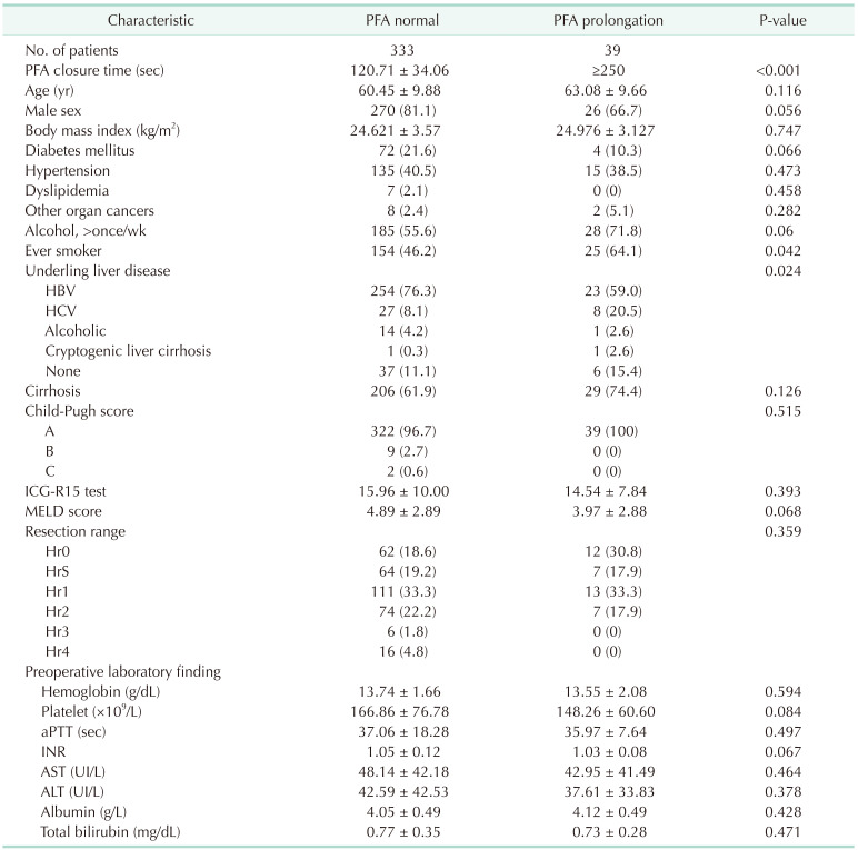
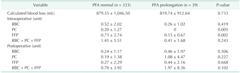
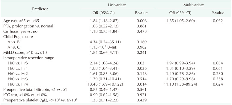
In our study, 10.5% of all patients exhibited prolonged PFA, and these patients were compared with the normal PFA group. However, there were no differences regarding calculated blood loss, intraoperative and postoperative RBC transfusions between the 2 groups. Further, multivariate analysis revealed that PFA did not impact a calculated blood loss. Resection range and age was the only predictive factor for calculated blood loss. Also regarding FFP, the normal PFA group showed a tendency to receive more transfusions than the prolonged PFA group. We supposed the reason is that the normal PFA group included more patients with extended sectionectomy than the PFA prolongation group. In liver cirrhosis patients, calculated blood loss and blood transfusion were similar to that of the total cohort. Also, hepatic resection was the only predictive risk factor for calculated blood loss. However, postoperative PC was more transfused in the PFA prolongation group. In liver cirrhosis patients, platelet function could be more dysfunctional and affect the postoperative needed PC transfusion. To prove it, further study was needed due to the small size of our study.
Other Korean studies on the usefulness of PFA before surgery reported similar results. Lee and Kim [12] did not confirm the usefulness of PFA in endoscopic sinus surgery and concluded that the routine use of PFA is not recommended in endoscopic sinus surgery. Jeon et al. [13] reported that PFA abnormalities in total knee arthroplasty were not related to postoperative bleeding. Yu et al. [14] argued that PFA was useful for various surgeries in 703 patients; however, in this study, although patients with increased PFA closure time exhibited a higher rate of transfusion than those without increased PFA closure time, this was not confirmed by multivariate analysis. In this study, surgery was divided according to the surgical department, but we believe that even if it is an operation in the same area, the effect on a calculated blood loss may be different depending on the type and severity. Therefore, to evaluate PFA, its usefulness should be investigated for each operation. Therefore, we investigated only patients who underwent liver resection for HCC. A strength of our study is that the sample size is larger than that of other studies investigating PFA in one type of surgery.
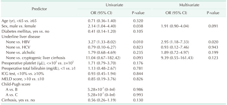
Our data did not confirm an effect of preoperative liver function on PFA values. The reason was that most of the patients had a satisfactory liver function, and most were classified as Child-Pugh A patients. In addition, each LFT value was within the normal range on average. Due to the nature of these cohorts, it was difficult to directly identify the factors that influence LFT on PFA. In multivariate analysis, hepatitis B was associated with PFA closure time. Hepatitis B was reported that it may associate with platelet production and dysfunction [15]. However, in our data, the rate of hepatitis B is very high and the number of normal patients is small. Further study was needed for evaluating the association with hepatitis B and PFA closure time.
To confirm that LFT including ICG, INR, and bilirubin levels affect PFA, patients with a wider range of LFT values should be included. The effect of liver function on PFA may be better confirmed if patients who undergo transplantation due to hepatic failure are included. We did not include these patients in the cohort because one of our primary objectives was to determine whether PFA could predict intraoperative and postoperative bleeding. Because transplantation involves vascular surgery such as artery or portal vein anastomosis, the amount of bleeding is much higher than that of liver resection. Therefore, we believed that the effect of PFA on postoperative bleeding would be greater than that of other liver surgeries. Therefore, as a future study, the usefulness of PFA only for liver transplant patients with various liver functions should be investigated.
The limitations of our study are as follows. First, our cohort included a small number of hepatectomies involving more than 3 segments. Therefore, we were unable to compare the usefulness of PFA by distinguishing patients with a large number of liver resections compared to those with a small number of resections. Second, we could not confirm the change in PFA after surgery because we only determined the PFA preceding surgery. By determining the change in PFA after liver preservation in a future study, we would be able to better confirm the effect of liver resection on platelet function. Third, as mentioned above, it was difficult to confirm the effect of preoperative liver function on PFA because the rate of abnormal liver function was low.
In liver resection, we could not confirm the effect of PFA on a calculated blood loss or RBC transfusion. In addition, laboratory findings or assessments of tool-related liver function were not associated with prolonged PFA. In patients undergoing liver resection who are not managed on antiplatelet agents or do not have chronic kidney disease, the use of routine PFA is not recommended.

Notes

Fund/Grant Support: None.

Conflict of Interest: No potential conflict of interest relevant to this article was reported.

Author Contribution:

  • Conceptualization: JGG.

  • Formal Analysis: All authors.

  • Investigation: JGG, SYK.

  • Methodology: HK.

  • Writing – Original Draft: HK.

  • Writing – Review & Editing: All authors.

References

1. Romano F, Garancini M, Uggeri F, Degrate L, Nespoli L, Gianotti L, et al. Bleeding in hepatic surgery: sorting through methods to prevent it. HPB Surg. 2012; 2012:169351. PMID: 23213268.
crossref
2. Rodgers RP, Levin J. A critical reappraisal of the bleeding time. Semin Thromb Hemost. 1990; 16:1–20.
crossref
3. Harrison P. Progress in the assessment of platelet function. Br J Haematol. 2000; 111:733–744. PMID: 11122132.
4. Favaloro EJ. Clinical utility of the PFA-100. Semin Thromb Hemost. 2008; 34:709–733. PMID: 19214910.
crossref
5. Paniccia R, Priora R, Liotta AA, Abbate R. Platelet function tests: a comparative review. Vasc Health Risk Manag. 2015; 11:133–148. PMID: 25733843.
crossref
6. Mahla E, Tantry US, Prüller F, Gurbel PA. Is there a role for preoperative platelet function testing in patients undergoing cardiac surgery during antiplatelet therapy? Circulation. 2018; 138:2145–2159. PMID: 30474416.
crossref
7. Malm CJ, Hansson EC, Åkesson J, Andersson M, Hesse C, Shams Hakimi C, et al. Preoperative platelet function predicts perioperative bleeding complications in ticagrelor-treated cardiac surgery patients: a prospective observational study. Br J Anaesth. 2016; 117:309–315. PMID: 27543525.
crossref
8. Ranucci M, Pistuddi V, Di Dedda U, Menicanti L, De Vincentiis C, Baryshnikova E. Platelet function after cardiac surgery and its association with severe postoperative bleeding: the PLATFORM study. Platelets. 2019; 30:908–914. PMID: 30365338.
crossref
9. Kudo M, Kitano M, Sakurai T, Nishida N. General rules for the clinical and pathological study of primary liver cancer, nationwide follow-up survey and clinical practice guidelines: the outstanding achievements of the Liver Cancer Study Group of Japan. Dig Dis. 2015; 33:765–770. PMID: 26488173.
crossref
10. Chee YL, Crawford JC, Watson HG, Greaves M. Guidelines on the assessment of bleeding risk prior to surgery or invasive procedures. British Committee for Standards in Haematology. Br J Haematol. 2008; 140:496–504. PMID: 18275427.
crossref
11. Wada H, Usui M, Sakuragawa N. Hemostatic abnormalities and liver diseases. Semin Thromb Hemost. 2008; 34:772–778. PMID: 19214915.
crossref
12. Lee AJ, Kim SG. Utility of preoperative in vitro platelet function tests for predicting bleeding risk in patients undergoing functional endoscopic sinus surgery. J Blood Med. 2016; 7:235–238. PMID: 27799837.
crossref
13. Jeon Y, Lee WI, Kang SY, Kim MH. Limitations of preoperative PFA-200 as a predictor of postoperative blood loss in total knee arthroplasty: according to use of tranexamic acid. Clin Lab. 2017; 63:1121–1128. PMID: 28792707.
crossref
14. Yu ES, Jeon MJ, Kang KW, Lee BH, Kang EJ, Park Y, et al. The role of platelet function analyzer-200 in predicting perioperative bleeding risk. Korean J Intern Med. 2020; 35:1199–1209. PMID: 32019292.
crossref
15. Jiang H, Li Y, Sheng Q, Dou X. Relationship between Hepatitis B virus infection and platelet production and dysfunction. Platelets. 2022; 33:212–218. PMID: 34806523.
crossref

SUPPLEMENTARY MATERIALS

Supplementary Tables 1–3 can be found via https://doi.org/10.4174/astr.2022.103.4.227.

Supplementary Table 1

Baseline characteristics of subjects with cirrhosis
astr-103-227-s001.pdf

Supplementary Table 2

A calculated blood loss and blood transfusion in cirrhosis patients
astr-103-227-s002.pdf

Supplementary Table 3

Multivariate analysis for a calculated blood loss (>400 mL)
astr-103-227-s003.pdf
Fig. 1

Enrollment flowchart. HCC, hepatocellular carcinoma; PFA, platelet function analyzer; ICG, indocyanine green; CVD, cardiovascular disease; CKD, chronic kidney disease.

astr-103-227-g001
Fig. 2

Platelet function analyzer (PFA) values according to liver function-related markers. (A) ICG-R15 test, (B) platelet count, (C) PT/INR, and (D) total bilirubin. ICG-R15, indocyanine green 15-minute clearance retention rate; INR, international normalized ratio.

astr-103-227-g002
Table 1

Baseline characteristics of subjects

astr-103-227-i001

Values are presented as number only, mean ± standard deviation, or number (%).

ICG-R15, indocyanine green 15-minute clearance retention rate; MELD, Model for End-stage Liver Disease; Hr0, resection of less than one segment; HrS, resection of one segment; Hr1, resection of one section (anterior, posterior, medial, or left lateral segmentectomy); Hr2, resection of 2 sections (right or left lobectomy or central bisegmentectomy); Hr3, resection of 3 sections (right or left trisegmentectomy); Hr4, resection of 4 sections; INR, international normalized ratio.

Table 2

A calculated blood loss and blood transfusion

astr-103-227-i002

Values are presented as mean ± standard deviation.

PC, platelet concentrate; FFP, fresh frozen plasma.

Table 3

Multivariate analysis for a calculated blood loss (>400 mL)

astr-103-227-i003

OR, odds ratio; CI, confidence interval; PFA, platelet function analyzer; Inf, infinite; MELD, Model for End-stage Liver Disease; Hr0, resection of less than one segment; HrS, resection of one segment; Hr1, resection of one section (anterior, posterior, medial, or left lateral segmentectomy); Hr2, resection of 2 sections (right or left lobectomy or central bisegmentectomy); Hr3, resection of 3 sections (right or left trisegmentectomy); Hr4, resection of 4 sections; ICG, indocyanine green.

Table 4

Multivariate analysis for prolonged PFA closure time

astr-103-227-i004

PFA, platelet function analyzer; OR, odds ratio; CI, confidence interval; ICG, indocyanine green; MELD, Model for End-stage Liver Disease; Inf, infinite.

TOOLS
Similar articles