Case Report
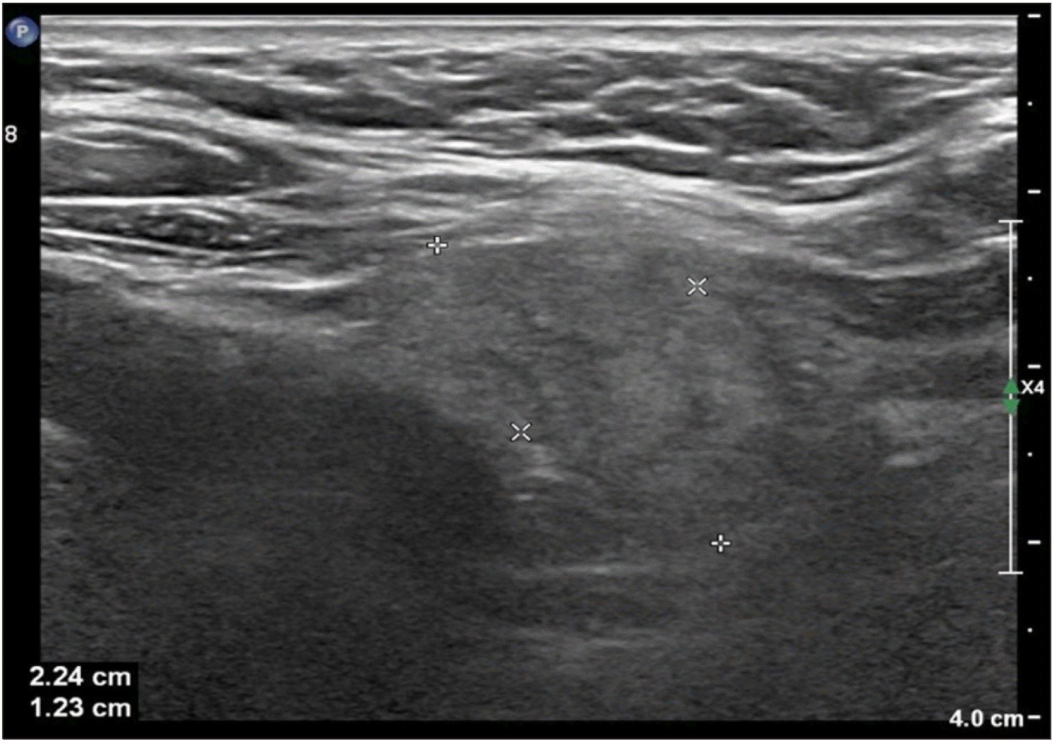
A 69-year-old woman visited our clinic with cervical compressive discomfort. On physical examination, there was no palpable mass such as a lesion at the anterior neck, and there was no definite abnormal finding in the larynx upon laryngoscopic examination. She had once undergone radiofrequency ablation of a thyroid nodule on the right side at another hospital. To investigate the cervical compressive discomfort and follow-up the thyroid nodule, we performed neck ultrasonography. There was a C5 nodular lesion measuring 1.5×1.0×0.6 cm with punctate echogenic foci and a heterogeneous echoic mass with an irregular surface at the suprasternal notch (
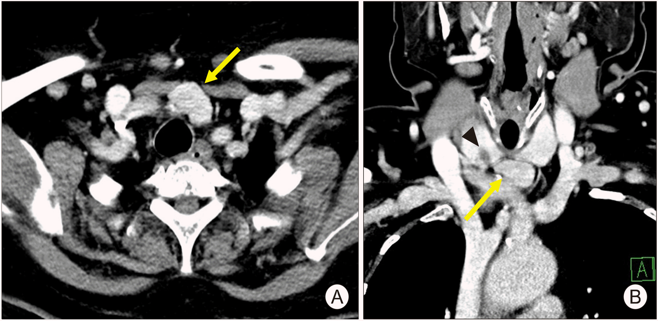
Fig. 1). Following fine needle aspiration of the thyroid nodule, cytological evaluation revealed atypical cells of undermined significance. We performed neck enhanced computed tomography (CT) to obscure adjacent anatomical structures of the thyroid gland and suprasternal notch mass. There was a relatively highly attenuated mass at the suprasternal notch with a well-bounded circumference, and a hypo-attenuated thyroid nodular lesion was found (
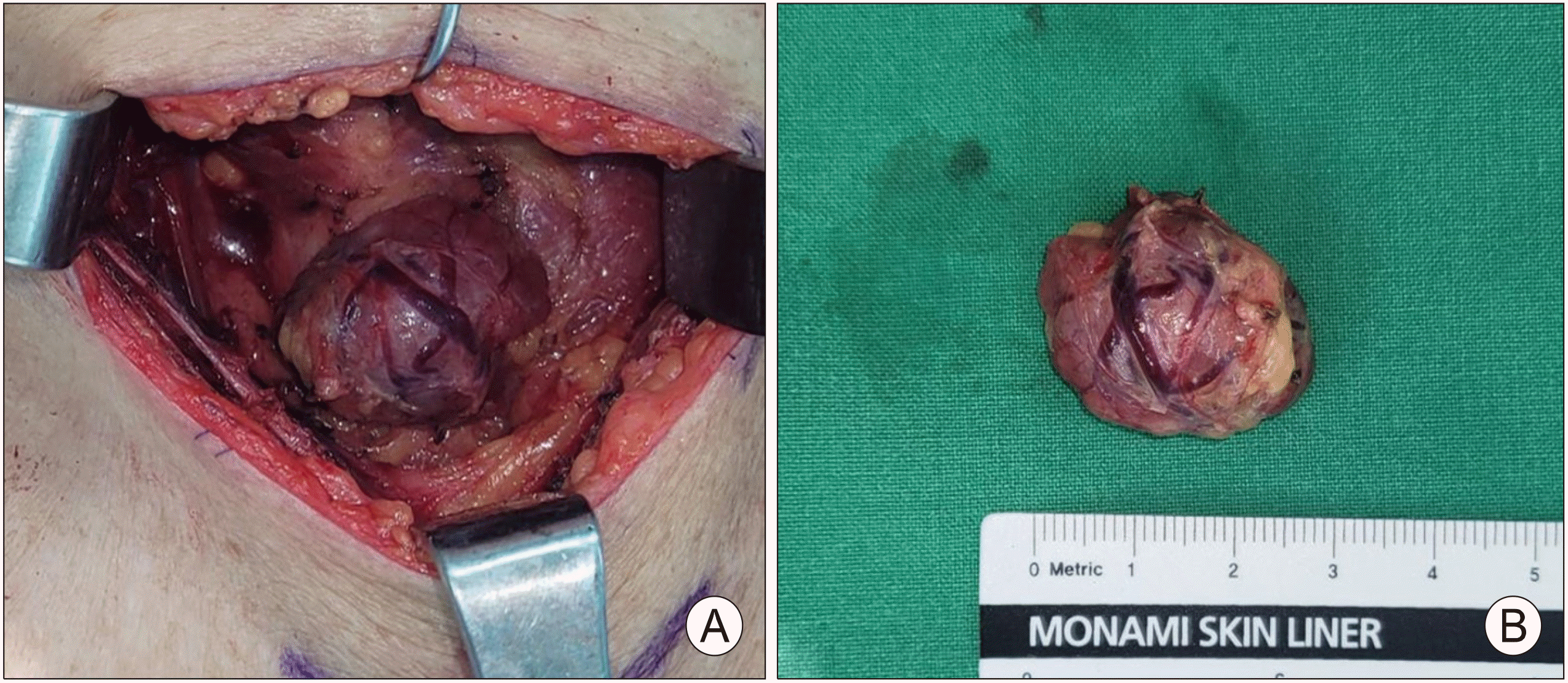
Fig. 2). We performed diagnostic right unilateral thyroidectomy with mass excision under general anesthesia. Preoperative serum thyroid hormone, thyroid-stimulating hormone, and calcium levels were within the normal limits. The suprasternal notch mass was encapsulated and well dissected without any connection to the thyroid gland (
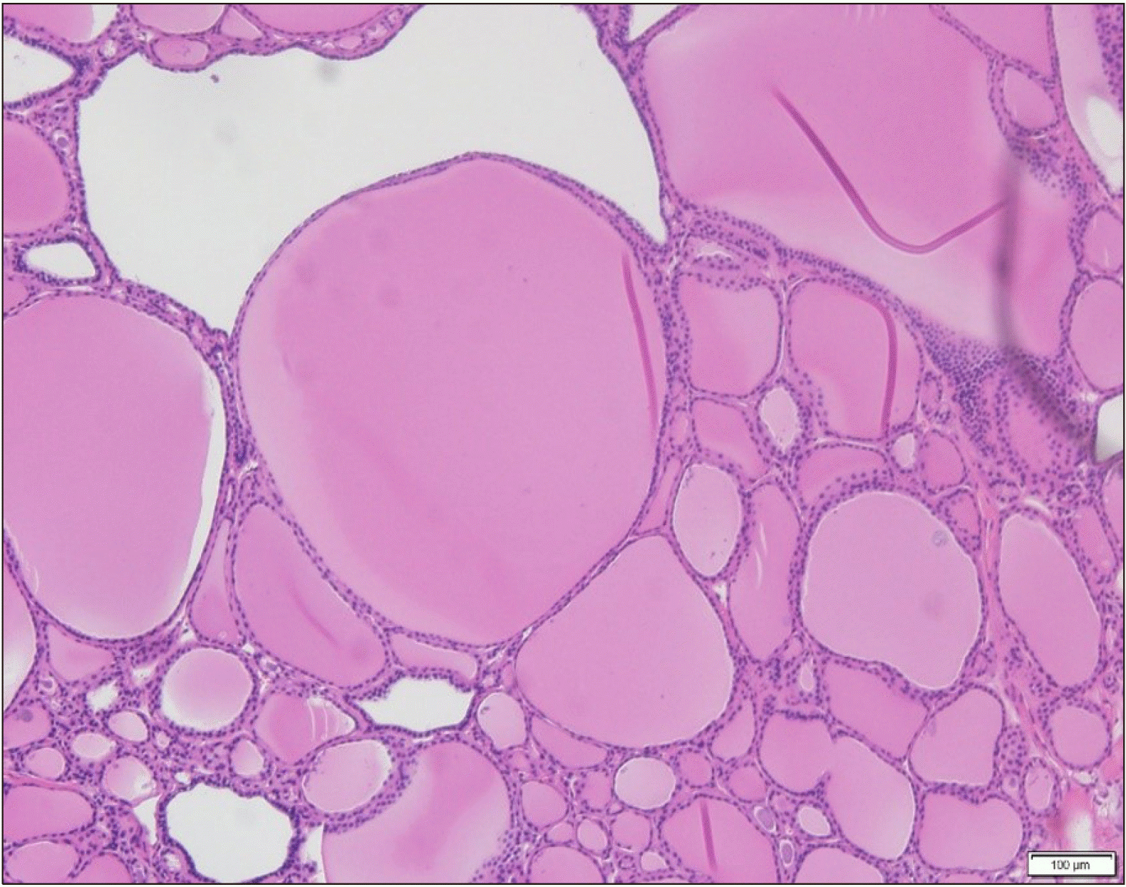
Fig. 3). It was confirmed to a nodular goiter and diagnosed as ectopic thyroid tissue (
Fig. 4). The right thyroid lobe was confirmed to be a nodular goiter. The patient was discharged 6 days postoperatively and has remained free of disease without cervical compressive discomfort for 3 months after surgery.
 | Fig. 1Representative sonographic image of the neck. A heterogeneous echoic mass measuring 2.24×1.23 cm with an irregular surface. 
|
 | Fig. 2Representative neck computed tomography images. A well-circumscribed irregular mass measuring 2.6 cm at the suprasternal notch (yellow arrow) and a thyroid nodule with low attenuation (black arrow-head) (A: axial, B: coronal). 
|
 | Fig. 3Operative findings. (A) A round well-circumscribed mass at the suprasternal notch without any connection to the thyroid gland. (B) A firm mass measuring 2.6×2 cm was removed. 
|
 | Fig. 4Representative pathologic section. A section of the suprasternal mass showing follicles of various sizes surrounded by flattened epithelium and containing colloid (Hematoxylin & Eosin staining, original magnification ×200). 
|
Go to :

Discussion
Ectopic thyroid is a rare form of thyroid dysgenesis and defined as thyroid tissue not located at an orthotropic site.
1) It is rare, and its prevalence is approximately 1 per 100,000-300,000 persons.
2) It arises mainly along the descending path of the normal thyroid gland during the gestational period and is usually found at the base of the tongue.
3) Ectopic thyroid at rare locations, including the trachea, mediastinum, heart, and esophagus, has been reported.
4)
The primitive thyroid arises from the pharyngeal base between the first and second pharyngeal pouches.
5) As an endodermal proliferation of the pharyngeal base, it forms a diverticulum. Around the fifth week of gestation, the thyroid diverticulum migrates caudally along the midline and passes through the hyoid bone and laryngeal cartilage.
1) During this migration, the hollow thyroid diverticulum solidifies with follicular elements.
6) At the seventh week of gestation, the thyroid finally locates in the anterior neck, while the fourth and fifth pharyngeal pouches contact and conglomerate at the thyroid with filling of para-follicular elements. If the primitive thyroid gland moves lower than the orthotropic location, an ectopic thyroid gland can appear at the suprasternal notch, mediastinum, and heart.
7)
Ectopic thyroid is usually asymptomatic and often found incidentally on radiological examinations.
6) If the ectopic thyroid grows to a considerable size, it can cause various symptoms depending on its location.
2) Lingual thyroid can cause throat discomfort, hoarseness, and dysphagia. Cervical ectopic thyroid can be a palpable mass and even cause dyspnea if it presses on the airway.
4)
Neck ultrasonography, CT, and radionuclide thyroid scanning are useful to diagnose a cervical midline mass suspected to be ectopic thyroid.
4) Neck ultrasonography is noninvasive and rapid, and can provide information about the properties of a mass, such as its size, cystic changes, and vascularity.
8) Ectopic thyroid has similar sonographic features as the normal thyroid gland with the exception of its shape and size. Variable echotexture of ectopic thyroid has been reported. CT can clarify the size and extension of the mass, and the relationship between ectopic thyroid tissue and adjacent structures.
3) Ectopic thyroid is a vessel-rich tissue and is therefore slightly attenuated on CT.
9,10) CT can help to determine a resection range before surgery.
10) Radionuclide thyroid scanning is a noninvasive diagnostic modality and useful to identify all the sites of ectopic thyroid tissue.
6,11)
Patients who are asymptomatic and have normal thyroid function do not require treatment, but patients with any symptoms or complications should be treated in a customized manner with consideration of the size and location of ectopic thyroid, and the thyroid function level.
11) Patients with hypothyroidism can be treated with levothyroxine. Levothyroxine suppresses thyroid-stimulating hormone secretion and can decrease the size of ectopic thyroid.
3,6) Ectopic thyroid that continues to cause symptoms despite medical treatment or is suspected to be malignant should be managed surgically. Before surgery, it is necessary to confirm the presence of a normal thyroid gland. The ectopic thyroid gland may be the only gland that makes thyroid hormones if there is no orthotropic thyroid gland.
9) Without confirmation of the presence of a normal thyroid gland, excision of ectopic thyroid may lead to permanent hypothyroidism.
6)
In our case, the suprasternal mass compressed the anterior neck, which led to compressive discomfort, and was suspected to be a metastasis of thyroid malignancy; therefore, surgical resection was necessary for diagnosis and treatment. If a mass at the suprasternal site is clinically found with symptoms, ectopic thyroid should be considered, and treatment should be decided based on the mass properties, thyroid function, whether a normal thyroid gland exists, and patient age.
Go to :


 PDF
PDF Citation
 Print
Print


 XML Download