Abstract
Zenker’s diverticulum is rarely known to be an impediment to the diagnosis and surgical procedure for thyroid disease. In this report, we discuss a rare case of pre-operative diagnosis of asymptomatic Zenker’s diverticulum and surgical treatment of the disease during thyroidectomy. A 48-year-old male was referred to our hospital with multiple thyroid nodules confirmed to be papillary thyroid cancer. Pre-operative ultrasonography and computed tomography scan findings suggested the presence of a left Zenker’s diverticulum. The left pharyngeal pouch was treated by reinforcing the overlying soft tissue during thyroidectomy, without structural injury to the recurrent laryngeal nerve. We report this case with a focus on the considerations during the pre-operative diagnosis and precautions to be taken during surgery, such as the distorted path of the recurrent laryngeal nerve and manipulation of the diverticulum.
Go to : 
Zenker’s diverticulum was first mentioned by Zenker and Von Ziemssen in 1877, but its correlation with other diseases began to emerge relatively recently. It is a rare disease caused by the formation of a posterior pharyngoesophageal sac above the hypopharyngeal wall.1) It usually occurs at the Killian dehiscence, which is a weak portion of the inferior pharyngeal constrictor muscle. The main symptoms of the disease include transient dysphagia, weight loss, pulmonary aspiration, neck mass, regurgitation, and esophageal obstruction.1)
Symptomatic Zenker’s diverticulum can be treated with surgical procedures using both open surgical and endoscopic approaches.2-4) An asymptomatic Zenker diverticulum does not necessarily require surgical treatment.1) However, according to previous studies, Zenker’s diverticulum could occasionally be misinterpreted as a thyroid nodule.5-10) As ultrasound techniques and equipment are being developed and the number of patients for screening thyroid disease is increasing, anatomically adjacent Zenker’s diverticulum may be accidentally diagnosed or mistaken for a thyroid nodule.5-10) However, in patients considering surgical treatments for thyroid cancer, little is known about the diagnosis and operation of asymptomatic Zenker’s diverticulum accompanying the thyroid cancer.
Due to the anatomical relationship between the thyroid and diverticulum, comorbid Zenker’s diverticulum can be a risk factor for structural injuries of surrounding tissues, such as the recurrent laryngeal nerve, during thyroidectomy. In addition, because of the rare incidence of Zenker’s diverticulum accompanying thyroid disease and a lack of previous studies on simultaneous surgery for both diseases, surgeons can easily overlook the risk of surgery. Moreover, an elective surgical plan is difficult to make, not only in terms of successfully preserving the important structures but also in successfully resecting the thyroid and diverticulum at the same time.
In this report, we introduce a case of left asymptomatic Zenker’s diverticulum that was found on pre-operative ultrasonography and computed tomography (CT) scan, and treated during left hemi-thyroidectomy and selective neck dissection (Level II-Vb) for papillary thyroid cancer. We discuss the pre-operative radiographic findings related to Zenker’s diverticulum with thyroid cancer, notable intra-operative points regarding the surgical procedures, and precautions to be taken during surgery to preserve the important structures close to the diverticulum.
Go to : 
A 48-year-old male presented with multiple thyroid nodules that were incidentally detected. The patient was referred to our hospital for further evaluation and surgical treatment of suspected malignant thyroid lesions. On pre-operative cervical ultrasonography, an isoechoic ill-marginated heterogeneous mass sized approximately 3.8×1.8 cm was observed in the left thyroid lobe. In the right lobe, a mass sized approximately 1.2×1.0 cm showing a similar aspect to the mass of the left lobe was found. Moreover, at least 40 multiple conglomerated hypoechoic lymph nodes with microcalcification were observed on both sides of the lateral and central neck. In addition, a 1.4×1.0 cm-sized nodule with a halo was observed, showing heterogeneous internal echogenicity (Fig. 1). The inside of the halo showed hyperechogenicity with posterior acoustic shadowing, but no clear findings suggesting microcalcification were found.
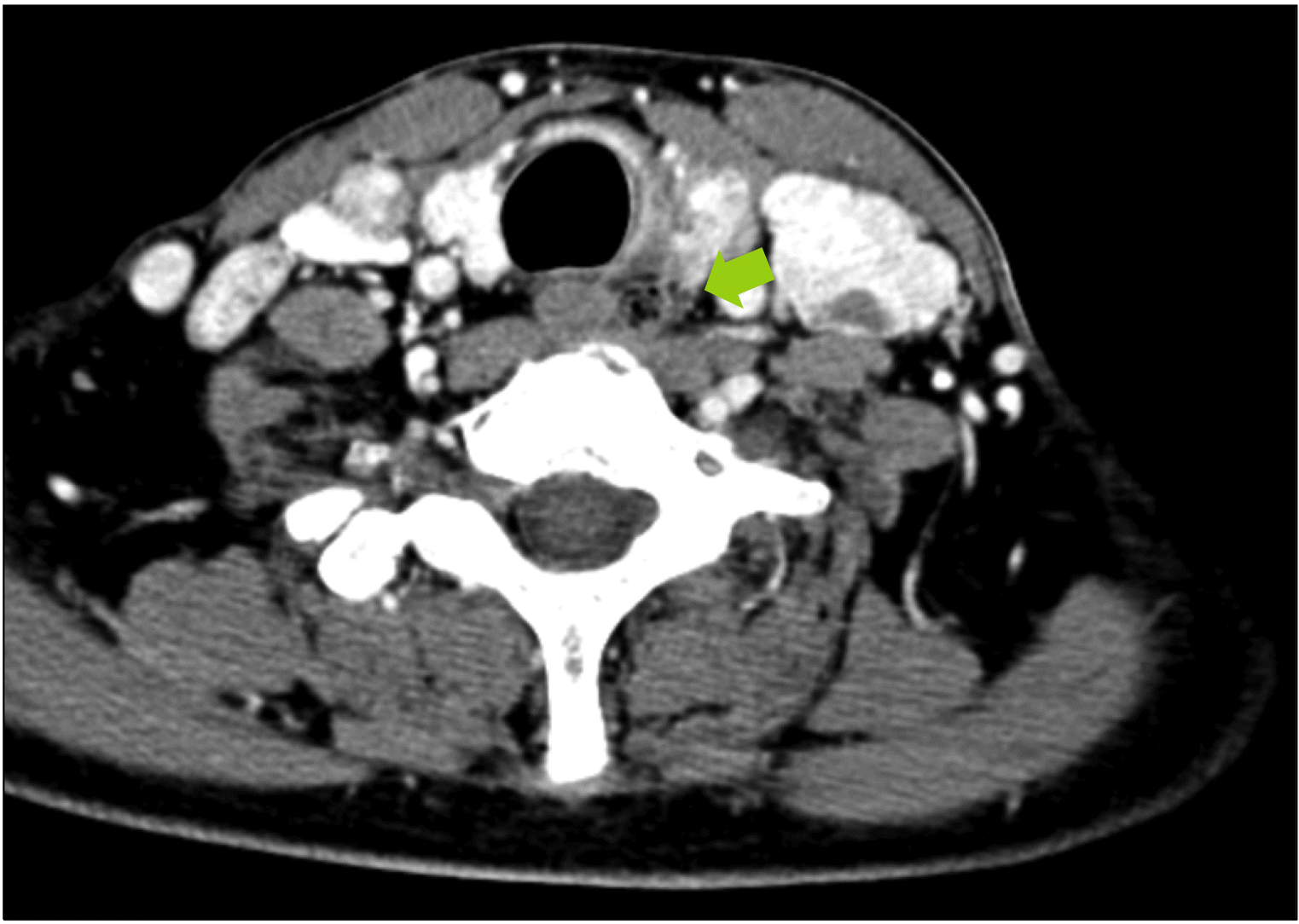
On the CT scan, neck masses suspected of multiple metastatic lymphadenopathy of both lateral and central neck areas and a collapsed left internal jugular vein due to lymph node metastasis were observed. Moreover, soft tissue density (indicating the presence of a 1.0×0.9 cm-sized mass), including multi-focal air shadows, was observed posterior to the left superior thyroid lobe and left to the esophagus (Fig. 2). Both ultrasonography and CT findings suggested that the lesion was a Zenker’s diverticulum. The largest nodule of the left thyroid lobe was confirmed to be papillary thyroid cancer by core needle biopsy, and suspected lymph nodes of both cervical level IV areas were confirmed to be metastases by fine needle aspiration biopsy.
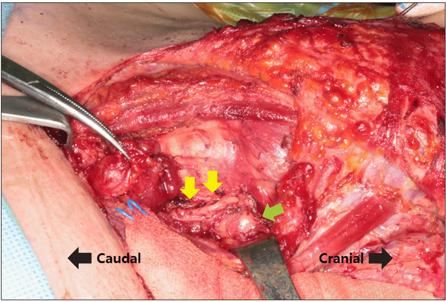
Thyroidectomy and selective neck dissection (Level II-Vb) were also performed. Simultaneously, we planned to treat Zenker’s diverticulum in collaboration with the thoracic surgery department during thyroidectomy. During left hemi-thyroidectomy, to preserve the recurrent laryngeal nerve, the nerve path was mapped using an intra-operative nerve monitor (NIM-Response 3.0 System; Medtronic, Jacksonville, FL, USA) using an electromyogram (EMG) endotracheal tube (TriVantage EMG tube 7; Medtronic, MN, USA). Using this method, we could predict the path of the dorsally distorted recurrent laryngeal nerve at the left middle-upper pole of the thyroid due to the mass effect of the Zenker’s diverticulum (Fig. 3). However, during the process, it was confirmed that the recurrent laryngeal nerve was superficially invaded by the tumor. Accordingly, the recurrent laryngeal nerve was shaved and R1 resection was performed. Immediately after removal, the amplitude of the nerve stimulation decreased, but the amplitude gradually increased during surgery. Although mild recovery of the amplitude was identified, it was not sufficient to ensure intact movement of the vocal cords. Therefore, we decided to perform a staged surgery. Postoperative fiberscopic images did not reveal vocal cord palsy.
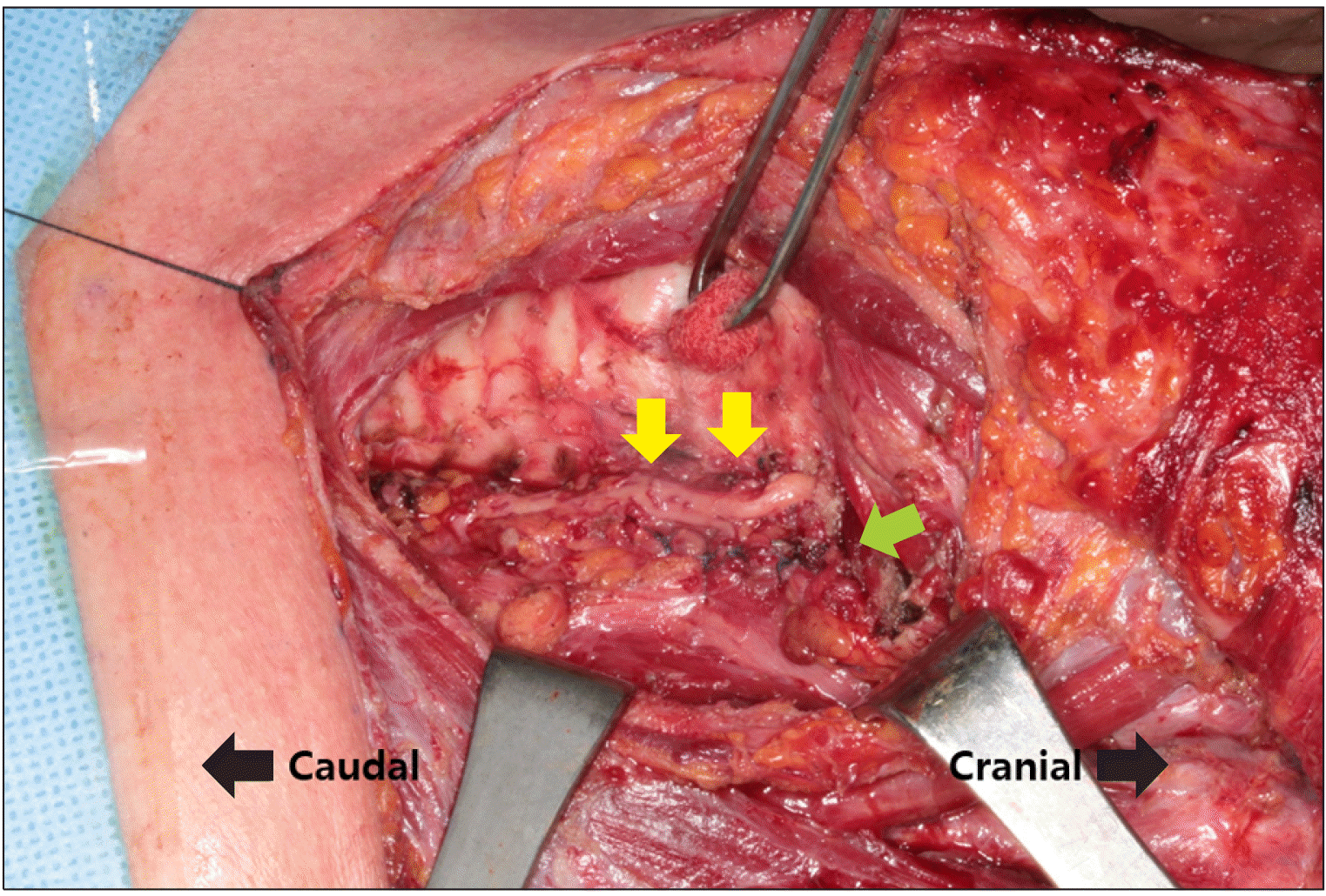
After thyroidectomy and neck dissection, Zenker’s diverticulum was treated. To avoid damage to the recurrent laryngeal nerve and diverticulum, the soft tissue around the diverticulum, which was deep in the recurrent laryngeal nerve, was undermined parallel to the nerve. The diverticulum was then pushed into the space beneath the soft tissue, and the surrounding soft tissue was sutured with an absorbable string (vicryl 4-0) (Fig. 4). Right completion thyroidectomy and right selective neck dissection (Level II-Vb) were performed 3 months after the first surgery, and radioactive iodine therapy was planned. Since then, the patient has been under follow-up for 2 months without any notable findings.
Go to : 
Owing to the recent development of techniques for ultrasound equipment, the correlation between Zenker’s diverticulum and thyroid disease has begun to emerge.5-10) In Korea, there are three reports on the discriminative diagnosis between Zenker’s diverticulum and thyroid nodules. This report describes the pre-operative diagnosis and the actual elective surgical process of the disease.
For thyroid nodules without histopathological information, the preferred diagnostic approaches are ultrasonography-guided fine needle aspiration biopsy and core needle biopsy. Zenker’s diverticulum is known to have a high risk of perforation during invasive procedures such as gastroesophageal endoscopy or endotracheal intubation.1) Thus, if a clinician performs a biopsy without being aware of risk and ultrasonographic findings of Zenker’s diverticulum, complications such as deep neck infection or mediastinitis may occur due to diverticular perforation.8-10) For patients suspected of diverticulum on ultrasonography, clinical and radiologic diagnosis via CT scans and esophagograms should be preceded to biopsy.8-10)
There are two notable points in this case report. First, it is possible to diagnose Zenker’s diverticulum using radiological findings at the stage of surgery planning. The ultrasonographic findings related to Zenker’s diverticulum showed an isoechoic or hypoechoic mass with a borderline hypoechoic zone, which was described as a halo in our report, and internal or peripheral echogenic foci.11,12) These lesions are usually found on the posterior side of the left thyroid gland and are accompanied by posterior acoustic shadows.11,12) In the sagittal view, connectivity between the lesion and posterior structure may be found.11,12) As mentioned earlier, additional CT scans and esophagograms can be used to help correlate the findings with ultrasonography and diagnose Zenker’s diverticulum.11,12)
Second, anatomical distortion of the recurrent laryngeal nerve due to the mass effect of Zenker’s diverticulum may lead to a high risk of injury. To avoid the injury of the recurrent laryngeal nerve, a pre-operative awareness on Zenker’s diverticulum must be establi-shed. In addition, it is important to map the pathway of the recurrent laryngeal nerve using intraoperative neuromonitoring. In this case, the recurrent laryngeal nerve was located between the thyroid cancer extending toward the tracheoesophageal groove and the Zenker’s diverticulum, deviating the nerve laterally. Therefore, special care was given not to injure the nerve during dissection and to shave off the tumor from the nerve. Moreover, because superficial recurrent laryngeal nerve invasion of the tumor was observed, shaving of the recurrent laryngeal nerve and R1 resection of the tumor were performed. Therefore, caution must be exercised under these conditions.
Generally, an asymptomatic Zenker’s diverticulum does not require surgical treatment. However, we decided on surgical treatment because the residual diverticulum may be confused as recurrent cancer during follow-up. Considering that Zenker’s diverticulum is an age-related disease, we hypothesized that if the diverticulum increases in size, or if an infection develops, it may affect the adjacent nerve.
In conclusion, Zenker’s diverticulum may be diagnosed using pre-operative ultrasonography or CT in patients who undergo thyroidectomy. In such cases, possible lateral dislocation of the recurrent laryngeal nerve by the diverticulum should be considered, and care should be taken to preserve the nerve.
Go to : 
References
1. Law R, Katzka DA, Baron TH. 2014; Zenker's diverticulum. Clin Gastroenterol Hepatol. 12(11):1773–82. quiz e111–2. DOI: 10.1016/j.cgh.2013.09.016. PMID: 24055983.

2. Verdonck J, Morton RP. 2015; Systematic review on treatment of Zenker's diverticulum. Eur Arch Otorhinolaryngol. 272(11):3095–107. DOI: 10.1007/s00405-014-3267-0. PMID: 25194579.

3. Bizzotto A, Iacopini F, Landi R, Costamagna G. 2013; Zenker's diverticulum: exploring treatment options. Acta Otorhinolaryngol Ital. 33(4):219–29.
4. Morton RP, Bartley JR. 1993; Inversion of Zenker's diverticulum: the preferred option. Head Neck. 15(3):253–6. DOI: 10.1002/hed.2880150315. PMID: 8491590.

5. Kang HC. 2004; A case of Zenker's diverticulum masquerading as a thyroid nodule. Korean J Med. 67:S757–S760.
6. Ip E, Yong C, Cheng M. 2020; Close encounter of the right-sided Zenker's diverticulum masquerading as a thyroid nodule. ANZ J Surg. 90(11):E112–E3. DOI: 10.1111/ans.15870.

7. Shanker BA, Davidov T, Young J, Chang EI, Trooskin SZ. 2010; Zenker's diverticulum presenting as a thyroid nodule. Thyroid. 20(4):439–40. DOI: 10.1089/thy.2009.0177. PMID: 20373989.

8. Kim JH, Choi YS, Kim BK, Lee JS, Park YH, Hur B. 2012; Zenker's diverticulum suspected to be a thyroid nodule diagnosed on fine needle aspiration: a case report. J Med Cases. 3(4):261–3. DOI: 10.4021/jmc635e.

9. Walts AE, Braunstein G. 2006; Fine-needle aspiration of a paraesophageal diverticulum masquerading as a thyroid nodule. Diagn Cytopathol. 34(12):843–5. DOI: 10.1002/dc.20570. PMID: 17115442.

10. Oertel YC, Khedmati F, Bernanke AD. 2009; Esophageal diverticulum presenting as a thyroid nodule and diagnosed on fine-needle aspiration. Thyroid. 19(10):1121–3. DOI: 10.1089/thy.2009.0136. PMID: 19772399.

11. Kwak JY, Kim EK. 2006; Sonographic findings of Zenker diverticula. J Ultrasound Med. 25(5):639–42. DOI: 10.7863/jum.2006.25.5.639. PMID: 16632788.

12. Lixin J, Bing H, Zhigang W, Binghui Z. 2011; Sonographic diagnosis features of Zenker diverticulum. Eur J Radiol. 80(2):e13–9. DOI: 10.1016/j.ejrad.2010.05.028. PMID: 20576383.

Go to : 


 PDF
PDF Citation
 Print
Print


 XML Download