

 PDF
PDF Citation
 Print
Print


Introduction
Multiple endocrine neoplasia type 2A (MEN 2A) is autosomal dominantly inherited disease with RET oncogene mutation. Typical presentations of MEN 2A are medullary thyroid carcinoma, pheochromocytoma and primary hyperparathyroidism (PHPT). Medullary thyroid carcinoma is the most common phenotype of MEN 2A and determined the prognosis of MEN 2A because of its malignancy.1) Pheochromocytoma and PHPT are less commonly presented in MEN 2A. Peptic ulcer disease and hypercalcemia are usually associated with multiple endocrine neoplasia type 1 (MEN 1) because PHPT is most common disease of MEN 1 and peptic ulcer disease can be caused by gastrinoma.1) However we experienced a case of MEN 2A with peptic ulcer perforation and hypercalcemia as first manifestations. We presented this case because it was extremely rare and not reported previously.
Go to : 
Case Report
A 38-year-old Korean-Chinese man was admitted to the emergency room for sudden abdominal pain. He had bilateral adrenalectomy 10 years ago at China but we could not obtain the pathologic report of adrenalectomy. In the chest X-ray and abdominal computed tomography (CT), pneumoperitoneum with panperitonitis was found without abnormality in the adrenalectomy site. After diagnosis of peptic ulcer perforation, laparoscopic primary closure and omentopexy was performed with steroid replacement due to previous bilateral adrenalectomy.
After operation, high ionized calcium; 1.54 mmol/L (1.13-1.32 mmol/L) and high alkaline phosphatase; 556 IU/L (40-129 IU/L) were noticed. The 24 hour urinary calcium was also elevated; 494 mg/day (100-300 mg/day) and the parathyroid hormone level was highly elevated; 668.6 pg/mL (10-57 pg/mL). We diagnosed him as PHPT and checked sonography, CT and sestamibi scan for localization during postoperative recovery period.
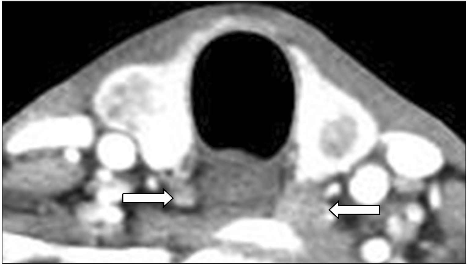
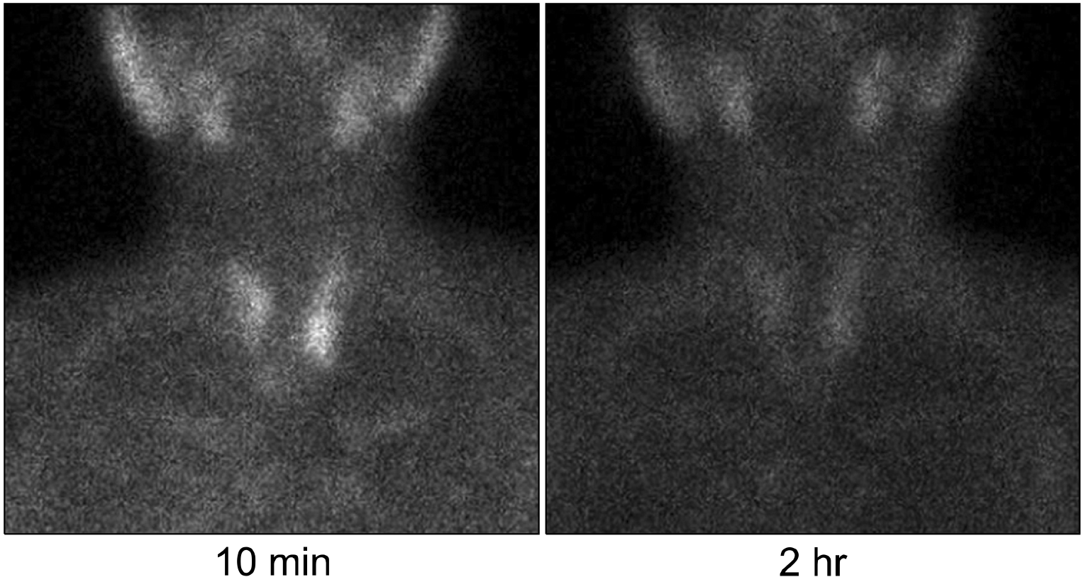
The thyroid sonography showed bilateral 1.4 cm sized heterogeneous low echoic solid nodules with multiple low echoic nodules around thyroid gland (Fig. 1). In the neck CT scan, bilateral thyroid masses with suspected parathyroid hyperplasia were observed (Fig. 2). The parathyroid hyperplasia was also suspected in the Tc-99m sestamibi scan without ectopic parathyroid gland (Fig. 3). In the fine needle aspiration cytology of thyroid nodule, the result was bilateral medullary thyroid carcinoma. Calcitonin level was highly elevated; 390 pg/mL (0-20 pg/mL) but gastrin level was normal; 83.5 pg/mL (0-90 pg/mL). From the above results, we can diagnose him as MEN 2A and a previous adrenal diagnosis could be presumed to be bilateral pheochromocytoma.
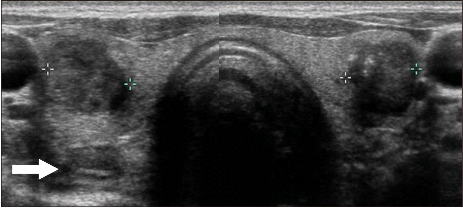 | Fig. 1Thyroid sonography shows both 1.4-cm-sized heterogeneous low echoic solid nodules with multiple low echoic nodules around thyroid which is suspected of parathyroid hyperplasia (arrow).
|
We checked patient’s family history again, but there was no history of thyroid cancer or endocrine disease. In the hormonal studies, 24 hour urinary metanephrine, vanillylmandelic acid (VMA) and cortisol was normal as 0.97 mg/day (0-1.3 mg/day), 6.8 mg/day (0-8 mg/day) and 163.6 ug/day (55.5-286 ug/day). Patients had severe osteoporosis in bone densitometry; T-score −4.6 in femur neck and −5.4 in lumbar spine.
After diagnosis of medullary thyroid carcinoma with parathyroid hyperplasia, we performed total thyroidectomy with prophylactic central node dissection and total parathyroidectomy with auto-transplantation of right inferior parathyroid gland to left forearm. In the operational field, right inferior parathyroid is smallest one as 7 mm. We minced into 1 mm3 pieces and transplanted 15 pieces to brachioradialis muscle of non-dominant left forearm (Fig. 4). The pathological diagnosis was also medullary thyroid carcinoma with multiple lymph node metastasis and parathyroid hyperplasia. In the 4th postoperative day, the calcitonin level was normal as 5.5 pg/mL but parathyroid hormone level was decreased as 4.4 pg/mL. The parathyroid hormone was normalized as 24.5 pg/mL one month later.
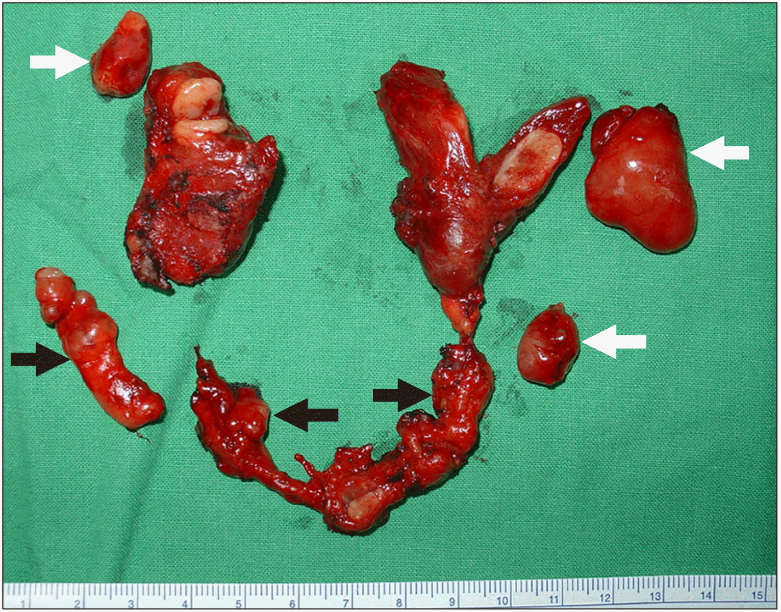 | Fig. 4The resected specimen of upper thyroid lobes shows whitish tan cut surface which is characteristic finding of medullary thyroid carcinoma. Dissected lymph nodes of central compartment are included in the lower part of photo (black arrows). The sizes of excised parathyroid are 3 cm of left upper one, 1.5 cm of right upper and 1.2 cm of left lower ones (white arrows). The right lower parathyroid is about 7 mm sized which is auto-transplanted to left forearm.
|
We tested germline mutation of RET oncogene with patient’s informed consent. Germline mutation was analyzed by direct sequencing of exons 10, 11, 13, 14, 15, 16 of RET oncogene. At the codon 634 of exon 11, point mutation of 1900th DNA from T to C (c.1900T>C) was found. It resulted amino acid change from Cysteine to Arginine (p.Cys634Arg) which was previously known as C634R (TGC>CGC).
Go to :
Discussion
PHPT is uncommon and delayed presentation in MEN 2A. The EUROMEN study group reported that PHPT was diagnosed earlier than medullary thyroid carcinoma in only 4% of MEN 2A.2) The French Calcitonin Tumors Study Group also reported only 4 cases (2%) out of 249 MEN 2A patients whose PHPT was diagnosed as first syndrome.3) Recent an international multicenter study reported that the prevalence of PHPT as first manifestation of MEN 2A was only 0.9% (10 cases out of 1085 MEN 2A).4) In this case, we diagnosed PHPT firstly because of incidental finding of hypercalcemia and then we could confirm medullary thyroid carcinoma during localization study of PHPT. Coexisting thyroid tumor is easily found because parathyroid and thyroid are in adjacent location. So thyroid tumor must be searched in case of PHPT and vice versa.
PHPT and hypercalcemia is possible cause of peptic ulcer disease but the exact pathophysiologic mechanism is not certain especially in MEN 2A. In MEN 1, PHPT and gastrinoma are relatively common phenotype, and elevated gastrin level cause peptic ulcer disease. We also checked gastrin but the level was normal. In one theory, the activation of calcium sensing receptor on the gastric parietal cell by acutely elevated calcium can cause gastric acid secretion, but there was controversy in chronic elevation of calcium, so further study is necessary.5) We could not find any report about peptic ulcer disease with hypercalcemia in MEN 2A, so this would be first case report about peptic ulcer disease in MEN 2A.
According to Revised American Thyroid Association Guidelines,6) PHPT in MEN 2A could be operated three options; 1) subtotal parathyroidectomy leaving one gland or a piece of one gland, 2) total parathyroidectomy with auto-transplantation; or 3) resection of only enlarged gland with intraoperative parathyroid hormone monitoring. Resection of only enlarged gland was usually recommended because most of PHPT in MEN 2A are asymptomatic or mild symptomatic. However we performed total parathyroidectomy with auto-transplantation to non-dominant left forearm because patients had severe osteoporosis and very high level of calcium and parathyroid hormone. We used brachioradialis muscle because parathyroid hormone monitoring and re-excision with local anesthesia would be easy and safely performed in case of recurrence.
In the germline mutation test of RET oncogene, mutation of c.1900T>C was found in this case which was previously described as C634R. Codon 634 of exon 11 is most frequent mutational hot spot in MEN 2A. About half of RET mutation of Korea was reported in codon 634 and C634R was also most frequent type of mutation in Korea.7) C634R mutation is related with high risk level of medullary thyroid carcinoma and the incidences of pheochromocytoma is more than 50%. The incidence of PHPT is about 20-30% which is highest among MEN 2A.6) In this case, the patient had a history of surgery for bilateral adrenal tumors in his twenties. Although the pathologic result could not be confirmed, we can be sure of bilateral pheochromocytoma from the results of mutational test.
In conclusion, a patients with peptic ulcer perforation and hypercalcemia was diagnosed as MEN 2A. Patients had total thyroidectomy with prophylactic central neck node dissection for medullary thyroid carcinoma and total parathyroidectomy with auto-transplantation for PHPT. Although peptic ulcer disease with hypercalcemia is very rare in MEN 2A, clinical suspicion of multiple endocrine neoplasia and germline mutational test would be important for accurate diagnosis and treatment.
Go to :
 XML Download