This article has been
cited by other articles in ScienceCentral.
Abstract
Although a pancreaticojejunostomy (PJ) is not required after a distal pancreatectomy in most cases, it needs to be performed to prevent atrophy of the remnant pancreas when the proximal duct is obstructed by a tumor, stone, or etc. In these conditions, the critical postoperative pancreatic fistula (POPF) gives surgeons cause to hesitate before performing a PJ. We previously presented the modified technique of Mattress PJ named “inverted mattress PJ” (IM-PJ) and published improved outcomes in the aspects of POPF after a pancreaticoduodenectomy and a central pancreatectomy. Recently, we had a case of a patient who has chronic pancreatitis with a proximal pancreatic duct obstruction, requiring a distal pancreatectomy and PJ. Based on the previous report, we decided to apply the “inverted mattress PJ” (IM-PJ) technique for a Roux-en Y PJ after a distal pancreatectomy. The patient was discharged after surgery without complications. We reviewed a case of a patient requiring PJ following a distal pancreatectomy and discussed the safety of our technique.
Go to :

Keywords: Pancreatitis, chronic, Pancreatectomy, Pancreaticojejunostomy, Pancreatic fistula
INTRODUCTION
A partial pancreatectomy, such as a pancreaticoduodenectomy and a central pancreatectomy for malignant and benign disease of the pancreas, require safe anastomosis between the pancreas and intestinal tract. Although there have many efforts to improve the safety of the procedures, the results have not been satisfactory. Despite various techniques being devised to anastomose the pancreatic duct and intestinal tract to avoid pancreatic fistula, the critical pancreatic fistula still remains the Achilles’ heel of the partial pancreatectomy requiring a pancreatoenterostomy [
1].
Although a pancreaticojejunostomy (PJ) is not usually required after a distal pancreatectomy, it needs to be performed to prevent atrophy of the remnant pancreas in rare conditions, in which the drainage of pancreatic juice through the proximal duct is impaired by stricture, tumor, or stone etc. The drainage procedure combined with distal pancreatectomy was known as the Duval procedure. The Duval procedure has been reported for stricture of the pancreatic duct and chronic pancreatitis to relieve ductal hypertension [
2]. However, despite the necessity of a Roux-en Y distal PJ in these conditions, the high probability of critical postoperative pancreatic fistula (POPF) causes hesitation in surgeons to perform the PJ [
3].
Our previous published papers report the use of “inverted mattress PJ” (IM-PJ) as an effort to increase the safety of a PJ. Compared to the conventional “duct to mucosa” technique after a pancreaticoduodenectmy, the IM-PJ, effectively reduced the risk of grade C POPF and serious complications such as a gastroduodenal artery pseudoaneurysm [
4]. Similarly, no pancreatic fistula occurred from IM-PJ after a central pancreatectomy [
5]. Based on the previous reports of low morbidity following IM-PJ, we decided to apply this technique after a distal pancreatectomy for a patient with chronic pancreatitis with proximal pancreatic duct obstruction, caused by an impacted stone, requiring the Duval procedure.
In this paper, we review a case of a patient requiring a distal pancreatectomy and PJ, for which we utilized “Roux-en Y inverted mattress PJ” after a distal pancreatectomy, and discuss the safety of this technique. This study has been approved by the research ethics committee of Kyungpook National University Chilgok Hospital (KNUCH 2021-07-009).
Go to :

CASE
Initially, a 51-year-old female, a known heavy alcoholic, was admitted for abdominal pain in October 2015. Abdominal computed tomography (CT) showed overall atrophy of the pancreas parenchyma and dilatation of the pancreas duct, with pancreatoliths in the proximal duct (
Fig. 1). Under the diagnosis of acute onset of chronic pancreatitis, the patient was admitted and treated conservatively. After the symptoms improved, the patient was discharged and followed up.
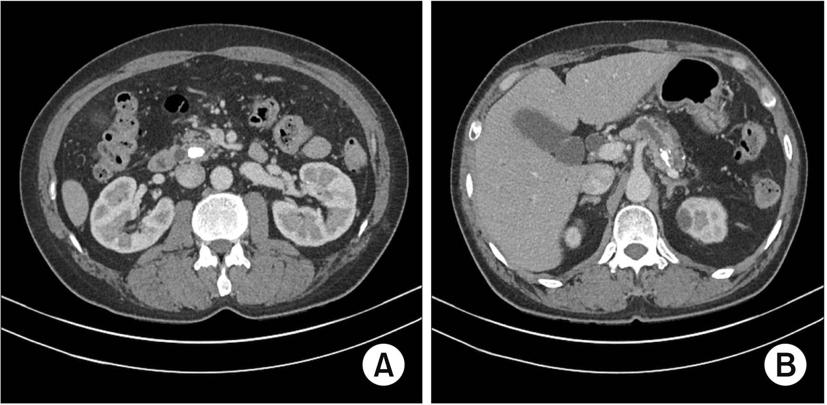 | Fig. 1(A) Abdominal contrast enhanced computed tomography images show a pancreatic stone that occludes the proximal pancreas. (B) Pancreatic duct dilatation due to proximal pancreatic duct obstruction is identified. 
|
During the period of follow-up for two years, the patient complained of several episodes of abdominal pain. In January 2017, balloon dilatation and stone removal were tried to relieve the symptoms. Four months later, carbohydrate antigen 19-9 was elevated, and a heterogeneous mass was identified on endoscopic ultrasonography. A fine needle aspiration biopsy showed only the infiltration of neutrophils.
The patient was referred for surgery for a possible hidden malignancy and persistent abdominal pain. The decision was made to surgically resect the pancreas, which was harboring a mass, and perform a drainage procedure to relieve the pain.
On open exploration, the pancreas showed atrophy with calcified nodules and the parenchyma had a hard texture. A distal pancreatectomy was performed to remove the suspected lesion harboring mass and the spleen was also removed due to dense fibrosis caused by repeated pancreatitis. The pancreatic duct of resection margin was dilated (
Fig. 2). The presence of an impacted stone in the proximal duct and dilatation of the entire pancreatic duct raised the possibility of a proximal pancreatic duct obstruction causing persisting recurrent abdominal pain. Therefore, the Duval procedure was performed. Based on our experience of reporting the safety of IM-PJ for pancreaticoduodenectmy and central pancreatectomy, we decided to apply the same technique for this patient. The anastomosis of the distal pancreatic stump and jejunal limb of the Roux-en-Y loop was performed utilizing the IM-PJ (
Fig. 3).
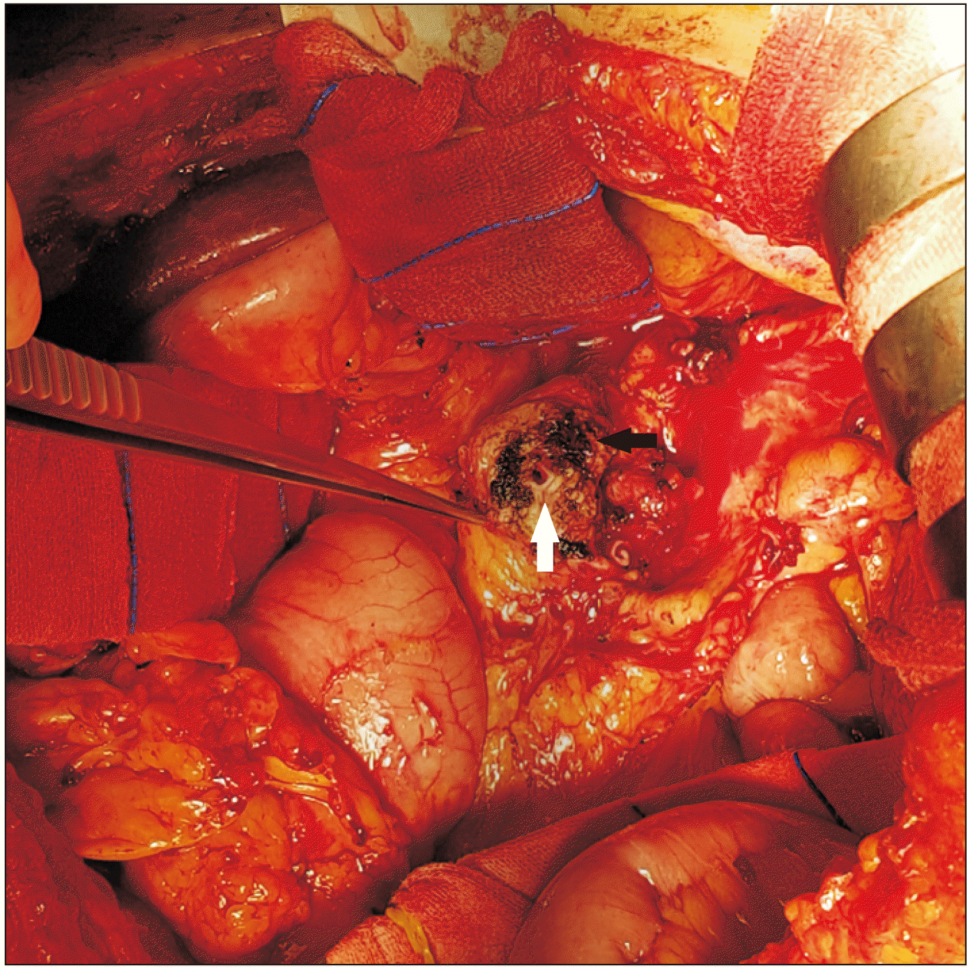 | Fig. 2The black arrow shows the resection margin of the pancreas, and the white arrow shows the enlarged pancreatic duct found on preoperative computed tomography. 
|
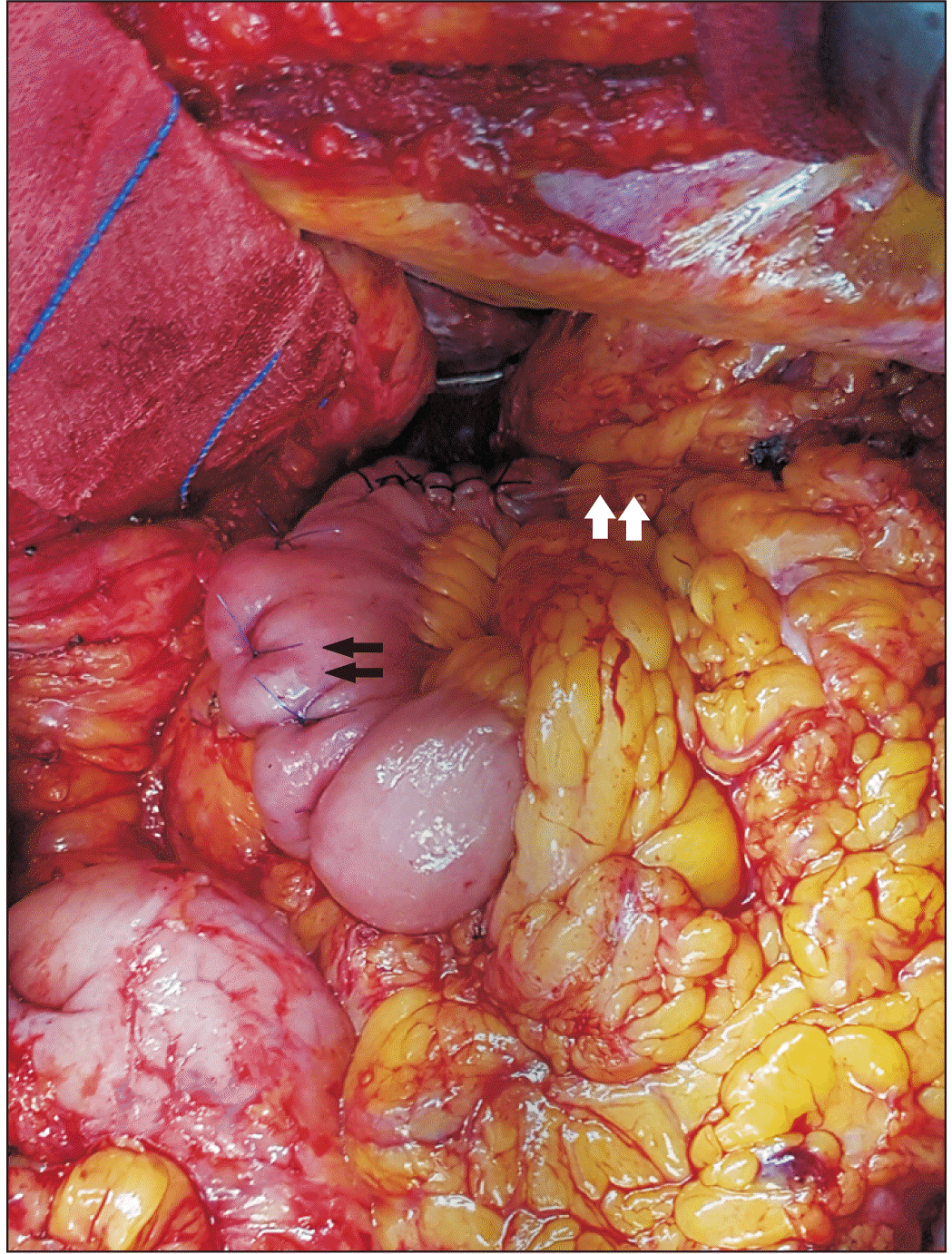 | Fig. 3The black arrows show the pancreaticojejunostomy and the white arrows show an external stent inserted into the pancreatic duct. 
|
The surgical procedure of the “IM-PJ” for this patient was as follows (
Fig. 4):
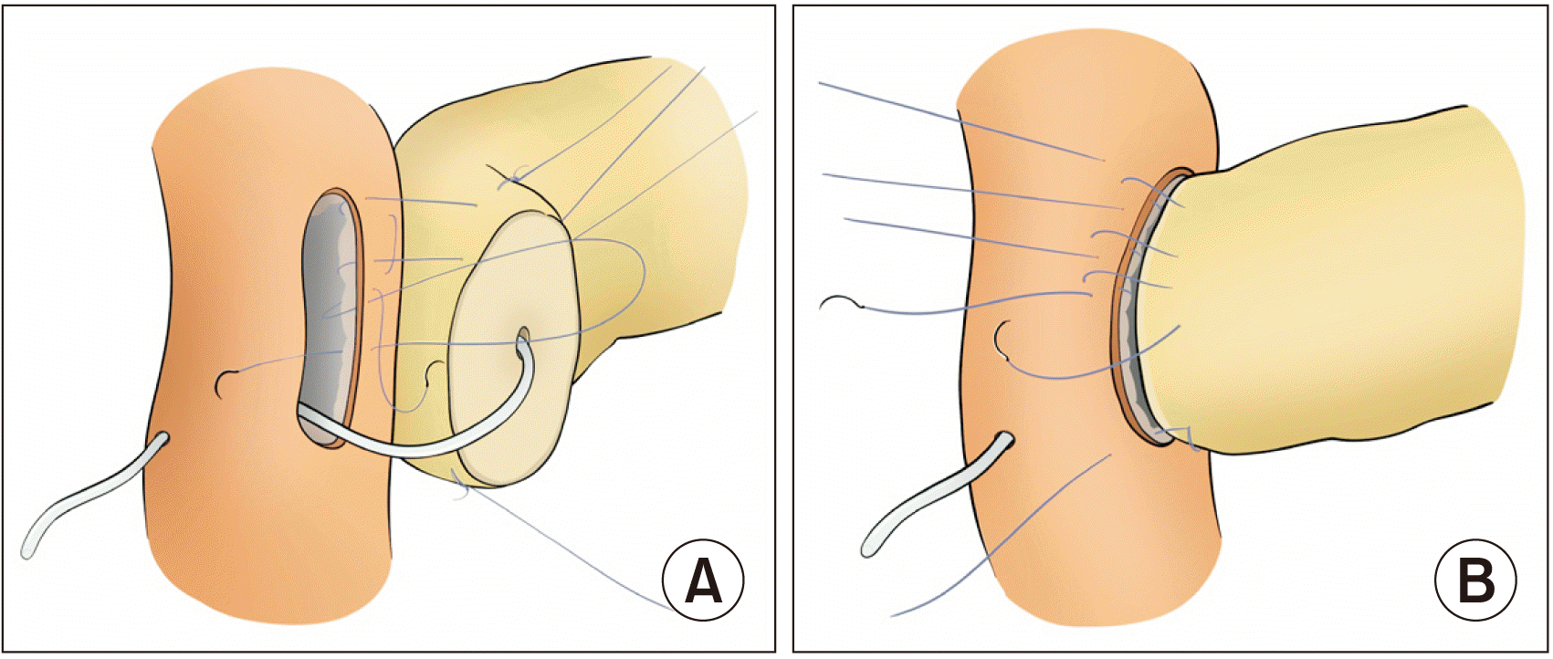 | Fig. 4(A) Three to four U-shaped mattress sutures (4-0 Prolene) were placed starting at the serosa of the posterior jejunal wall. The inverted seromuscular stitches were made going in-out. (B) After penetrating the pancreatic parenchyma with a needle, the seromuscular layer of the anterior jejunal wall was inverted with the sutures going out-in, followed by a full thickness stitch of the anterior jejunal wall going in-out. 
|
1. An antimesenteric border of the distal jejunum, measuring the same size as the pancreatic cut surface, was longitudinally opened.
2. An external stent was inserted into the pancreatic duct and distal jejunum before the start of the inverted mattress technique procedure. Three-to-four U-shaped mattress sutures (4-0 Prolene) were placed starting at the serosa of the posterior jejunal wall and going out-in through the full thickness of the jejunum. Then, inverted seromuscular stitches were placed going in-out.
3. These sutures penetrated the pancreatic remnant in a straight manner.
4. The seromuscular layer of the anterior jejunal wall was inverted by the transpancreatic U-shaped sutures going out-in followed by a full thickness stitch in the anterior jejunal wall going in-out, thereby invaginating the pancreatic remnant into the lumen of the jejunum.
5. A U-shaped suture was placed in a corner of the jejunum starting at the serosa of the jejunal wall and going out-in through the full thickness of the jejunum. Then, an inverted seromuscular stitch was placed going in-out. The suture penetrated an end of the pancreas stump in a straight manner, then the seromuscular layer of the other jejunal wall going in-out. Finally, in the same way, a U-shaped suture at the other corner of the jejunum fixed the other end of the pancreas.
6. The 5-6 U-shaped sutures, including the sutures at both corners of the jejunum, were pulled overusing adequate tension and tied at the anterior wall and at both corners of the jejunum, thereby enclosing the jejunal opening around the remnant pancreas. An external stent was fixed on the Roux limb stump using three or four sutures for the prevention of spontaneous removal.
Before abdominal closure, a closed suction drain was placed near the IM-PJ anastomosis to monitor leakage of pancreatic juice. The surgery was uneventful and the patient recovered from general anesthesia.
Postoperatively, the patient did not have any specific surgical complications during the recovery period. To find out the effect of IM-PJ on the pancreatic fistula, we checked the amount of fluid drainage, the amylase level of the drained fluid, and performed a weekly abdominal CT. The abdominal CT scan on the seventh day after surgery showed no fluid collections around the PJ and the drainage catheter was removed. We determined no pancreatic fistula occurred based on the definition by the 2016 International Study Group on Pancreatic Surgery (ISGPS). The patient was discharged on the 14th day after the surgery. Histopathological findings showed chronic pancreatitis with dystrophic calcification in a focal area.
The patient was followed up regularly for four years after surgery in the outpatient department. During this follow-up period, the patient visited the outpatient clinic in a healthy state, with no need for analgesics. The abdominal CT scan during the period showed no significant changes in pancreas parenchyma caused by PJ.
Go to :

DISCUSSION
The continuity of the pancreatic duct should be established after partial pancreatectomy such as a pancreaticoduodenectomy and central pancreatectomy. Although a PJ is not required after distal pancreatectomy in most cases, it may need to be performed in rare conditions, in which the drainage of pancreatic juice through the proximal duct is impaired by stricture, tumors, stone, or chronic pancreatitis etc., and the procedure has been known as the Duval procedure. The purpose of the procedure is to reduce the pressure of the pancreatic duct thereby preventing atrophy of the remnant pancreas. In cases of chronic pancreatitis, this procedure has been reported to relieve abdominal pain.
Chronic pancreatitis is a progressive inflammatory disease characterized by irreversible destruction of the pancreas. Surgical treatment should be considered in cases of intractable pain after the failure of conservative treatment, the possible presence of neoplasm, and local complications including common bile duct stenosis, pseudoaneurysm or erosion of the large vessels, large pancreatic pseudocysts, and internal pancreatic fistula [
6]. Several surgical strategies for the treatment of symptomatic chronic pancreatitis can be categorized into three procedures: a drainage procedure focusing on ductal hypertension, resection focusing on inflammatory masses in the pancreatic parenchyma, and mixed techniques [
7]. Intractable pain that does not respond to medical treatment is the main reason for attempting surgery for pancreatitis. The mechanism of abdominal pain in chronic pancreatitis patients is either perineural inflammation or ductal hypertension. Because the mechanisms usually coexist in chronic pancreatitis, a surgical strategy should focus on the elimination of both mechanisms. Markowitz et al. [
8] reported a case of failure to relieve symptoms by simple pancreatic decompression in chronic pancreatitis. In this case, the salvage strategy for recurred pain was resection of the pancreatic lesion such as a pylorus-preserving pancreaticoduodenectomy or Whipple surgery. They explained that the reason for the failure of the simple pancreaticojejunal decompression could be that the duct pressure was not the sole mechanism causing pain. Coexisting neuritis, not eliminated by the decompression procedure, might be a significant cause of pain. For these reasons, pure drainage procedures have been replaced by techniques that combine both resection and drainage, for the majority of patients. Surgical options for chronic pancreatitis include a pancreaticoduodenectomy and a mixed type of drainage and resection, such as Frey, Beger, or Berne.
For chronic pancreatitis, limited to the tail of the pancreas, a distal pancreatectomy is indicated. As presented in our case, for chronic pancreatitis with a suspicious mass lesion on the pancreas, the mixed techniques of a distal pancreatectomy and the Duval drainage procedure may be the most appropriate option. In our case, the patient, a heavy alcoholic for several years, complained of persisting recurrent pain for which analgesic medications, lifestyle modification, and endoscopic intervention were performed. Despite medical treatment, she complained about recurrent pain and imaging studies showed the possibility of a malignant tumor in the pancreas body. Based on persisting recurrent pain for more than two years and the possibility of hidden malignancy, surgical resection of the distal pancreas harboring the mass and a drainage procedure to relieve the pain was planned. Usually, a PJ is not required after a conventional distal pancreatectomy. However, for this patient, we thought that drainage was necessary because the pancreatic duct obstruction, due to the impacted stone, could be a cause of recurrent abdominal pain. Despite the necessity of a Roux-en Y PJ in this condition, the incidence of critical POPF makes even experienced surgeons hesitate to perform the PJ. Surgeons may be much more reluctant to perform a PJ after distal pancreatectomy because the rarity of such cases makes the risk of POPF more unpredictable.
As previously published, our data showed that IM-PJ reduced the risk of Grade C POPF and serious complications such as a gastroduodenal artery pseudoaneurysm after pancreaticoduodenectmy [
4]. No pancreatic fistula occurred from IM-PJ after a central pancreatectomy [
5]. Based on our experiences of reporting the safety of IM-PJ for a pancreaticoduodenectmy and central pancreatectomy, we decided to apply the same technique for this patient. The anastomosis of the distal pancreatic stump and jejunal limb of the Roux-en-Y loop was performed utilizing the IM-PJ. After the operation, the patient was discharged two weeks after surgery without complications. We determined no pancreatic fistula occurred based on the definition by the 2016 ISGPS because the drainage fluid was only a small amount, with a normal amylase level, on the third day after surgery [
3]. The abdominal CT scan on the seventh day after surgery showed no fluid collections around the PJ. Compared to the diameter of the pancreatic duct on the abdominal CT before surgery, it was reduced after surgery indicating the adequate drainage of pancreatic juice through the PJ (
Fig. 5).
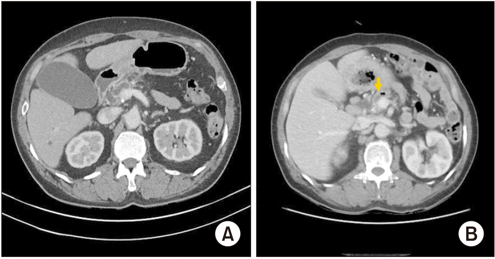 | Fig. 5Abdominal contrast enhanced computed tomography (CT) images. (A) The pancreas duct remained dilated seven days after surgery. (B) When the CT was performed at follow-up, three years after the surgery, the diameter of the pancreatic duct (arrow) was reduced when compared with the perioperative period. 
|
Our study showed that the inverted mattress end-to-side PJ is a safe technique and can be applied for the Duval procedure. Furthermore, IM-PJ may be safely applied for cases in which a distal pancreatectomy is indicated in the presence of a proximal pancreatic duct obstruction caused by a stricture, tumor or stone etc., in addition to chronic pancreatitis.
Go to :


 PDF
PDF Citation
 Print
Print


 XML Download