Journal List > Ann Hepatobiliary Pancreat Surg > v.26(4) > 1516080334

Lee, Hong, Kim, Hwang, Lee, and Kang: Laparoscopic distal pancreatosplenectomy for left-sided pancreatic cancer in patients with radical subtotal gastrectomy for gastric cancer

Abstract

After radical subtotal gastrectomy (RSTG) for stomach cancer, the remnant stomach is supposed to be perfused through the short gastric vessels. What if a patient who received previous RSTG is diagnosed with resectable distal pancreatic cancer? Can radical distal pancreatosplenectomy (DPS) be performed safely without ischemic damage to the remnant stomach? Unfortunately, there are limited studies on this specific clinical issue. Notably, in spite of rare clinical presentation, it is expected to increase due to prolonged survival of patients with resected gastric cancer. Therefore, we aimed to demonstrate the safety and feasibility of the radical DPS in patients with previous RSTG. In this study, we investigated perioperative and long-term survival outcomes of DPS for left-sided pancreatic cancer in patients with previous RSTG.

INTRODUCTION

The incidence of pancreatic cancer is relatively low among gastrointestinal tract cancers, however, 5-year survival is still poor and there has been no improvement in the last 20 years [1]. At the time of diagnosis, most pancreatic cancer cases are already locally advanced or metastatic with the resectable disease accounting for only 10% of the total cases [2]. The 5-year survival of pancreatic cancer ranges from 2% to 9% in published literature, which is the most dismal prognosis among all the gastrointestinal tract cancers [1,3,4].
For patients diagnosed with resectable disease, radical surgery with a negative resection margin is the only treatment that provides a chance of cure. Unfortunately, up to 75% of patients experience recurrence after surgery, thus, adjuvant chemotherapy after surgery is recommended.
Minimally invasive (laparoscopic or robot-assisted) distal pancreatectomy (DP) has been increasingly adopted in surgical practice for pancreatic cancer over the last decade [5]. However, without randomized controlled trials, the oncologic efficacy of minimally invasive distal pancreatectomy (MIDP) remains controversial. Nonetheless, MIDP, when performed by an experienced expert surgeon, can be regarded as a preferred treatment approach for pancreatic body or tail cancer [6]. Outcomes are improved after MIDP as compared to open DP, without obvious downsides in high-risk subgroups [7].
Gastric cancer is one of the most frequent cancers in Korea [8]. Early diagnosis of gastric cancer is consistently increasing due to the recent expansion of endoscopic evaluation for routine medical check-ups, and long-term survival is expected after curative treatment. Pancreatic cancer often occurs later during the long-term follow-up period after radical subtotal gastrectomy (RSTG) for gastric cancer. In clinical circumstances of resectable pancreatic body or tail cancer in patients with previous RSTG for gastric cancer, is laparoscopic distal pancreatosplenectomy (DPS) feasible and appropriate treatment option? Is blood perfusion in the remnant stomach well preserved after DPS?
Herein, we present two consecutive patients who underwent successful laparoscopic DPS for left-sided pancreatic cancer after RSTG for gastric cancer. The purpose of this study was to prove the technical feasibility and safety of laparoscopic DPS after RSTG for gastric cancer.

CASES

Case 1

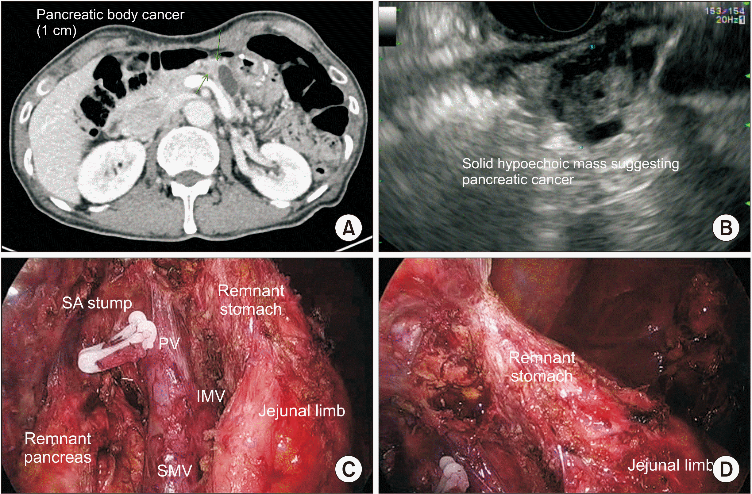
A 78-year-old male patient with abdominal discomfort was diagnosed with advanced gastric cancer, Borrmann type II, after an esophagogastroduodenoscopy (EGD). He underwent RSTG with Billroth II anastomosis (with incidental cholecystectomy) in 2011. A pathologic report confirmed moderate differentiated tubular adenocarcinoma with submucosal invasion without lymph node metastasis (T1bN0M0-stage la). The patient had regular postoperative follow-up without adjuvant chemotherapy. A follow-up computed tomography (CT) scan in 2016, showed a 1cm-sized pancreatic mass with distal pancreatic duct dilatation (Fig. 1A), and subsequent endoscopic ultrasonography and fine-needle aspiration (Fig. 1B) was performed, showing a few clusters of highly dysplastic epithelial cells, suggestive of malignancy.
The patient underwent laparoscopic DPS with splenectomy. Intraoperative findings showed prominent adhesion around the left-sided pancreas under previous gastrojejunostomy (GJ) site without major vascular structure invasion. The gastrosplenic ligament was securely divided and after the division of the pancreatic neck, both splenic artery and vein were controlled at the origin. After meticulous adhesio-bandlysis, retroperitoneal dissection was performed to obtain a safe tangential margin of the pancreas (Fig. 1C). After completion of the laparoscopic DPS, it was confirmed that remnant gastric perfusion was well preserved (Fig. 1D).
The patient recovered without complications and was discharged on a postoperative day (POD) #9. On pathologic examination, the lesion on the pancreas body was confirmed as moderately differentiated adenocarcinoma of 2.1 cm × 1.3 cm (pT2) in size. There was no lymphovascular invasion, however, perineural invasion was noted. The resected pancreatic margin was reported to be free from tumor. Lymphatic metastasis was not noted (0/6, pN0). The patient was treated with 6 cycles of adjuvant chemotherapy (Gemcitabine) and is on regular follow-up without evidence of tumor recurrence.

Case 2

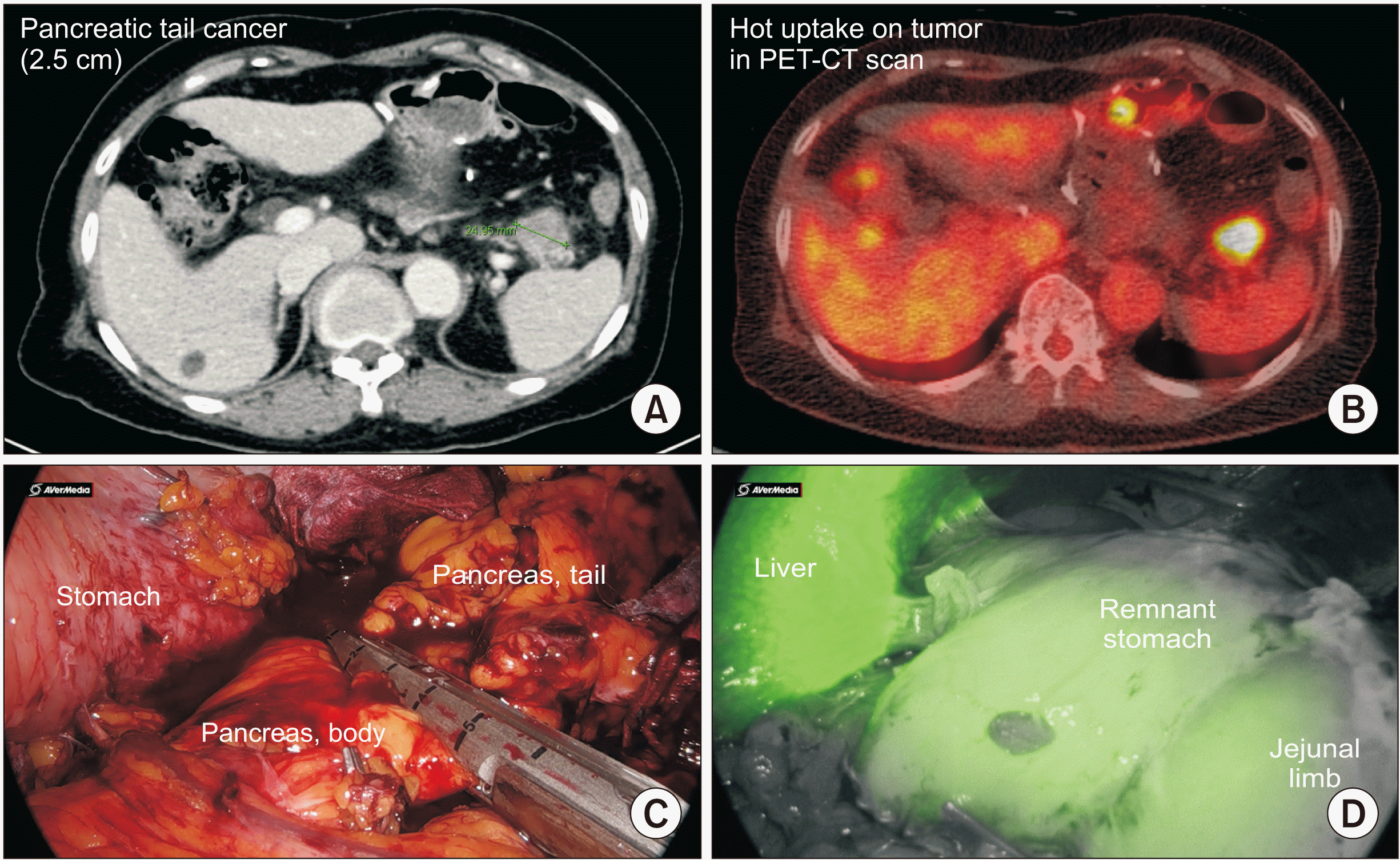
A 75-year-old female patient with recurrent epigastric pain underwent evaluation and an EGD biopsy showed early gastric cancer on the anterior wall of the lesser curvature. The patient underwent an endoscopic submucosal dissection procedure, and the resection margin was basal 200 µm (submucosal 2 invasion). Subsequently, the patient received laparoscopic RSTG with Billroth II anastomosis. The patient was on regular follow-up without evidence of recurrence for 12 years postoperatively. Later, due to elevated tumor markers (carcinoembryonic antigen 9.02 ng/mL, carbohydrate antigen 19-9 146.0 U/mL), the patient underwent a PET-CT scan (Fig. 2B) and a mass with intense FDG uptake in the pancreatic tail was found.
A biliary CT (Fig. 2A) scan was done to check the major vessel anatomy and variation, and a laparoscopic DPS was performed. In the operative findings, there was moderate adhesion around the distal pancreas between the remnant stomach without invasion of the splenic artery or vein. The gastrosplenic ligament and splenocolic ligament were divided gently, and the pancreas was divided and dissected from the retroperitoneum. DPS was completed by modified lasso technique after ligating the splenic artery (Fig. 2C) and the perfusion of the remnant stomach was confirmed by indocyanine green (ICG) technology (Fig. 2D). After the surgery, the patient had uneventful progress on POD without surgical complications, and was discharged on POD #7.
Pathologic report revealed pT2 cancer with perineural invasion without lymphovascular invasion. The pancreatic parenchymal and duct resection margin was free of carcinoma (safety margin: 0.4 cm), and the circumferential margin showed an extension of carcinoma. One out of 5 lymph nodes was positive for metastatic carcinoma (pN1). The patient is scheduled for adjuvant chemotherapy (considering low-dose Gemcitabine/Xeloda).

Institutional experiences of DP in patients with previous radical gastrectomy

Table 1 shows 6 serial cases of our experiences of DP in patients with previous RSTG. The median age at the time of DP was 66.5-year-old (95% confidence interval [CI], 58.41–74.59 years). Four patients were male and two patients were female. The operative methods were RSTG with Billroth I anastomosis in 1 patient, RSTG with Billroth II anastomosis in 3 patients, laparoscopic radical total gastrectomy in 1 patient, and laparoscopic RSTG with Billroth II anastomosis in 1 patient. The median interval between radical gastrectomy and DP was 122.67 months (95% CI, 35.27–210.06 months). In the second operation (DP), the median estimated blood loss was 591.67 mL (95% CI: –163.21 to 1,346.54 mL) and intraoperative transfusion was done in 2 patients. In the perioperative outcomes, the median operation time was 369 minutes (95% CI, 126.63–611.37 minutes) and the length of hospital stay was 17 days (95% CI, –1.67 to 35.67 days). All the patients received postoperative adjuvant chemotherapy. The mean survival time after radical DP was 48.2 months (95% CI, 35.53–60.87 months) (Fig. 3).

DISCUSSION

According to our previous reports [9,10], the bloodless and margin-negative resection are the most important factors in the surgical treatment of left-sided pancreatic cancer. Based on this observation, we developed the potential tumor conditions that can lead to the bloodless and margin-negative resection in left-sided pancreatic cancer using the “Yonsei criteria” [11]. Based on these criteria, minimally invasive radical DPS has been carefully applied to well-selected patients for left-sided pancreatic cancer [12]. In particular, the Yonsei criteria were set as the inclusion criteria for pancreatic cancer in the recent multicenter prospective randomized control study comparing open and laparoscopic DPS [12], suggesting the Yonsei criteria as a valuable surgical indication for safe and effective laparoscopic radical DPS. With the validation of our surgical techniques and evidence of the oncological effects of neoadjuvant chemotherapy, currently, laparoscopic DPS has been expanded to tumors exceeding Yonsei criteria [13]. To date, no prospective randomized controlled studies are comparing laparoscopic and open DPS for left-sided pancreatic cancer; however, laparoscopic DPS is carefully regarded as a safe and effective treatment option in well-selected pancreatic cancer patients.
Clinical investigation of pancreatectomy in patients with previous RSTG has been reported, and most literature are related to the technical feasibility and safety of pancreaticoduodenectomy (PD) after RSTG [13-15]. In fact, our group also published technical notes for PD in patients with previous radical gastrectomy for gastric cancer [16]. However, there are no reports of subsequent DPS in patients with previous RSTG. To the best of our knowledge, this is the first report showing the technical feasibility and safety of laparoscopic DPS for left-sided pancreatic cancer in patients with previous RSTG for gastric cancer.
Is laparoscopic DPS in patients with previous RSTG technically feasible and safe? Two issues should be considered; intra-abdominal adhesion and the potential risk of ischemic gastropathy in the remnant stomach after DPS.
Since the pancreas is located behind the stomach, considering the location of GJ after RSTG, adhesions that occur after RSTG is an important factor in determining the success of laparoscopic DPS for resectable pancreatic cancer. In particular, the anatomic area around the common hepatic artery, left gastric artery, and splenic artery, which are the areas for lymph node dissection during previous RSTG, and near the GJ can be accompanied by severe adhesion. Therefore, the relationship between the potential pancreatic division line for margin-negative resection and this adhesion area could be an important factor in predicting successful laparoscopic DPS in patients with previous RSTG. Unlike the first case, the second case was clearly less adherent near the GJ site and the potential division line of the pancreas was left to the previous GJ. In this case, the pancreas was transected safely via a modified lasso technique [17].
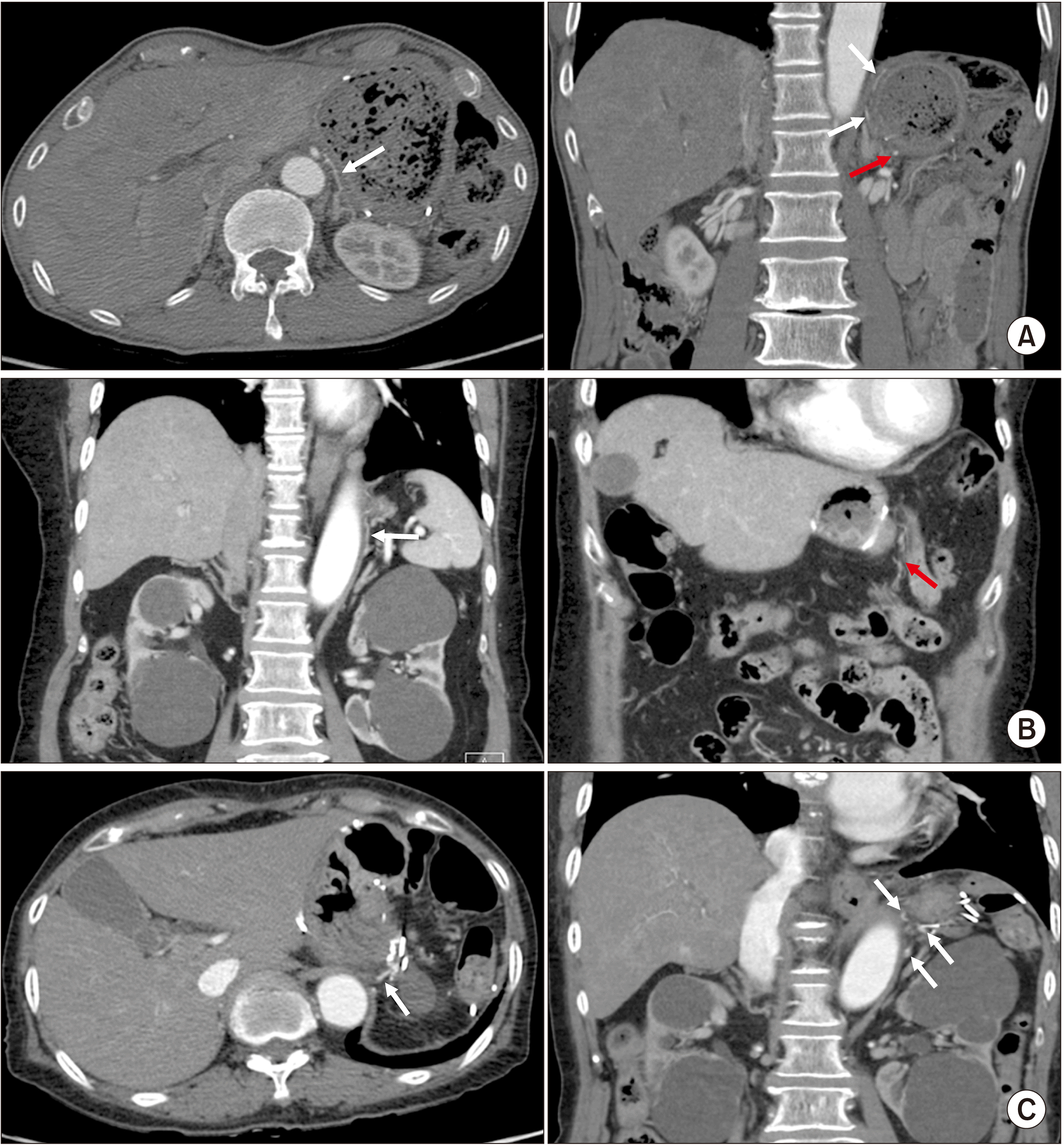
Regarding potential postoperative ischemic gastropathy, we retrospectively reviewed the radiological evidence of collateral blood supply to the remnant stomach in pre- and post-distal pancreatectomy state (Table 2). On the preoperative CT scan, the patients showed thin enhancing collateral vessels from the left inferior phrenic artery, and SMA and these collaterals got more prominent after DP. Fig. 4. Shows the examples of prominent collateral vessel formation after DP (Fig. 4A, 4B), and one of the patients (case 2) already had prominent collateral vessels even in pre-distal pancreatectomy state (Fig. 4C). These observations suggest that the remnant stomach can be consistently vascularized by the enforced collateral vessels after DP despite the thin or absence of the collateral vessels in the remnant stomach before DP. Recently, ICG has been demonstrated as an emerging technology for confirming organ perfusion during operation [18,19]. For instance, a Japanese group reported the use of digital subtraction angiography of the remnant stomach to evaluate vessel perfusion after DP [20]. When applied to all of the present cases, the ICG showed perfusion of the remnant stomach to be well preserved despite dividing all gastrosplenic ligaments including short gastric vessels. The preservation of the remnant stomach perfusion was also confirmed by the absence of complications related to ischemia of the remnant stomach after laparoscopic DPS. According to our institution’s experience, collateral vessels excepting left gastric artery and short gastric vessel are developed during the interval period after RSTG, and they maintain the perfusion of the stomach without ischemic gastropathy in spite of DPS after RSTG.
In conclusion, laparoscopic DPS in patients with previous RSTG is feasible and safe. In this study, improved long-term oncologic outcomes were reported. Considering long-term survival after radical gastrectomy for gastric cancer, cases of second primary left-sided pancreatic cancer found during follow-up are also expected to increase in near future, which will be one of the challenges for pancreatic surgeons. Individualized pancreatectomy should be conducted considering not only oncologic safety but also the patient’s general safety. Further study based on accumulating clinical experiences is needed.

Notes

FUNDING

None.

CONFLICT OF INTEREST

No potential conflict of interest relevant to this article was reported.

AUTHOR CONTRIBUTIONS

Conceptualization: WJL. Data curation: KHL. Methodology: SK. Visualization: HKH. Writing - original draft: SSH. Writing - review & editing: CMK.

REFERENCES

1. Rawla P, Sunkara T, Gaduputi V. 2019; Epidemiology of pancreatic cancer: global trends, etiology and risk factors. World J Oncol. 10:10–27. DOI: 10.14740/wjon1166. PMID: 30834048. PMCID: PMC6396775.
2. Vincent A, Herman J, Schulick R, Hruban RH, Goggins M. 2011; Pancreatic cancer. Lancet. 378:607–620. DOI: 10.1016/S0140-6736(10)62307-0. PMID: 21620466.
3. Luo J, Xiao L, Wu C, Zheng Y, Zhao N. 2013; The incidence and survival rate of population-based pancreatic cancer patients: Shanghai Cancer Registry 2004-2009. PLoS One. 8:e76052. Erratum in: PLoS One 2014;9. DOI: 10.1371/journal.pone.0076052. PMID: 24130758. PMCID: PMC3794034.
4. McGuigan A, Kelly P, Turkington RC, Jones C, Coleman HG, Mc-Cain RS. 2018; Pancreatic cancer: a review of clinical diagnosis, epidemiology, treatment and outcomes. World J Gastroenterol. 24:4846–4861. DOI: 10.3748/wjg.v24.i43.4846. PMID: 30487695. PMCID: PMC6250924.
5. Moekotte AL, Rawashdeh A, Asbun HJ, Coimbra FJ, Edil BH, Jarufe N, et al. 2020; Safe implementation of minimally invasive pancreas resection: a systematic review. HPB (Oxford). 22:637–648. DOI: 10.1016/j.hpb.2019.11.005. PMID: 31836284.
6. de Rooij T, van Hilst J, van Santvoort H, Boerma D, van den Boezem P, Daams F, et al. 2019; Minimally invasive versus open distal pancreatectomy (LEOPARD): a multicenter patient-blinded randomized controlled trial. Ann Surg. 269:2–9. DOI: 10.1097/SLA.0000000000002979. PMID: 30080726.
7. Korrel M, Vissers FL, van Hilst J, de Rooij T, Dijkgraaf MG, Festen S, et al. 2021; Minimally invasive versus open distal pancreatectomy: an individual patient data meta-analysis of two randomized controlled trials. HPB (Oxford). 23:323–330. DOI: 10.1016/j.hpb.2020.10.022. PMID: 33250330.
8. Ko KP. 2019; Epidemiology of gastric cancer in Korea. J Korean Med Assoc. 62:398–406. DOI: 10.5124/jkma.2019.62.8.398.
9. Hong SS, Hwang HK, Lee WJ, Kang CM. 2020; Feasibility and safety of laparoscopic radical distal pancreatosplenectomy with adrenalectomy in advanced pancreatic cancer. Ann Surg Oncol. 27:5235–5236. DOI: 10.1245/s10434-020-08670-9. PMID: 32474822.
10. Kang CM. 2020; ASO author reflections: from concept to real clinical practice of laparoscopic distal pancreatectomy for left-sided pancreatic cancer. Ann Surg Oncol. 27:5237–5238. DOI: 10.1245/s10434-020-08676-3. PMID: 32462529.
11. Chong JU, Kim SH, Hwang HK, Kang CM, Lee WJ. 2017; Yonsei criteria: a clinical reflection of stage I left-sided pancreatic cancer. Oncotarget. 8:110830–110836. DOI: 10.18632/oncotarget.22734. PMID: 29340019. PMCID: PMC5762287.
12. Lee SH, Kang CM, Hwang HK, Choi SH, Lee WJ, Chi HS. 2014; Minimally invasive RAMPS in well-selected left-sided pancreatic cancer within Yonsei criteria: long-term (>median 3 years) oncologic outcomes. Surg Endosc. 28:2848–2855. DOI: 10.1007/s00464-014-3537-3. PMID: 24853839.
13. Kim CY, Lee SH, Jeon HM, Kim HK, Kang CM, Lee WJ. 2014; AFP-producing acinar cell carcinoma treated by pancreaticoduodenectomy in a patient with a previous radical subtotal gastrectomy by gastric cancer. Korean J Hepatobiliary Pancreat Surg. 18:33–37. DOI: 10.14701/kjhbps.2014.18.1.33. PMID: 26155245. PMCID: PMC4492330.
14. Lee D, Lee JH, Choi D, Kang CM, Chong JU, Kim SC, et al. 2017; Surgical strategy and outcome in patients undergoing pancreaticoduodenectomy after gastric resection: a three-center experience with 39 patients. World J Surg. 41:552–558. DOI: 10.1007/s00268-016-3729-1. PMID: 27730351.
15. Fukuta M, Tomibayashi A, Tsuneki T, Nishioka K, Matsuo Y, Mori O, et al. 2020; Pancreaticoduodenectomy after distal gastrectomy: a case series. Int J Surg Case Rep. 76:240–246. DOI: 10.1016/j.ijscr.2020.09.169. PMID: 33053481. PMCID: PMC7566198.
16. Kim SH, Hwang HK, Kang CM, Lee WJ. 2012; Pancreatoduodenectomy in patients with periampullary cancer after radical subtotal gastrectomy for gastric cancer. Am Surg. 78:E164–E167. DOI: 10.1177/000313481207800319.
17. Kawasaki Y, Hwang HK, Kang CM, Natsugoe S, Lee WJ. 2018; Improved perioperative outcomes of laparoscopic distal pancreatosplenectomy: modified lasso technique. ANZ J Surg. 88:886–890. DOI: 10.1111/ans.14351. PMID: 29266719.
18. De Nardi P, Elmore U, Maggi G, Maggiore R, Boni L, Cassinotti E, et al. 2020; Intraoperative angiography with indocyanine green to assess anastomosis perfusion in patients undergoing laparoscopic colorectal resection: results of a multicenter randomized controlled trial. Surg Endosc. 34:53–60. DOI: 10.1007/s00464-019-06730-0. PMID: 30903276.
19. Abdelwahab M, Spataro EA, Kandathil CK, Most SP. 2019; Neovascularization perfusion of melolabial flaps using intraoperative indocyanine green angiography. JAMA Facial Plast Surg. 21:230–236. DOI: 10.1001/jamafacial.2018.1874. PMID: 30730539. PMCID: PMC6537835.
20. Asari S, Toyama H, Goto T, Yamashita H, Nanno Y, Ishida J, et al. 2021; Indocyanine green (ICG) fluorography and digital subtraction angiography (DSA) of vessels supplying the remnant stomach that were performed during distal pancreatectomy in a patient with a history of distal gastrectomy: a case report. Clin J Gastroenterol. 14:1749–1755. DOI: 10.1007/s12328-021-01493-5. PMID: 34342840.
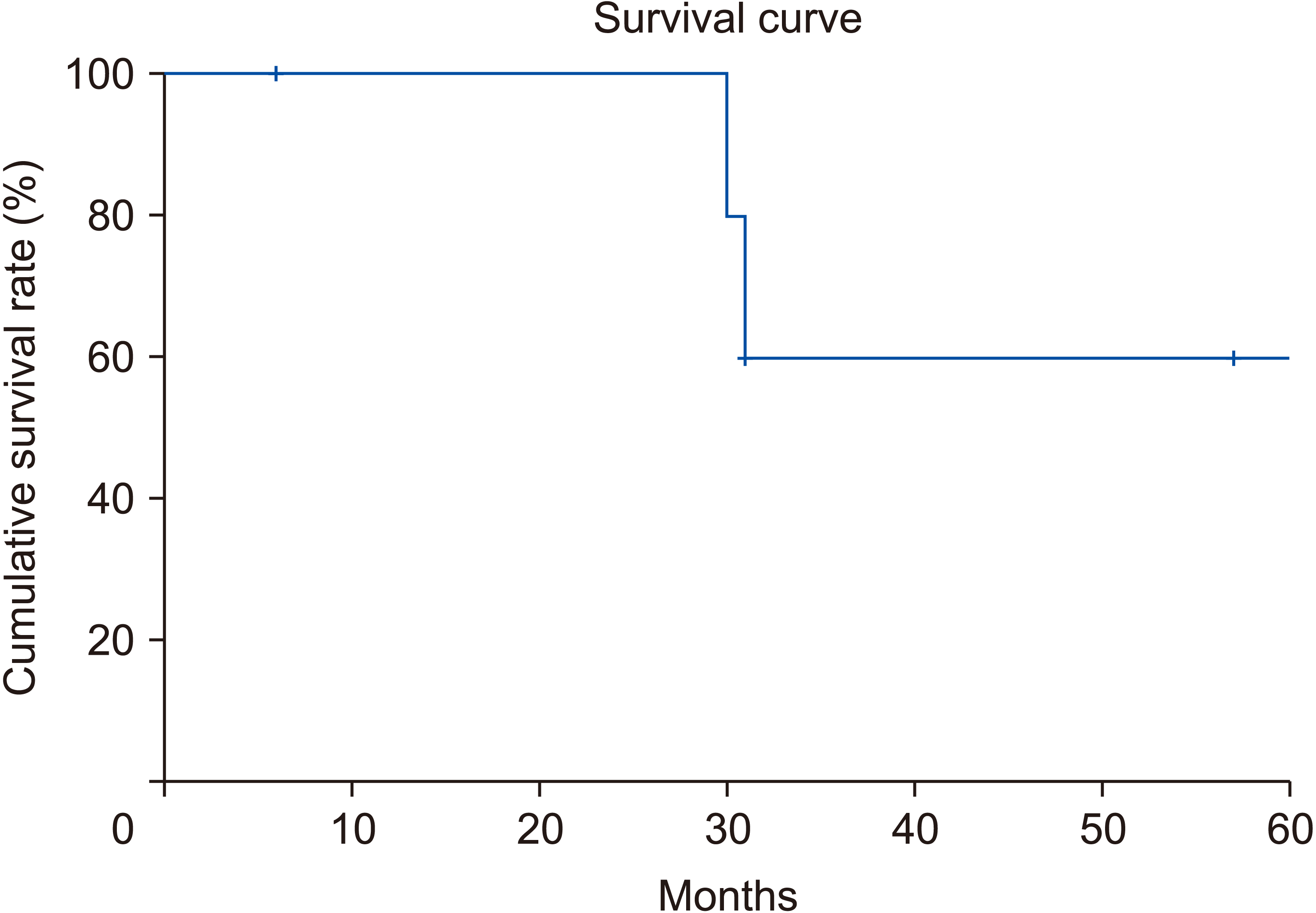
Fig. 1
(A) P reoperat ive computed tomography, (B) preoperative endoscopic ultrasound, (C) f inal view of the distal pancreatosplenectomy, (D) well preserved perfusion of the remnant stomach. SA, splenic artery; PV, portal vein; IMV, inferior mesenteric vein; SMV, superior mesenteric vein.
ahbps-26-4-395-f1.tif
Fig. 2
(A) Preoperative computed tomography (CT), (B) preoperative positron emission tomography-computed tomography (PET-CT), (C) division of the pancreas by modified lasso technique, (D) well preserved perfusion of the remnant stomach confirmed by indocyanine green technology.
ahbps-26-4-395-f2.tif
Fig. 3
Long-term oncologic outcomes of patients who underwent radical distal pancreatectomy for pancreatic cancer following previous radical gastrectomy for gastric cancer.
ahbps-26-4-395-f3.tif
Fig. 4
Radiological (axial and coronal) evidence of the collateral vessels supplies to the remnant stomach. (A) Case 1: after distal pancreatectomy. Case 2: before (B) and after (C) distal pancreatectomy. White arrows indicate left inferior phrenic artery; red arrows, jejunal branch from superior mesenteric vein.
ahbps-26-4-395-f4.tif
Table 1
Cases of distal pancreatosplenectomy (DPS) for pancreatic cancer after radical gastrectomy for gastric cancer
No. of patients Age (yr)/sex Previous gastrectomy Time interval (mon) Approach for DP Splenectomy Stage of PDAC EBL (mL) Op. time (min) LOH (day) Complication Postop. adjuvant chemotherapy Survival since DPS
1 71/Male STG B2 240 Open O pT2N0 Stage IIa 1,700 790 53 DGE O 30 mon Death
2 59/Male STG B2 196 Open O pT1N0 Stage Ia 250 475 14 None O 57 mon Alive
3 62/Female Lap TG 48 Open O pT3N1 Stage IIb 1,300 270 9 None O 31 mon Death
4 58/Male STG B1 48 Open X pT1N1 Stage IIb 50 240 10 None O 31 mon F/U loss
5 (case 1) 74/Male STG B2 60 Laparoscopic O pT2N0 Stage Ib 50 279 9 None O 60 mon Alive
6 (case 2) 75/Female Lap STG B2 144 Laparoscopic O pT2N1 Stage IIb 200 160 7 None O 6 mon Alive

STG, subtotal gastrectomy; TG, total gastrectomy; DP, distal pancreatectomy; PDAC, pancreatic ductal adenocarcinoma; EBL, estimated blood loss; Op., operative; Postop., postoperative; LOH, length of hospitalization; DGE, delayed gastric emptying, stage of PDAC was based on AJCC 8th staging; F/U, follow-up.

Time interval: mean = 122.67 mon, 95% confidence interval = 35.27–210.06 mon; Op. time: mean = 369.00 min, 95% confidence interval = 126.63–611.37 min.

Table 2
Radiological evidence of collateral blood supply to the remnant stomach on pre- and post-distal pancreatectomy state
No. of patients Age (yr)/Sex Previous gastrectomy Time interval (mon) Radiological evidence of collateral blood supply to the remnant stomach

Pre-distal pancreatectomy Post-distal pancreatectomy
1 71/Male STG B2 240 No evident enhance of collaterals Collateral arteries from CHA and left IPA
2 59/Male STG B2 196 Thin enhanced collateral artery from left IPA Evident collateral arteries from left IPA and SMA
3 62/Female Lap TG 48 Total gastrectomy status Total gastrectomy status
4a) 58/Male STG B1 48 Thin enhanced collateral artery from left IPA Evident collateral arteries from left IPA and SMA
5 (case 1) 74/Male STG B2 60 Thin enhanced collateral artery from left IPA Evident collateral arteries from left IPA and SMA
6 (case 2) 75/Female Lap STG B2 144 Evident collateral arteries from left IPA and SMA Evident collateral arteries from left IPA

STG, subtotal gastrectomy; TG, total gastrectomy; IPA, inferior phrenic artery; CHA, common hepatic artery; SMA, superior mesenteric artery.

a)Spleen preserving distal pancreatectomy.

TOOLS
Similar articles