Abstract
Backgrounds/Aims
To analyze relationships of hepatic histopathological findings and bile microbiological profiles with perioperative outcomes and risk of late biliary stricture in individuals undergoing surgical bile duct injury (BDI) repair.
Methods
A historical cohort study was carried out at a tertiary university hospital. Fifty-six individuals who underwent surgical BDI repair from 2014–2018 with a minimal follow-up of 24 months were enrolled. Liver biopsies were performed to analyze histopathology. Bile samples were collected during repair procedures. Hepatic histopathological findings and bile microbiological profiles were then correlated with perioperative and late outcomes through uni- and multi-variate analyses.
Results
Forty-three individuals (76.8%) were females and average age was 47.2 ± 13.2 years; mean follow-up was 38.1 ± 18.6 months. The commonest histopathological finding was hepatic fibrosis (87.5%). Bile cultures were positive in 53.5%. The main surgical technique was Roux-en-Y hepaticojejunostomy (96.4%). Overall morbidity was 35.7%. In univariate analysis, liver fibrosis correlated with the duration of the operation (R = 0.3; p = 0.02). In multivariate analysis, fibrosis (R = 0.36; p = 0.02) and cholestasis (R = 0.34; p = 0.02) independently correlated with operative time. Strasberg classification independently correlated with estimated bleeding (R = 0.31; p = 0.049). The time elapsed between primary cholecystectomy and BDI repair correlated with hepatic fibrosis (R = 0.4; p = 0.01).
Laparoscopic cholecystectomy was firstly performed by Erich Mühe in 1985 [1]. Since then, it has been performed more than an open approach because it can lead to benefits such as reduction of postoperative pain, shorter hospital stays, and earlier return to daily activities [2]. Nonetheless, Renz et al. [3] have reported that the rate of clinically relevant iatrogenic bile duct injury (BDI) is around 3% in laparoscopic cholecystectomy, higher than that (0.1%–0.5%) in an open surgery. BDI can cause greater operative morbidity, reduce quality of life, and lead to complications such as biliary strictures, which in turn can lead to biliary cirrhosis. It can also generate higher costs associated with hospital admissions and procedures, in addition to costs associated with legal issues [4-7]. BDI is a potentially life-threatening condition, with long-term mortality rate that can reach 1.8%–4.6% [8]. Some factors such as injury site, previous repair attempt, sepsis, postoperative leaks, and corrective operation by a surgeon without required skills are associated with a worse prognosis after BDI correction [9-12]. Factors that can lead to a worse prognosis are still under debate. Some studies have reported that vascular injuries and timing of repair are associated with negative outcomes and primary treatment failure [9-11]. However, according to a study by Stewart et al. [12], the incidence of right hepatic artery injury does not influence the mortality or repair success rate. The timing of repair, when performed by a hepatobiliary surgeon, has no impact on outcomes either according to another study carried out by Stewart and Way [13].
At the facility in which this study was conducted, bilobar liver biopsies were routinely performed during surgical treatment of BDI and bile was collected for microbiological analysis. Little is currently known on the potential associations of hepatic histopathological findings, bile microbiological profiles, and biochemical examinations with perioperative outcomes, late outcomes, and risk of late biliary stricture in individuals undergoing surgical BDI repair. Analysis of these associations could predict patients at higher risk of surgical complications and late anastomotic strictures.
Therefore, the aim of this study was to analyze associations of hepatic histopathological findings and bile microbiological profiles with perioperative outcomes and the risk of late biliary stricture in individuals undergoing surgical BDI repair.
This was a retrospective, descriptive, observational, and longitudinal study (historical cohort). Individuals undergoing surgical BDI repair by an advanced biliary surgery unit at a tertiary university hospital were enrolled. This study was approved by the local institutional review board under the reference number 3.510.604/UNICAMP.
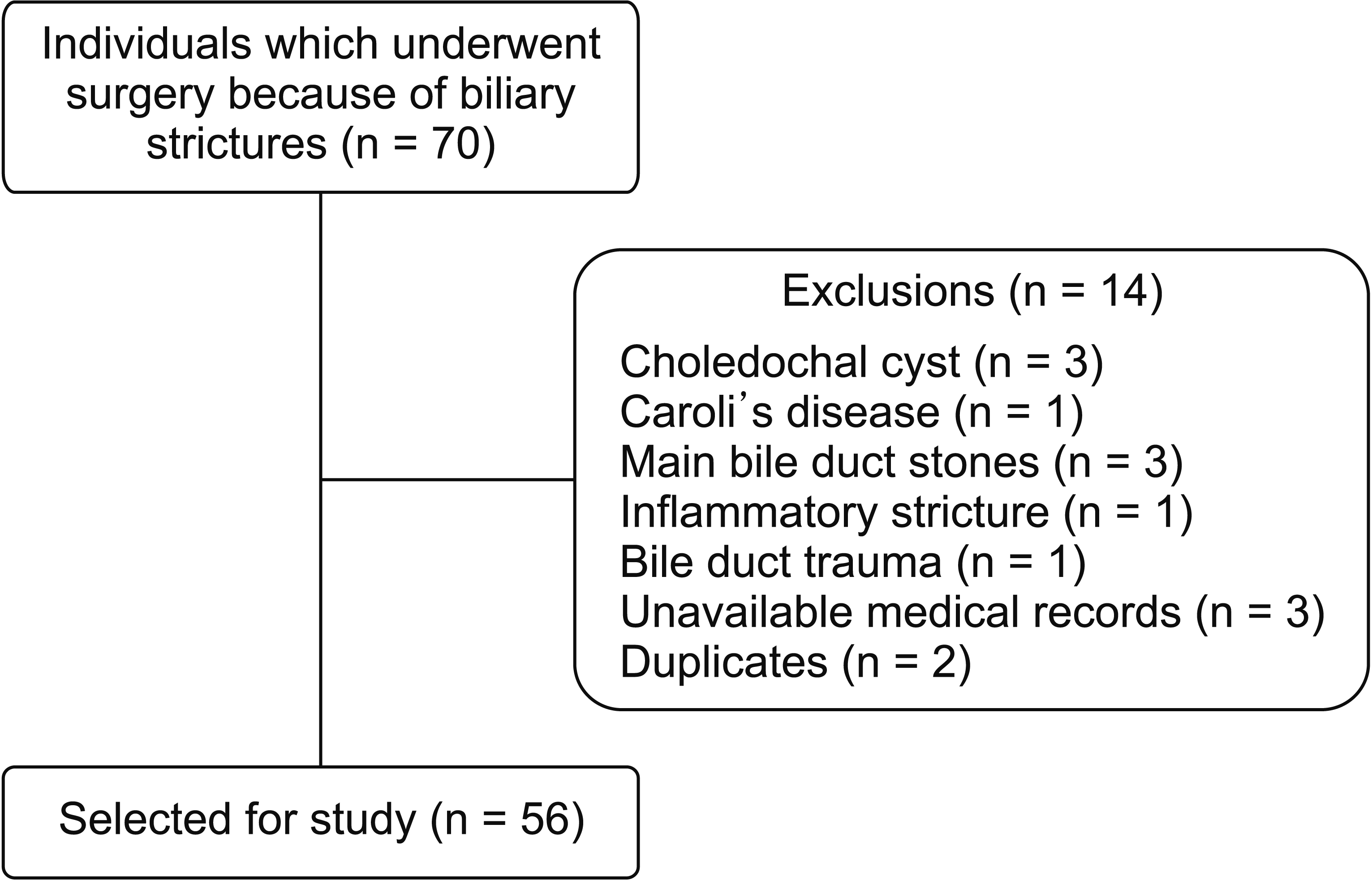
Study participants were identified through electronic outpatient spreadsheets. All individuals who consecutively underwent surgical BDI repair from January 2014 to December 2018 regardless of sex with age of at least 18 years were included. Patients who belonged to vulnerable groups, those who had incomplete medical records, and those who were followed-up for less than 24 months were excluded. The flow chart showing the selection of the study population is shown in Fig. 1.
The following variables were analyzed: age at repair surgery, sex, weight, body mass index (BMI), symptoms, comorbidities, previous data related to primary surgery (elective or emergency procedure; open or laparoscopic approach; reinterventions at the facility of origin), time elapsed between admission and definitive surgical correction, preoperative laboratory tests, type of BDI according to the Strasberg classification, presence of associated vascular injury, histopathological findings (presence of elementary liver traits in bilobar liver biopsy performed during surgery), bilobar heterogeneity, microbiological aspects (culture of bile collected in the intraoperative period), surgical technique adopted, estimated bleeding, surgical time, length of stay in the intensive care unit (ICU), total length of postoperative stay, 30-day postoperative complications, 30-day mortality, 90-day mortality, and occurrence of biliary stricture detected at least after 24 months (confirmed through imaging or scintigraphy scans).
Elementary hepatic characteristics considered were steatosis (occurrence of more than 5% of fat deposition in hepatocytes), fibrosis (classified according to the Kleiner classification in grades 0 to 4), cholestasis (classified according to its severity in absent, mild, moderate, and severe), acute and chronic cholangitis (classified as absent, mild, moderate, and severe), ductular proliferation (classified as absent, mild, moderate, and severe), and ductopenia (classified as absent or present).
Descriptive analysis was performed with presentation of frequency tables for categorical variables and position and dispersion measurements for numerical variables. Data normality was assessed using the Shapiro-Wilk test. To compare proportions, the chi-squared test or Fisher’s exact test was used. To compare continuous or ordinal variables between two independent groups, the Mann–Whitney test was used. Continuous variables were compared between three or more groups using the Kruskal–Wallis test. To analyze correlations between continuous variables and continuous or ordinal outcomes, single and multiple logistic regression analyses were performed. The significance level was set at p < 0.05. All statistical analyses were performed using SAS System for Windows (Statistic Analysis System), version 9.2 (2002–2008; SAS Institute Inc., Cary, NC, USA).
Of 56 individuals included, 43 (76.8%) were females. Of all subjects, the average age was 47.2 ± 13.2 years and the mean follow-up time was 38.1 ± 18.6 months. The average time elapsed between primary cholecystectomy and definitive surgical repair was 30.1 ± 40.4 months. Complete demographic, anthropometric, and clinical characteristics of participants are summarized in Table 1.
Surgical procedures were performed at the origin facility after cholecystectomy in 30 (53.5%) individuals, including 16 (28.5%) who underwent abdominal drainage and 14 (25.0%) who underwent bilioenterostomy. For 16 patients who underwent abdominal drainage, six (37.5%) cases had a T-tube placed, four (25.0%) cases had a simple closure, and the remaining six (37.5%) cases had no other reported procedures. Endoscopic retrograde choangiopancreatography (ERCP) was performed in 50% of the individuals who underwent abdominal drainage. With regard to individuals who underwent bilioenterostomy, four (28.6%) patients underwent choledocoduodenostomy and the remaining ten (71.4%) underwent Roux-en-Y hepaticojejunostomy.
Main histopathological findings identified were hepatic fibrosis (87.5%) and cholestasis (62.5%). Bilobar heterogeneity occurred in five (8.9%) patients, of which three had more severe alterations on the right lobe. The Kappa coefficient of agreement between findings of the two lobes was 0.82. Nine (16.1%) individuals had associated vascular injury. In all cases, the right hepatic artery was affected.
Positive bile cultures with one or more identified microorganisms were present in 30 (53.5%) patients. Among positive cultures, 18 (60.0%) had two or more microorganisms. Microorganisms from 12 (40.0%) cultures were considered multiresistant. The most prevalent bacteria are presented in Table 1.
The main surgical technique used was the Roux-en-Y hepaticojejunostomy (96.4%). One patient underwent choledocojejunostomy and another underwent left hepatectomy with Roux-en-Y right hepaticojejunostomy. Mean intraoperative bleeding was 509.1 ± 405.5 mL. The mean operative time was 213.4 ± 49.6 minutes.
The mean length of stay in the ICU was 2.9 ± 6.4 days. The average total postoperative hospital stay was 6.8 ± 3.8 days. Postoperative complications are summarized in Table 2. Overall morbidity was 35.7% (20 patients). The most frequent complications were intracavitary abscesses and biliary fistulas in five (8.9%) patients each. There was one (1.7%) death within 90 days due to multiple organ dysfunction secondary to abdominal sepsis. Late biliary stricture occurred in 12 (21.4%) patients during the follow-up period.
Univariate analysis of perioperative outcomes and continuous variables identified a positive relationship between the value of alanine aminotransferase (ALT) and the length of postoperative hospital stay (R = 0.3; p = 0.04). The degree of BDI estimated by the Strasberg classification (R = 0.3; p = 0.02) and ALT levels (R = 0.3; p = 0.04) were significantly associated with a higher estimated intraoperative bleeding. The degree of liver fibrosis was associated with a longer duration of the surgical procedure (R = 0.3; p = 0.02). Alkaline phosphatase (ALP) levels were associated with a higher rate of late stricture (p = 0.01). Detailed results of univariate analyses are shown in Tables 3 and 4.
Multivariate analyses of factors associated with length of postoperative and ICU stay, estimated bleeding, and operative time were performed. No variable showed a significant association with the length of postoperative hospital stay or ICU stay. The intensity of liver fibrosis (R = 0.36; p = 0.02) and cholestasis (R = 0.34; p = 0.02) were independently and positively correlated with operative time. Bismuth classification was independently and positively correlated with the estimated bleeding (R = 0.31; p = 0.049). Complete results of multivariate analyses are shown in Table 5.
The time elapsed between primary cholecystectomy and definitive surgical repair was significantly and positively correlated with the severity of hepatic fibrosis (R = 0.4; p = 0.01). The longer the waiting time for repair, the more severe the degree of liver damage. There was no significant correlation between waiting time and the degree of cholestasis (R = –0.1; p = 0.39).
The positivity of cultures and sensitivity profile in relation to surgical variables were analyzed. No statistically significant association was identified. Detailed results are shown in Table 6.
Although cholecystectomy-related BDI is considered an infrequent complication, it can have deleterious impacts on general health, physical condition, and social functioning compared to uncomplicated cholecystectomy [14]. In addition, it can lead to longer hospital stays, increase the risk of 30-day readmission, and result in higher costs [14,15]. Individuals with cholecystectomy-related BDI are subjected to recurrent cholangitis and liver cirrhosis. They may eventually require a liver transplant. Patients with bile leaks and BDI present higher one-year mortality than those who undergo uncomplicated cholecystectomy [7].
In the current study, 25% of individuals underwent an unsuccessful BDI repair at the facility of origin. Roy et al. [16] have found that patients undergoing repair by non-specialist surgeons have higher long-term complication rates, in addition to higher costs associated with treatment and legal litigation. Stewart and Way [13] observed that patients undergoing repair by the same surgeon who performed the cholecystectomy had significantly lower success rates than those reoperated on by a hepatobiliary surgeon.
In the current study, it was found that 53.5% of patients had positive bile culture for one or more microorganisms, of which 60% were polymicrobial and 40% of cultures were found to have multi-resistant microorganisms, albeit these findings were not statistically related to surgical outcomes. In the literature, few studies have analyzed intraoperative bile culture and its relationship with outcomes. Würstle et al. [17] have analyzed patients with bilomas mostly associated with surgical procedures (65.6%) and ERCP (21.9%). They found that 93.9% of samples were positive for any bacterial infection and 25% of study patients had bacterial infections with advanced resistance. The most frequently isolated microorganisms were Enterococcus faecium, Enterobacter spp., Escherichia coli, Klebsiella spp. and Candida spp. [17]. A 2020 Chinese study analyzed 1,339 patients whose bile samples were collected during endoscopic procedures by radiointervention or surgically. Samples were positive in 55.12% of cases. The most frequently isolated microorganisms were E. coli, Pseudomonas aeruginosa, Klebsiella pneumoniae and E. faecium. Age over 60 years, fever, benign diseases or a history of biliary tract diseases, and surgical procedures were associated with a higher positivity rate [18]. The same study detected high levels of bacterial resistance to ceftriaxone, quinolones, and ampicillin commonly used in the treatment of biliary tract infections. This finding could be due to inappropriate use of antibiotics. Routine microbiological analysis of intraoperatively collected bile can contribute to rational prescription of antimicrobials. These previous reported data and findings of the present study highlight the severity of BDI cases, considering the high rate of infections by multidrug-resistant germs. Thus, upon admission of patients with BDI, adequate infectious screening and careful evaluation of antibiotic therapy initiation are of great relevance.
In liver biopsies, more than 80% of patients had some degree of liver fibrosis, although only three (5.3%) individuals had liver cirrhosis on biopsy. Our findings are comparable with those of Negi et al. [19] who identified liver fibrosis or cirrhosis in 92% of patients. Although most histological changes are reversible after stricture repair in experimental studies, the presence of liver cirrhosis is an important risk factor for postoperative morbidity and mortality, in addition to being a predictor of surgical repair failure [20-24].
Despite the high overall morbidity rate (35.7%), complications generally did not cause serious repercussions. However, 21.4% of individuals had some degree of late stricture, a rate close to that found in the literature [8], with most being managed conservatively. There were no deaths in the early postoperative period. There was only one death within 90 days. One of the leading multicenter studies has reported a 30-day postoperative morbidity rate of 26.3% and a 30-day mortality rate of 2% [25].
ALT, but not aspartate aminotransferase, canalicular enzymes, or bilirubin, was associated with longer postoperative hospital stay. Lower albumin values had positive correlations with higher rates of severe complications. Serum albumin has been described in the literature as a predictor of morbidity and mortality within 30 days after surgery. It is an important marker of comorbidities and malnutrition [25,26]. ALP was related to higher rates of late strictures. There is an extensive literature analyzing preoperative clinical data (type of injury, timing of repair, presence of associated infections, use of abdominal drain) as predictors of repair success. However, the literature on the relationship between perioperative laboratory tests and surgical outcomes is scarce. Duration of biliary obstruction, baseline preoperative ALT level, and time until normalization of ALT levels after surgery have been identified as independent predictors of grades 2 and 3 liver fibrosis in patients with post-cholecystectomy stricture [19]. The timing of definitive repair is highly important considering that the waiting time is significantly correlated with the degree of liver fibrosis, pointing that these patients should be referred to tertiary facilities as soon as possible.
The current study has some limitations that should be taken into consideration. First, this was a retrospective, single-center study with a small sample of patients. Data related to individuals’ previous hospitalizations were not always complete or detailed. BDI is usually underdiagnosed. It is often initially diagnosed at the facility of origin, which can contribute to a severity bias because only patients with more complex injuries are referred to the tertiary service. On the other hand, many patients who firstly underwent repair in the facility of origin and evolved with treatment failure and strictures were referred to this reference hospital with more severe and complex injuries than they would have initially. Likewise, as only patients undergoing surgical repair were included, individuals with partial injuries and leaks, treated exclusively by ERCP or radio intervention, and those who would have a better prognosis and lower late stricture rates were not included in the sample.
In conclusion, fibrosis and cholestasis were the most frequent histopathological abnormalities. Bacterial contamination of bile was observed in most cases. The Strasberg degree of injury was positively correlated with the estimated bleeding. Degrees of fibrosis and cholestasis were correlated with the operative time. The waiting time for definitive repair was directly correlated with the severity of liver fibrosis.
Notes
REFERENCES
1. Reynolds W Jr. 2001; The first laparoscopic cholecystectomy. JSLS. 5:89–94.
2. Pucher PH, Brunt LM, Davies N, Linsk A, Munshi A, Rodriguez HA, et al. 2018; Outcome trends and safety measures after 30 years of laparoscopic cholecystectomy: a systematic review and pooled data analysis. Surg Endosc. 32:2175–2183. DOI: 10.1007/s00464-017-5974-2. PMID: 29556977. PMCID: PMC5897463.
3. Renz BW, Bösch F, Angele MK. 2017; Bile duct injury after cholecystectomy: surgical therapy. Visc Med. 33:184–190. DOI: 10.1159/000471818. PMID: 28785565. PMCID: PMC5527188.
4. Boerma D, Rauws EA, Keulemans YC, Bergman JJ, Obertop H, Huibregtse K, et al. 2001; Impaired quality of life 5 years after bile duct injury during laparoscopic cholecystectomy: a prospective analysis. Ann Surg. 234:750–757. DOI: 10.1097/00000658-200112000-00006. PMID: 11729381. PMCID: PMC1422134.
5. Hariharan D, Psaltis E, Scholefield JH, Lobo DN. 2017; Quality of life and medico-legal implications following iatrogenic bile duct injuries. World J Surg. 41:90–99. DOI: 10.1007/s00268-016-3677-9. PMID: 27481349.
6. Berney CR. 2012; Major common bile duct injury and risk of litigation: a surgeon's perspective. Am J Surg. 204:800–802. DOI: 10.1016/j.amjsurg.2011.06.009. PMID: 21872206.
7. Fong Z, Pitt H, Strasberg S, Loehrer A, Sicklick J, Talamini M, et al. 2018; Diminished survival in patients with bile leak and ductal injury: management strategy and outcomes. J Am Coll Surg. 226:568–576.e1. DOI: 10.1016/j.jamcollsurg.2017.12.023. PMID: 29307612. PMCID: PMC6053912.
8. Schreuder A, Busch O, Besselink M, Ignatavicius P, Gulbinas A, Barauskas G, et al. 2020; Long-term impact of iatrogenic bile duct injury. Dig Surg. 37:10–21. DOI: 10.1159/000496432. PMID: 30654363. PMCID: PMC7026941.
9. Bansal V, Krishna A, Misra M, Prakash P, Kumar S, Rajan K, et al. 2015; Factors affecting short-term and long-term outcomes after bilioenteric reconstruction for post-cholecystectomy bile duct injury: experience at a tertiary care centre. Indian J Surg. 77(Suppl 2):472–479. DOI: 10.1007/s12262-013-0880-x. PMID: 26730048. PMCID: PMC4692855.
10. Ekmekcigil E, Ünalp Ö, Uğuz A, Hasanov R, Bozkaya H, Köse T, et al. 2018; Management of iatrogenic bile duct injuries: Multiple logistic regression analysis of predictive factors affecting morbidity and mortality. Turk J Surg. 34:264–270. DOI: 10.5152/turkjsurg.2018.3888. PMID: 30216168. PMCID: PMC6340667.
11. Koffron A, Ferrario M, Parsons W, Nemcek A, Saker M, Abecassis M. 2001; Failed primary management of iatrogenic biliary injury: incidence and significance of concomitant hepatic arterial disruption. Surgery. 130:722–728. discussion 728–731. DOI: 10.1067/msy.2001.116682. PMID: 11602904.
12. Stewart L, Robinson TN, Lee CM, Liu K, Whang K, Way LW. 2004; Right hepatic artery injury associated with laparoscopic bile duct injury: incidence, mechanism, and consequences. J Gastrointest Surg. 8:523–530. discussion 530–531. DOI: 10.1016/j.gassur.2004.02.010. PMID: 15239985.
13. Stewart L, Way LW. 2009; Laparoscopic bile duct injuries: timing of surgical repair does not influence success rate. A multivariate analysis of factors influencing surgical outcomes. HPB (Oxford). 11:516–522. DOI: 10.1111/j.1477-2574.2009.00096.x. PMID: 19816617. PMCID: PMC2756640.
14. Flores-Rangel G, Chapa-Azuela O, Rosales A, Roca-Vasquez C, Böhm-González S. 2018; Quality of life in patients with background of iatrogenic bile duct injury. World J Surg. 42:2987–2991. DOI: 10.1007/s00268-018-4564-3. PMID: 29520485.
15. O'Brien S, Wei D, Bhutiani N, Rao MK, Johnston SS, Patkar A, et al. 2020; Adverse outcomes and short-term cost implications of bile duct injury during cholecystectomy. Surg Endosc. 34:628–635. DOI: 10.1007/s00464-019-06809-8. PMID: 31286250.
16. Roy P, Soonawalla Z, Grant H. 2009; Medicolegal costs of bile duct injuries incurred during laparoscopic cholecystectomy. HPB (Oxford). 11:130–134. DOI: 10.1111/j.1477-2574.2008.00023.x. PMID: 19590636. PMCID: PMC2697871.
17. Würstle S, Göß A, Spinner CD, Huber W, Algül H, Schlag C, et al. 2019; A retrospective clinical and microbial analysis of 32 patients with bilomas. BMC Gastroenterol. 19:50. DOI: 10.1186/s12876-019-0968-2. PMID: 30947689. PMCID: PMC6450004.
18. Gu XX, Zhang MP, Zhao YF, Huang GM. 2020; Clinical and microbiological characteristics of patients with biliary disease. World J Gastroenterol. 26:1638–1646. DOI: 10.3748/wjg.v26.i14.1638. PMID: 32327912. PMCID: PMC7167412.
19. Negi SS, Sakhuja P, Malhotra V, Chaudhary A. 2004; Factors predicting advanced hepatic fibrosis in patients with postcholecystectomy bile duct strictures. Arch Surg. 139:299–303. DOI: 10.1001/archsurg.139.3.299. PMID: 15006888.
20. Abdel-Aziz G, Lebeau G, Rescan PY, Clément B, Rissel M, Deugnier Y, et al. 1990; Reversibility of hepatic fibrosis in experimentally induced cholestasis in rat. Am J Pathol. 137:1333–1342.
21. Soares PFDC, Gestic MA, Utrini MP, Callejas-Neto F, Chaim EA, Cazzo E. 2019; Epidemiological profile, referral routes and diagnostic accuracy of cases of acute cholangitis among individuals with obstructive jaundice admitted to a tertiary-level university hospital: a cross-sectional study. Sao Paulo Med J. 137:491–497. DOI: 10.1590/1516-3180.2019.0109170919. PMID: 32159634.
22. Pellegrini C, Thomas M, Way L. 1984; Recurrent biliary stricture. Patterns of recurrence and outcome of surgical therapy. Am J Surg. 147:175–180. DOI: 10.1016/0002-9610(84)90054-0. PMID: 6691544.
23. Pottakkat B, Vijayahari R, Prakash A, Singh RK, Behari A, Kumar A, et al. 2010; Factors predicting failure following high bilio-enteric anastomosis for post-cholecystectomy benign biliary strictures. J Gastrointest Surg. 14:1389–1394. DOI: 10.1007/s11605-010-1241-8. PMID: 20589447.
24. Mitsunaga TM, Jimenez LS, Soares PFDC, Gestic MA, Utrini MP, Chaim FDM, et al. 2021; Effect of transient obstructive cholestasis on liver histology: a cross-sectional study. Sao Paulo Med J. 139:351–363. DOI: 10.1590/1516-3180.2020.0536.r1.1502021. PMID: 34161522. PMCID: PMC9615596.
25. Ismael H, Cox S, Cooper A, Narula N, Aloia T. 2017; The morbidity and mortality of hepaticojejunostomies for complex bile duct injuries: a multi-institutional analysis of risk factors and outcomes using NSQIP. HPB (Oxford). 19:352–358. DOI: 10.1016/j.hpb.2016.12.004. PMID: 28189346.
26. Gibbs J, Cull W, Henderson W, Daley J, Hur K, Khuri SF. 1999; Preoperative serum albumin level as a predictor of operative mortality and morbidity: results from the National VA Surgical Risk Study. Arch Surg. 134:36–42. DOI: 10.1001/archsurg.134.1.36. PMID: 9927128.
Table 1
Table 2
Table 3
Variable | Hospital stay | ICU stay | Estimated bleeding | Operative time | |||||||
---|---|---|---|---|---|---|---|---|---|---|---|
|
|
|
|
||||||||
R | p-value | R | p-value | R | p-value | R | p-value | ||||
Age | 0.2 | 0.30 | 0.2 | 0.29 | 0.07 | 0.59 | 0.07 | 0.63 | |||
Time elapsed between cholecystectomy and repair | 0.03 | 0.82 | 0.03 | 0.82 | –0.08 | 0.64 | 0.1 | 0.38 | |||
Aspartate aminotransferase | 0.3 | 0.06 | 0.2 | 0.18 | 0.3* | 0.05* | 0.1 | 0.32 | |||
Alanine aminotransferase | 0.3* | 0.04* | 0.2 | 0.15 | 0.3 | 0.04 | 0.2 | 0.25 | |||
Alkaline Phosphatase | –0.1 | 0.62 | 0.03 | 0.82 | –0.02 | 0.84 | 0.05 | 0.74 | |||
Gamma-glutamyl transferase | –0.1 | 0.58 | 0.06 | 0.74 | –0.08 | 0.63 | 0.1 | 0.26 | |||
Bilirrubin | –0.1 | 0.62 | 0.1 | 0.45 | –0.1 | 0.44 | –0.2 | 0.15 | |||
International normatized ratio | 0.03 | 0.83 | 0.1 | 0.51 | –0.1 | 0.39 | –0.004 | 0.99 | |||
Albumin | –0.1 | 0.62 | 0.1 | 0.49 | –0.06 | 0.64 | –0.04 | 0.84 | |||
Strasberg classification degree | 0.2 | 0.09 | 0.07 | 0.64 | 0.3* | 0.02* | 0.06 | 0.62 | |||
Body mass index | –0.1 | 0.39 | 0.1 | 0.29 | –0.02 | 0.93 | –0.3 | 0.07 | |||
Fibrosis severity | 0.06 | 0.64 | 0.002 | > 0.99 | 0.2 | 0.18 | 0.3* | 0.03* | |||
Cholestasis severity | 0.2 | 0.17 | 0.2 | 0.07 | 0.2 | 0.27 | –0.1 | 0.45 |
Table 4
Variable | Mild to moderate complication (Clavien-Dindo 1-2) | Severe complication (Clavien-Dindo ≥ 3) | p-value | Late stricture | No stricture | p-value |
---|---|---|---|---|---|---|
Number | 42 (75.0) | 14 (25.0) | NA | 12 (21.4) | 44 (78.6) | NA |
Age (yr) | 46.3 ± 13.0 | 50.0 ± 13.9 | 0.39 | 41.3 ± 12.5 | 48.8 ± 13.0 | 0.08 |
Sex |
M: 11 (26.2) F: 31 (73.8) |
M: 2 (14.3) F: 12 (85.7) |
0.42 |
M: 2 (16.7) F: 10 (83.3) |
M: 11 (25.0) F: 33 (75.0) |
0.49 |
Body mass index (kg/m2) | 26.8 ± 5.1 | 25.9 ± 4.9 | 0.58 | 27.1 ± 5.3 | 25.7 ± 4.7 | 0.72 |
Time elapsed between cholecystectomy and repair (mon) | 29.58 ± 43.91 | 22.6 ± 27.0 | 0.56 | 27.58 ± 39.45 | 29.66 ± 40.50 | 0.88 |
Aspartate aminotransferase (U/L) | 52.2 ± 36.7 | 72.9 ± 45.9 | 0.10 | 73.0 ± 57.3 | 66.3 ± 40.9 | 0.62 |
Alanine aminotransferase (U/L) | 69.3 ± 57.4 | 103.4 ± 78.4 | 0.12 | 91.6 ± 48.5 | 95.7 ± 80.9 | 0.87 |
Alkaline phosphatase(U/L) | 506.1 ± 595.9 | 433.4 ± 455.0 | 0.63 | 799.5 ± 832.6 | 402.9 ± 307.0 | 0.01* |
Gamma-glutamyl transferase (U/L) | 693.5 ± 856.0 | 697.1 ± 488.8 | 0.97 | 792.7 ± 853.4 | 667.6 ± 509.5 | 0.46 |
Bilirrubin (mg/dL) | 5.8 ± 8.1 | 6.0 ± 6.8 | 0.92 | 7.7 ± 7.8 | 5.4 ± 7.8 | 0.35 |
International normatized ratio | 1.1 ± 0.2 | 1.1 ± 0.2 | 0.48 | 1.1 ± 0.2 | 1.1 ± 0.1 | > 0.99 |
Albumin (g/dL) | 3.4 ± 0.8 | 3.8 ± 0.5 | 0.04* | 3.5 ± 0.7 | 3.8 ± 0.6 | 0.09 |
Strasberg E1-E2 vs. ≥ E3 |
1–2: 22 ≥ 3: 20 |
1–2: 7 ≥ 3: 7 |
0.88 |
1–2: 6 (50.0) ≥ 3: 6 (50.0) |
1–2: 22 (50.0) ≥ 3: 22 (50.0) |
> 0.99 |
Fibrosis |
Present: 37 (88.1) Ausente: 5 (11.9) |
Present: 12 (85.7) Absent: 2 (14.3) |
0.79 |
Present: 11 (91.7) Absent: 1 (8.3) |
Present: 38 (86.4) Absent: 6 (13.6) |
0.62 |
Cholestasis |
Present: 28 (66.7) Absent: 14 (33.3) |
Present: 7 (50.0) Absent: 7 (50.0) |
0.27 |
Present: 8 (66.7) Absent: 4 (33.3) |
Present: 27 (61.4) Absent: 17 (38.6) |
0.68 |
Bile culture |
Positive: 20 (47.6) Negative: 22 (52.4) |
Positive: 10 (71.5) Negative: 4 (28.5) |
0.23 |
Positive: 7 (58.3) Negative: 5 (41.7) |
Positive: 23 (52.3) Negative: 21 (47.7) |
0.68 |
Table 5
Variable | Hospital stay | ICU stay | Estimated bleeding | Operative time | |||||||
---|---|---|---|---|---|---|---|---|---|---|---|
|
|
|
|
||||||||
R | p-value | R | p-value | R | p-value | R | p-value | ||||
Age | –0.12 | 0.52 | –0.12 | 0.53 | 0.08 | 0.65 | –0.21 | 0.21 | |||
Body mass index | –0.20 | 0.21 | –0.25 | 0.12 | –0.04 | 0.76 | –0.23 | 0.09 | |||
Strasberg classification | 0.01 | 0.93 | –0.18 | 0.27 | 0.31* | < 0.05* | 0.16 | 0.23 | |||
AST | –0.26 | 0.40 | –0.41 | 0.19 | 0.10 | 0.73 | 0.17 | 0.50 | |||
ALT | –0.09 | 0.73 | 0.16 | 0.55 | –0.33 | 0.19 | –0.33 | 0.15 | |||
ALP | 0.20 | 0.37 | 0.18 | 0.41 | 0.13 | 0.54 | 0.008 | 0.96 | |||
GGT | 0.02 | 0.90 | –0.14 | 0.49 | 0.04 | 0.82 | 0.17 | 0.31 | |||
Totak bilirubin | 0.02 | 0.93 | 0.28 | 0.34 | –0.3 | 0.28 | –0.36 | 0.15 | |||
Fibrosis severity | 0.09 | 0.56 | 0.23 | 0.16 | –0.09 | 0.57 | 0.4* | 0.02* | |||
Steatosis severity | 0.21 | 0.19 | –0.02 | 0.89 | 0.3 | 0.05 | 0.10 | 0.44 | |||
Acute cholangitis | 0.009 | 0.96 | 0.12 | 0.48 | –0.04 | 0.77 | 0.01 | 0.91 | |||
Chronic cholangitis | –0.09 | 0.56 | –0.05 | 0.72 | 0.07 | 0.63 | 0.3* | 0.02* | |||
Cholestasis | –0.12 | 0.62 | –0.43 | 0.10 | 0.18 | 0.46 | 0.11 | 0.61 | |||
Ductular proliferation | 0.01 | 0.92 | 0.09 | 0.61 | 0.02 | 0.86 | –0.02 | 0.88 |