Journal List > Ann Hepatobiliary Pancreat Surg > v.26(4) > 1516080319

Bhavsar, Yadav, and Nundy: Portal cavernoma cholangiopathy: Update and recommendations on diagnosis and management

Abstract

Portal cavernoma cholangiopathy is defined as an obstruction of the biliary system due to distended veins surrounding bile ducts that mainly occur in patients with extrahepatic portal venous obstruction. The periductal venous plexuses encircling the ducts can cause morphological changes which may or may not become symptomatic. Currently, non-invasive techniques such as ultrasonography, computed tomography, magnetic resonance cholangiopancreatography, and dynamic contrast enhanced magnetic resonance images are being used to diagnose this disorder. Only a few patients who have symptoms of biliary obstruction require drainage which might be accomplished using endoscopic stenting, decompression of the portal venous system usually via a lienorenal shunt, a difficult direct hepaticojejunostomy, and rarely a liver transplant.

INTRODUCTION

Numerous studies have detailed biliary alterations found in connection with cavernomatous transformation of the portal vein since Fraser and Brown’s [1] 1944 description of symptomatic biliary obstruction owing to collateral vessels in a patient with extrahepatic portal venous obstruction (EHPVO). Other types of portal hypertension, such as cirrhosis and noncirrhotic portal fibrosis, have been associated with similar biliary alterations, but at a considerably lower incidence than EHPVO. A variety of names and criteria have been devised to describe these biliary changes [2-5]. The clinical relevance, natural history, and prognosis of these biliary alterations remain inadequately documented. Therapy has not been rationalised due to the lack of a defined diagnostic and standard inclusion criteria in these studies. In this paper, we will examine current studies on portal cavernomatous biliopathy and discuss different diagnostic and therapeutic options available to manage this uncommon entity.

DEFINITION

An abnormality in the extrahepatic biliary system in a patient with a portal cavernoma is known as portal cavernoma cholangiopathy (PCC). The following conditions must be met in order to establish a diagnosis: 1) a portal cavernoma, 2) characteristic cholangiographic alterations on endoscopic retrograde cholangiography (ERC) or magnetic resonance cholangiography (MRC), and 3) the lack of other biliary causes such as bile duct damage, primary sclerosing cholangitis, or cholangiocarcinoma [6].

ANATOMY AND RELATION TO THE VENOUS DRAINAGE

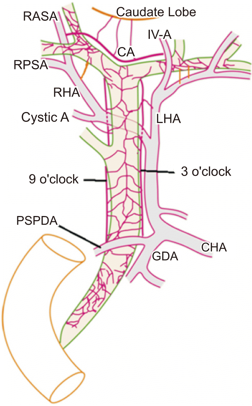
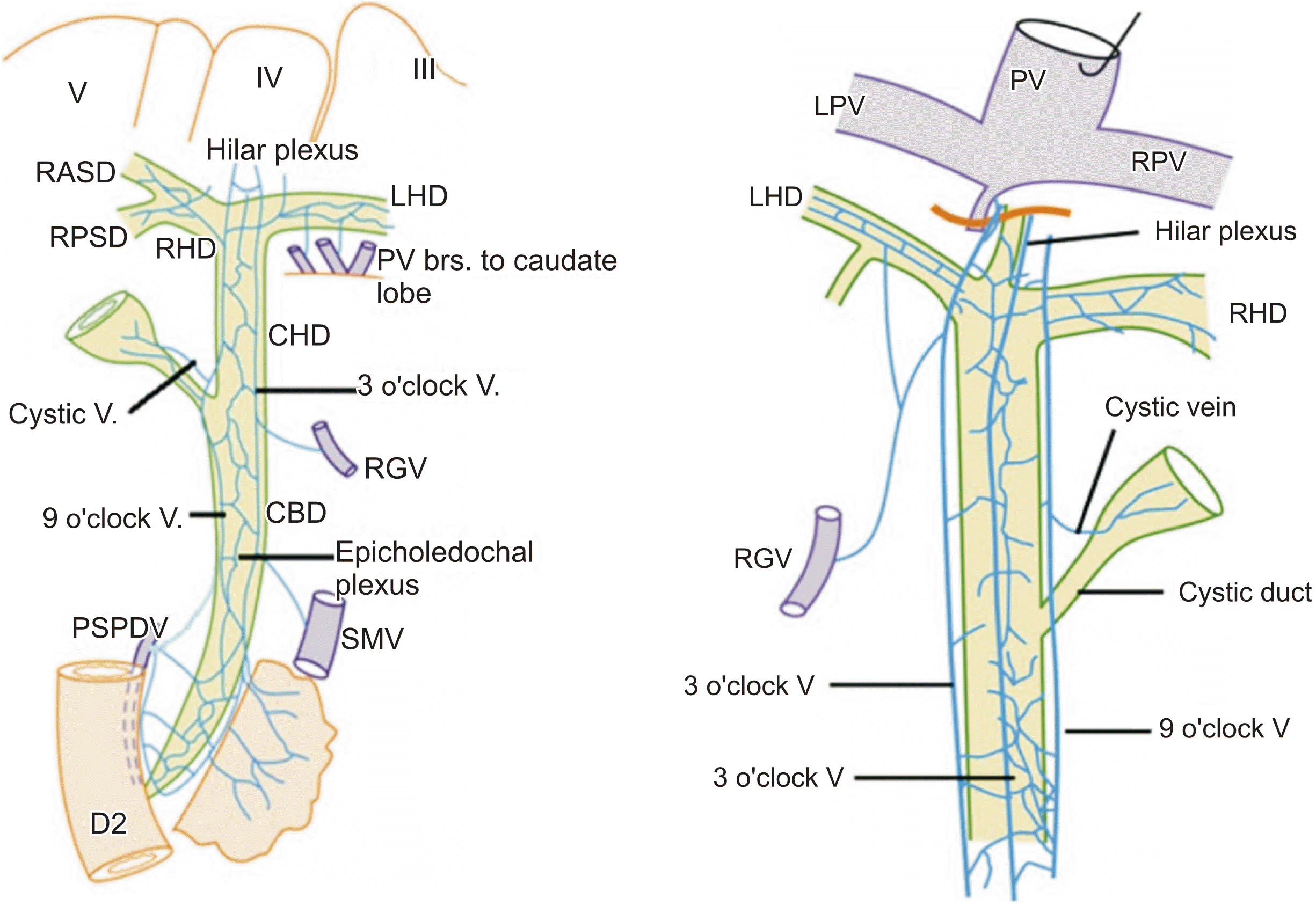
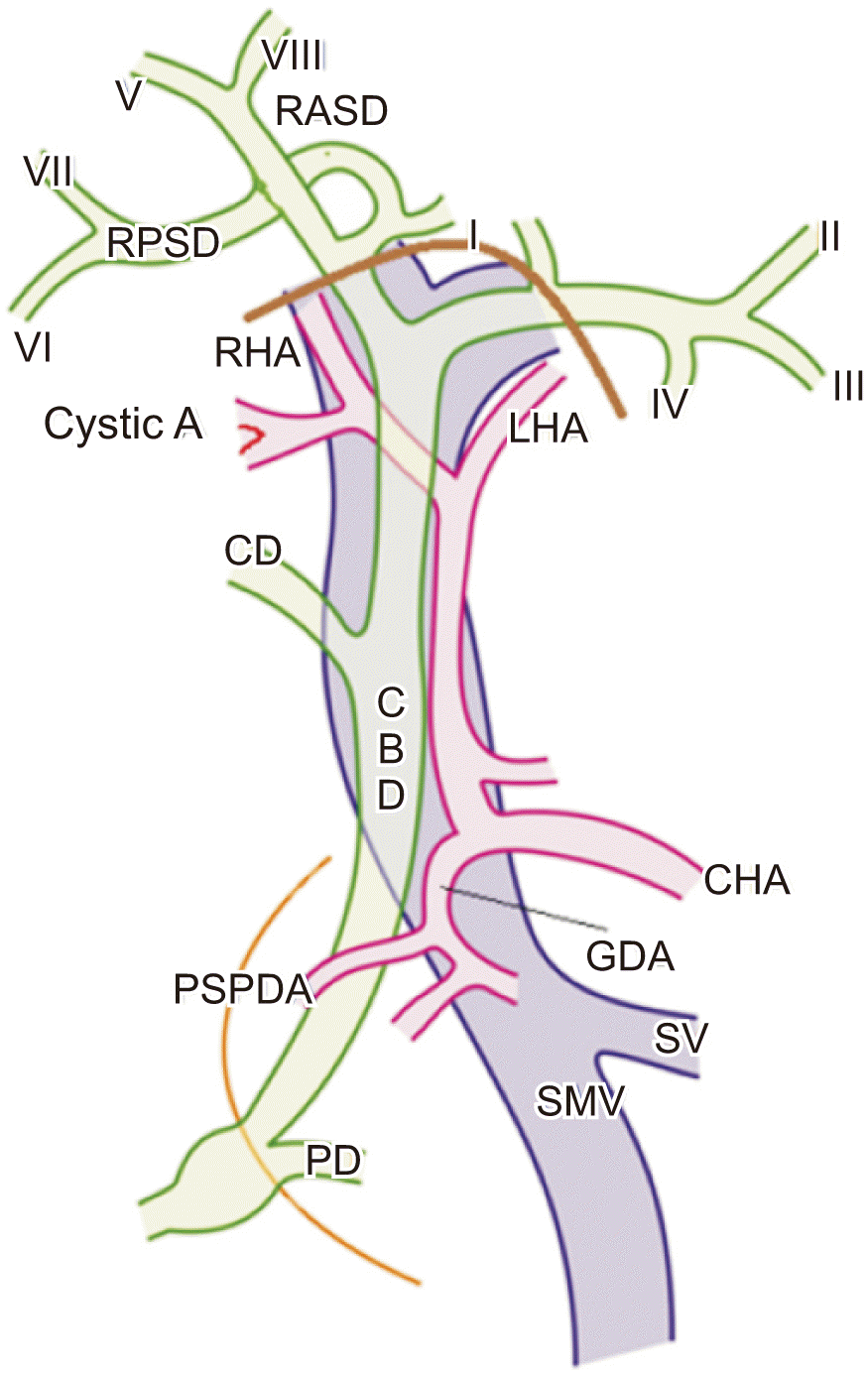
The arterial and venous supply and drainage of the bile ducts are generally typical in design. Two plexuses are created by fine branches from the posterior superior pancreaticoduodenal, retro portal, gastroduodenal, hepatic, and cystic arteries, which supply the ducts. The right and left marginal arteries, as well as the paracholedochal plexus, travel parallel to the bile duct, whereas the reticular epicholedochal plexus sits on its surface (Fig. 1, 2) [7]. The blood supply to the retropancreatic, hilar, and intrahepatic biliary tracts is substantial. However, the supraduodenal bile duct is the least vascularized. Hence, it is susceptible to ischemic injury [8,9]. The biliary tract is drained by two venous plexuses. The fine reticular epicholedochal venous plexus of Saint [10] on the bile duct wall drains into the paracholedochal venous plexus of Petren [11], which is connected to the posterior superior pancreaticoduodenal vein, gastrocolic trunk, right gastric vein, superior mesenteric vein inferiorly, and intrahepatic portal vein branches (Fig. 3). These plexuses expand to create a portal cavernoma encircling the bile duct in cases of porto-mesenteric venous blockage, causing morphological alterations seen in portal cavernomatous cholangiopathy.

PATHOGENESIS AND NOMENCLATURE

There are three primary theories on how portal biliopathy develops:
1. Compression with a reversible component
2. Ischaemia with an irreversible component
3. Infection

Compression

Compression by large collateral vessels next to the bile duct, as well as perhaps intracholedochal varices, causes the reversible component. Collaterals that develop after portal vein thrombosis have been linked to the reversible component of PCC, which may resolve when these collaterals are decompressed through a portosystemic shunt. Dilated venous collaterals in PCC can produce external pressure and protrusion over the thin and flexible common bile duct (CBD) and hepatic ducts [12-15]. Fine abnormalities in the biliary tract are caused by dilatation of the plexus of Saint [10], whereas dilatation of the plexus of Petren [11] produces extrinsic compression of the bile duct. PCC is more often associated with the left hepatic duct, which might be connected with the development of large collateral veins where the umbilical vein meets the left branch of the portal vein [16]. In addition to extrinsic collaterals compressing the bile duct, intra bile duct collaterals have been discovered, which might contribute to biliary obstruction.

Ischaemia

According to this theory, persistent PCC has various causes:
• Ischaemia of the bile duct due to thrombosis of the small venules
• Local ischaemia due to prolonged local wall compression by collaterals, and
• Encasement by a fibrous “solid tumor-like cavernomatous” structure which encases the bile duct
Long-term portal thrombosis also causes sclerosis in veins that drain bile ducts, which might affect capillaries and arterioles. The resolution of these cholangiographic anomalies is not always complete, suggesting that blaming it on collateral compression or a portal cavernoma alone is a simplistic view of the pathogenesis. After a decompressive portosystemic shunt surgery, smooth biliary strictures will open and the proximal dilatation will disappear in the majority of patients. Shunt surgery removes the indentations and calibre abnormalities. However, bile duct angulations and ectasias persist [12]. The ischaemia aetiology is also supported by the rigidity of strictures, which has been seen to cause more severe compression over the bile duct [17,18]. While some authors have demonstrated imaging evidence of collaterals causing biliary constriction in some individuals, this is not present in all cases. Dhiman et al. [15] have used MRC combined with MR portography to examine the relationship of biliary changes with portal cavernoma in 18 patients with EHPVO. They found that only in five of the nine patients with dominant strictures of the bile duct, the stricture was caused by compression from adjacent collaterals, while no such relationship was seen in the remaining four. Hence, the stricture was considered to be due to ischaemia in these four cases.

Infection

Jaundice in individuals with portal vein thrombosis was formerly thought to be caused by infection or cholangitis. Later, cholangiographic investigations have revealed that alterations in the biliary system can be observed even in asymptomatic individuals and that cholangitis develops late in the course of the disease [19-21]. Cholangitis can cause inflammation, bile duct neogenesis, fibrous tissue deposition, and persistence of strictures after shunt surgery.

CLINICAL CHARACTERISTICS AND SYMPTOMATOLOGY

Asymptomatic phase

Patients in their asymptomatic phase are discovered to have biliary abnormalities on ERC or MRC in the absence of any biliary symptoms. In the majority of investigations, biliary abnormalities were found in 78%–100% of patients who did not have symptoms, whereas symptoms were found in 5%–38% patients [14,22-29].

Symptomatic phase

Symptomatic individuals, on the other hand, have chronic cholestasis with or without biliary discomfort or acute cholangitis, which is usually caused by biliary strictures or stones. All patients with symptomatic biliopathy have a history of jaundice. However, this might be seen at the time of presentation in only around two-thirds of these individuals [17]. Cholangitis can affect half to two-thirds of people, with the frequency of cholangitic episodes varying from person to person. In addition, the majority of EHPVO with PCC patients had a history of variceal haemorrhage [30]. In a study of 97 individuals with EHPVO, Webb and Sherlock [31] discovered that 13 had elevated blood bilirubin levels, six had gastrointestinal bleeding, two had intermittent jaundice, and five had chronic jaundice. Before presenting with symptoms of PCC, patients with EHPVO generally have had a longstanding illness of 8–10 years. The majority of individuals with symptomatic PCC have jaundice, an enlarged spleen, and hepatomegaly. A report from the United Kingdom of 13 individuals with symptomatic PCC showed comparable findings [30]. All had jaundice, five had stomach discomfort, ten had a history of variceal bleeding, and 11 had splenomegaly (excluding two patients who had undergone a splenectomy previously) [17].

Complicated phase

Patients with severe and widespread biliary ductal alterations (e.g., long [> 2 cm] or multifocal, extrahepatic and/or intrahepatic strictures complicated by choledochal or intrahepatic calculi and biliopancreatic consequences of PCC) are considered to have a complicated phase.

End stage liver disease

According to Condat et al. [14], patients for whom therapeutic options are limited due to extensive venous thromboses show progressive liver dysfunction due to liver fibrosis, secondary biliary cirrhosis, eventually progressing to end stage liver disease are found in 2%–4% of individuals with biliary blockage that has been established for a long period.

EVALUATION

Vascular alterations in the form of portosystemic collaterals and biliary changes with extrinsic impressions and strictures are the most common radiographical findings. Conventional endoscopic cholangiography has been the gold standard for diagnosing PCC until recently. Because it is an invasive procedure that can lead to infection, duodenal perforation, and bleeding, non-invasive techniques such as ultrasonography (USG), computed tomography (CT), magnetic resonance cholangiopancreatography (MRCP), and dynamic contrast enhanced MR images, also known as MR portography, are being used more often nowadays. MR portography provides a good delineation of both the vascular and the biliary anatomy.

Blood investigations

Liver function tests are the most effective way to determine whether individuals may benefit from imaging studies. Hyperbilirubinaemia and cholestasis are both indications for biliary imaging. When secondary biliary cirrhosis develops, serum albumin levels and prothrombin time also become abnormal [32].

Ancillary findings

Splenomegaly is nearly always present. However, cirrhosis with nodularity of the liver surface, atrophic medial segments of the right lobe as well as hypertrophic caudate or lateral segments of the left lobe, expansion of fissures, and coarsened echotexture or changed signal intensity of the liver are infrequent [33]. Imaging can reveal any secondary diseases causing portal vein blockage, such as a large hepatic tumor and acute pancreatitis.

Abdominal ultrasound and Doppler examination (USG)

This should be the initial screening modality. It is also used for follow-up, although it is an operator dependent investigation. Color and spectral Doppler ultrasound can demonstrate the portal cavernoma and gall bladder varices. However, the exact delineation of the collaterals is not possible by ultrasound alone. It can assess biliary dilatation. However, it usually has difficulty in delineating biliary changes due to bowel gas. It may also detect associated lithiasis and splenomegaly. Gall bladder varices are seen in all cases according to some studies, while others have correlated colour Doppler examination with ERCP, revealing that only 54% patients with Doppler ultrasound having biliary abnormalities on ERCP [31,32].

Triple phase computed tomography

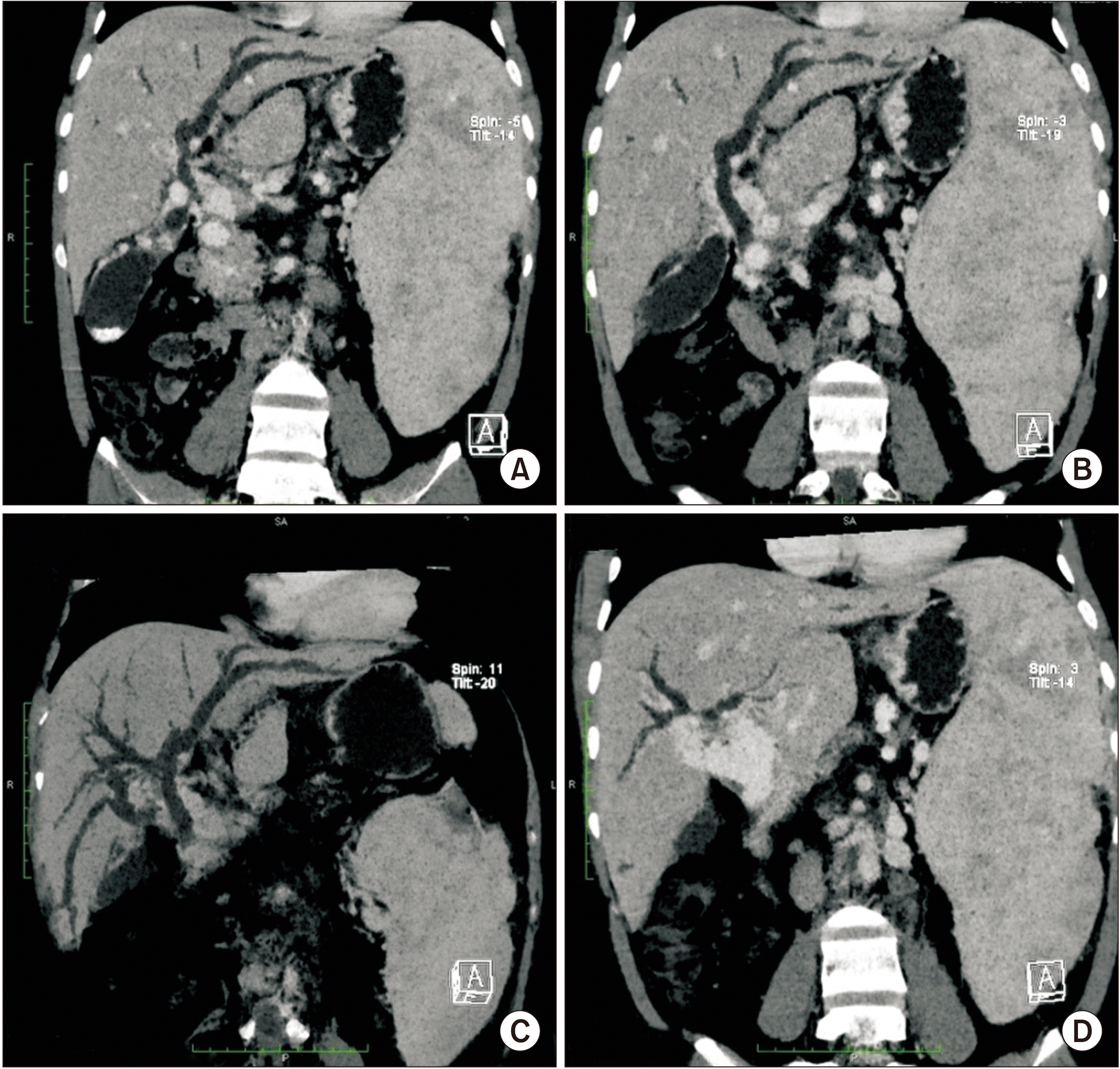
Triple phase CT scan can demonstrate vascular and biliary changes better by delineating the extent of vascular thrombosis (if present) and portosystemic collaterals. It can also assess biliary dilatation and CBD changes, although it is less accurate than MRCP (Fig. 4). It may detect the presence of cirrhosis and help rule out mimickers of PCC like malignant lesions. In a retrospective study of 11 patients after magnetic resonance imaging (MRI) (n = 7) and CT (n = 4), it was found that high resolution CT and MRI were comparable for providing anatomical details of the presence and severity of bile duct dilatation, portal vein obstruction, portal cavernoma, and portosystemic collaterals [33]. However, as the disease may require repeated imaging, CT is not the preferred technique because of the risk of radiation exposure.

Magnetic retrograde cholangiopancreatography with magnetic resonance portography

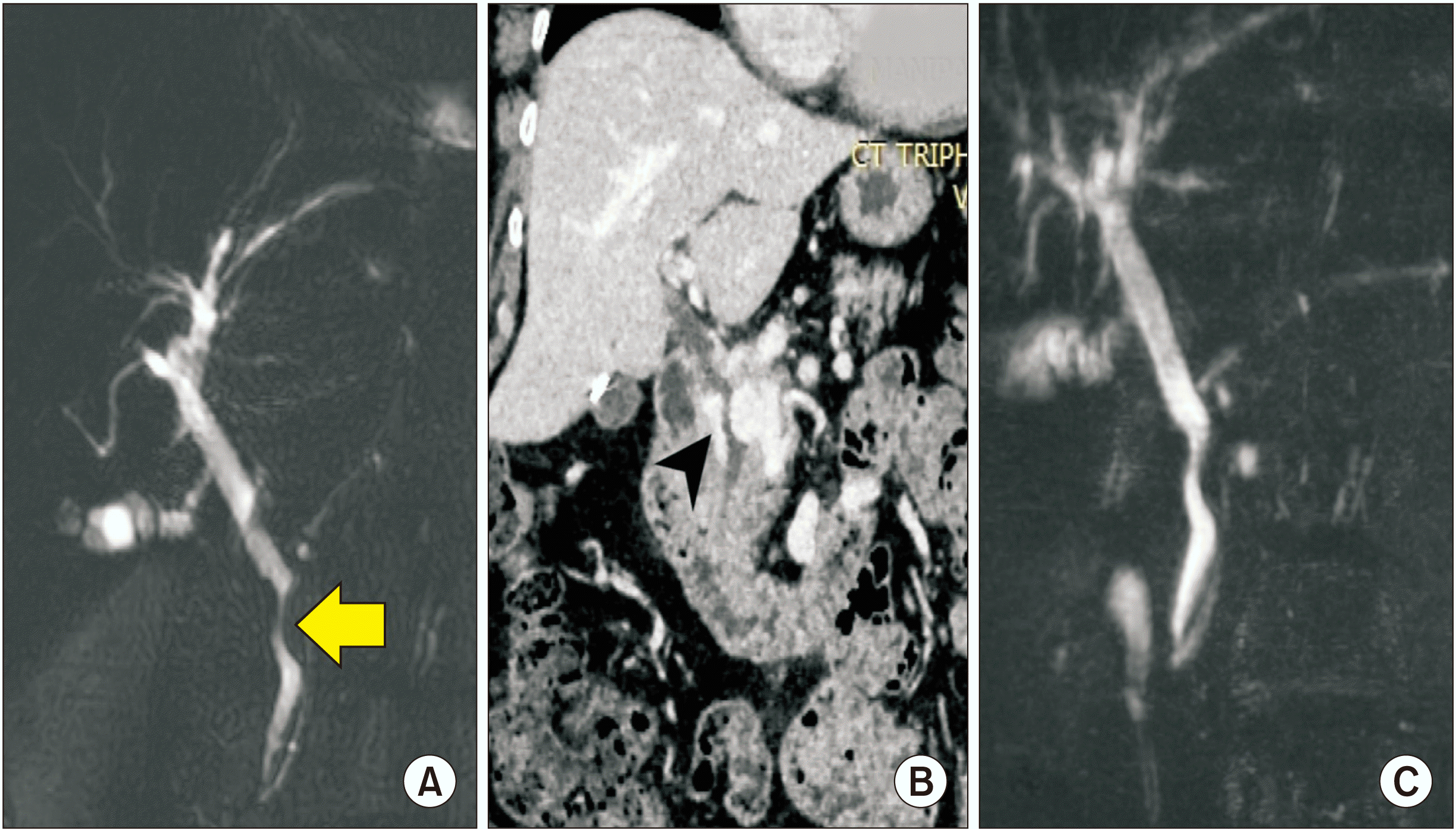
This is the gold standard for demonstrating PCC. It has replaced invasive ERC. It provides a near exact delineation of the extent of vascular thrombosis and portosystemic collaterals similar to CT (Fig. 5). It also provides a complete assessment of the biliary changes similar to direct cholangiography. It may also help differentiate between varicoid and fibrotic types of strictures and detect associated lithiasis. Chandra et al. [4] have proposed a classification system for PCC based on the location of narrowing after direct cholangiography: Type I) involvement of extrahepatic ducts, type II) involvement of intrahepatic bile ducts, type III a) involvement of extrahepatic bile ducts and unilateral intrahepatic bile ducts, and type III b) extrahepatic bile duct and bilateral intrahepatic bile duct involvement. This classification system can also be applied to MRCP image [34]. Recent reports of MRCP imaging in PCC show that either type I or type III are the most frequent, with extrahepatic bile ducts being the most common location of narrowing [35]. A retrospective study of 10 EHPVO patients had reported that MRCP can overestimate the bile duct stenosis compared to conventional cholangiographic studies [36]. Llop et al. [29] have classified cholangiographic findings of 67 patients with portal vein thrombosis using MRCP according to the severity of biliary dilatation where six patients had grade I (irregularities or angulations of the biliary tree), 12 patients had grade II (stenosis without dilation), and 34 patients had grade III (strictures with dilation) changes.

Endoscopic retrograde cholangiography

With the availability of MRC imaging, indications for ERC with diagnostic intent are becoming less common in patients with suspected PCC. Although ERC still remains the gold standard to define changes of PCC, its use in diagnosis is now limited. However, the second and third order intrahepatic bile ducts can be demonstrated in greater detail with ERC due to its greater spatial and contrast resolution, while MRC can provide a non-invasive ‘snapshot’ image of the biliary tree. Possible indications for ERC in patients with suspected portal biliopathy are shown below:
1. Cholangitis
2. Bile duct stones
3. Bile duct strictures (either symptomatic or persisting after portal decompression)
4. If there is diagnostic ambiguity

MANAGEMENT

If liver function tests are within the normal limits, no treatment is required. Imaging (MRCP/ERCP/USG) should be used to search for biliary tract abnormalities in patients with consistently elevated blood bilirubin and alkaline phosphatase levels [4]. There are a number of critical management questions that need to be addressed.

Who should treat?

For optimal therapy of symptomatic and complex PCC, competent endoscopic and surgical teams must work together to address problems in the biliary and portal vascular tree of each patient. The goal is to keep track of the patient until the biliary blockage is cleared, the patient is asymptomatic, and no additional treatment is required.

Whom to treat?

Any intervention should have a high barrier for initiation. In asymptomatic PCC with just cholangiographic alterations or mild biochemical abnormalities, intervention should be avoided. Endoscopic intervention is usually needed at first presentation for patients with cholestasis, choledocholithiasis with cholangitis, cholangiolar abscesses, and biliary strictures. It should be done as part of a strategy devised in collaboration with the surgical team [37].

How to treat?

This may be divided into four phases:

Medical

Llop et al. [29] have used ursodeoxycholic acid (UDCA) in 10 of 14 symptomatic PCC patients, including five patients with abdominal pain and cholestasis who had been treated with UDCA alone, two patients with strictures but no calculi, and three of six patients with choledochal stones after sphincterotomy and ductal clearance. They claimed that all treated patients had “disappeared symptoms” and “improved liver tests” during follow-up. Others, on the other hand, have only utilised UDCA infrequently, if at all. Because it is hard to tell whether apparent improvement is due to UDCA in the absence of rigorous controls, it is not suggested as a primary therapeutic modality. Its experience at many centres is limited [29].

Endoscopic treatment

The second phase of treatment is sphincterotomy and biliary drainage with or without stone extraction. Biliary blockage is commonly caused by biliary sludge and calculi. Clearing the bile duct can give long-term relief. In this case, UDCA may be advantageous. However, controlled research on its efficacy is required first. Sphincterotomy has been associated with an increased risk of bleeding in PCC patients. However, Dormia baskets and balloon extractors are considered better. Haemobilia was found after endoscopic treatment in three patients treated in Italy, compelling the authors to recommend surgical surgery over endotherapy [38]. During endoscopic clearance, intra-choledochal varices masquerading as filling defects are a source of bleeding. The bleeding could be controlled with terlipressin infusion [17,37,39,40]. Modern endoscopic treatments for ‘difficult’ biliary calculi, such as large balloon sphincteroplasty and cholangioscopy with intraductal lithotripsy employing laser or electrohydraulic probes have had minimal experience in symptomatic PCC.

Portal decompression/transjugular intrahepatic portosystemic shunts

The next phase in the treatment of symptomatic PCC is portal decompression with a portosystemic shunt or TIPSS (transjugular intrahepatic portosystemic shunt). Symptomatic PCC affects only 5%–38% of people with portal hypertension [23]. The most common symptoms of EHPVO, the main causes of PCC, are variceal haemorrhage and hypersplenism, which are also the most common reasons for surgery. To reverse the biliary blockage in those who have PCC, their portal venous system needs to be decompressed. After a portosystemic (usually splenorenal) shunt, inflated varices in and around the bile duct collapse and biliary abnormalities in most patients are corrected. Resulting changes can be sometimes observed within 7 days (Fig. 6). There is an increasing but uncertain role of TIPSS in the treatment of PCC. However, TIPSS shunts usually get blocked and require re-intervention at rates ranging from 70% to 90% [41,42]. In addition, 28%–50% patients are not relieved from PCC following a shunt procedure, requiring a direct biliary drainage to reverse the blockage. The drainage is achieved either by a stent or a bilio-enteric bypass [19,32,43-45]. These individuals are generally older. They have had many bouts of cholangitis and stent exchanges. While this approach is reasonable in the early course of the disease and relief can be obtained by endoscopic clearance treating sludge and microcalculi with sphincterotomy and UDCA, rapid recurrences are observed post stent removal in patients with advanced changes, complicated PCC, and fixed biliary tract obstruction. A portosystemic shunt has been shown to reverse alterations found in early cholangiopathy and to completely resolve biliary blockage in 60%–88% of patients, with only 25%–30% requiring further treatments for residual biliary obstruction [12,32,40,44-46]. By conducting a non-selective portosystemic shunt, the portal system can be decompressed. The kind of shunt varies depending on the surgeon’s preference. The most frequently used shunts are the proximal splenorenal shunt and the mesocaval shunt. Some surgeons conduct a side-to-side splenorenal shunt to preserve the spleen in young children. If the splenic vein is not suitable (being too small to anastomose or thrombosed), other less common makeshift shunts include the meso-gonadal vein shunt, the meso-renal shunt, and the shunt between a portal varix and cava [47-50]. In children with EHPVO, a Rex bypass (between the mesenteric vein and the left portal vein) can also be done. In the majority of individuals with PCC, a portosystemic shunt with Rex bypass might be the only surgery necessary. It may cause pericholedochal collaterals to regress and the PCC may no longer require treatment [51,52].

Biliary decompression

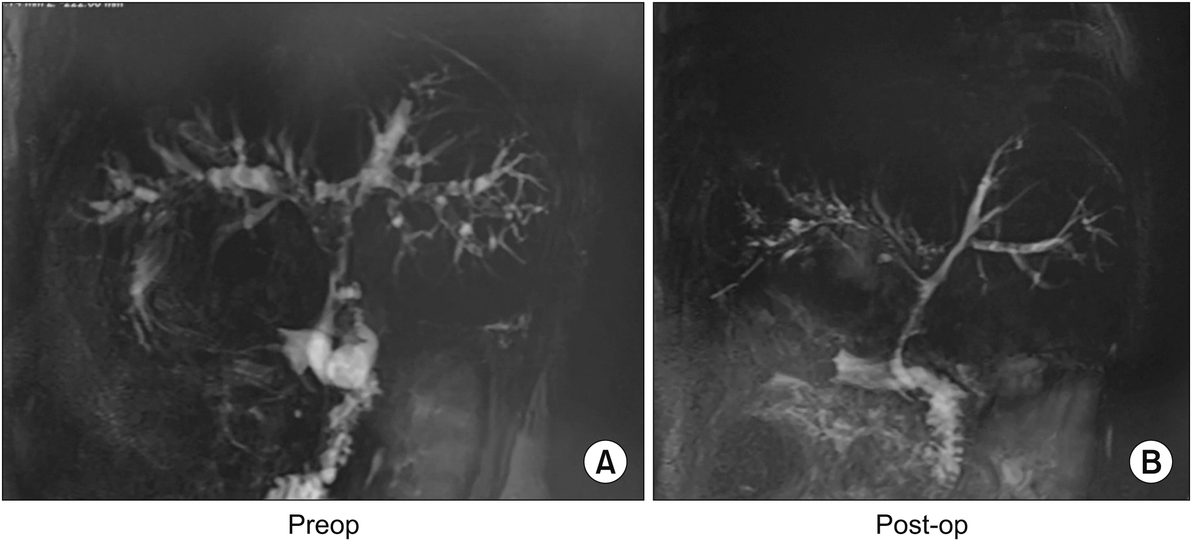
Although the use of second-stage surgery or an upfront single-stage Roux-en-Y hepaticojejunostomy for symptomatic biliary blockage has been documented in the literature, it is generally considered to be hazardous owing to severe bleeding during approaching a bile duct surrounded by large, high pressure venous collaterals. On-table mortality is not rare [53,54]. However, Cellich et al. [55] have reported excellent outcomes for three patients who had single-stage upfront biliary bypass without major bleeding or death. All three patients were asymptomatic after the treatment, indicating that biliary decompression without previous shunt surgery might be done effectively if necessary [56]. Single-stage biliary bypass, on the other hand, bears the risk of greater intraoperative blood loss and surgery time. Furthermore, according to a report by Perakath et al. [53] from South India, surgeons should be prepared to stop the surgery if significant bleeding occurs owing to the opening of collaterals. This variation, which involves executing the shunt first and then the biliary bypass with the Pringle manoeuvre to regulate portal inflow, can be utilised in patients with portal biliopathy who have biliary obstruction due to both pericholedochal collateral compression and ischaemic biliary strictures. This eliminates the need for a second operation [57]. However, in patients who have unshuntable veins, a blocked previously performed portosystemic shunt, or previous shunt is unable to relieve the biliary obstruction, bilioenteric decompression becomes necessary. We have performed 18 such procedures with one operative mortality and followed these patients between 11 months and 12 years. Symptoms returned in only two patients. These symptoms were relieved by percutaneous transhepatic dilatation (unpublished data) (Fig. 6, 7). There is a scarcity of information on the role of segment 3 bypass in portal biliopathy. A few studies have addressed it as a part of the treatment strategy [32,44,45,51,58]. However, they did not indicate what percentage of patients might benefit from this procedure. The majority of patients who had a conventional hepaticojejunostomy had type I biliary anomalies. They might be candidates for a sector 3 bypass according to a comprehensive study [44,45,51,58]. A long-term follow-up of four patients who underwent a segment 3 bypass revealed a significant rate of recurrent stone disease between seven and 40 months following surgery. Cholangioscopy through the afferent bowel loop aided biliary clearance. After an average of 8 to 9 years of follow-up, three (75%) patients were still alive and asymptomatic [19].

Devascularization

We have found that splenectomy devascularization alone in patients without ‘shuntable’ veins not only treats variceal bleeding and hypersplenism, but also lowers the pressure in the pericholedochal collaterals (reducing portal inflow and pressure). It might help those with symptomatic PCC [45].

Liver transplantation

A liver transplantation is another uncommon surgical option for patients with end stage liver disease due to PCC [52,55]. It is used for those who are more concerned with PCC than hypersplenism or variceal bleed. If the porto-mesenteric thrombosis is significant, the portal inflow to the graft will be via the systemic circulation as a cavoportal hemi-transposition or a reno-portal inflow. Unfortunately, this therapy does not decompress the splanchnic bed. Therefore, patients will still experience hypersplenism and esophagogastric varices.

CONCLUSIONS

Extrahepatic portal venous obstruction may result in PCC, which is distinguished by particular vascular, biliary, and ancillary features. The majority of patients with PCC are asymptomatic, with just around 20% of them experiencing symptoms of cholestatic jaundice. Patient’s age, the length of the history, the existence of gall stones, and the presence of CBD stones are all major risk factors for symptomatic PCC. Direct ERC was formerly the gold standard for delineation of the anatomy. However, non-invasive methods with excellent diagnostic accuracy are now being used more often. For suspected instances of PCC, ultrasound should be used as the first imaging modality. For mapping biliary and vascular anomalies, MRCP with MR portography is now the preferred method. Endoscopic drainage has been recommended as a non-surgical method for treating PCC. Unfortunately, it is only effective in a few patients. According to current evidence, using a portosystemic shunt decompression as the primary therapy for PCC patients is beneficial in the vast majority of cases. It is more likely to be effective in reversing the PCC if the shunt remains patent, if the patient does not have a dominant stricture, and if the patient has not had multiple bouts of cholangitis and stent exchanges. In the absence of a shuntable vein, splenectomy and devascularization can help some patients with PCC by lowering the portal pressure. Otherwise, a difficult hepaticojejunostomy should be carefully done by experienced surgeons. In a few individuals with PCC, liver transplantation is the last resort. However, there are patients who have unshuntable veins, in whom a previously performed portosystemic shunt becomes blocked or does not relieve the predominant symptoms of biliary obstruction, in whom a bilioenteric decompression becomes necessary. The sole acknowledged indication for liver transplantation is secondary biliary cirrhosis. However, due to difficulties in establishing a portal inflow, even this may not be possible in all patients.

ACKNOWLEDGEMENTS

Authors would like to acknowledge Dr. Kanika Dhawan for his perennial support in obtaining and maintaining patient records.

Notes

FUNDING

None.

CONFLICT OF INTEREST

No potential conflict of interest relevant to this article was reported.

AUTHOR CONTRIBUTIONS

Conceptualization: RB. Data curation: RB. Methodology: RB, AY. Visualization: All authors. Writing - original draft: RB. Writing - review & editing: All authors. Supervision: AY, SN.

REFERENCES

1. Fraser J, Brown AK. 1944; A clinical syndrome associated with a rare anomaly of vena portal system. Surg Gynecol Obstet. 78:520–524.
2. Malkan GH, Bhatia SJ, Bashir K, Khemani R, Abraham P, Gandhi MS, et al. 1999; Cholangiopathy associated with portal hypertension: diagnostic evaluation and clinical implications. Gastrointest Endosc. 49(3 Pt 1):344–348. DOI: 10.1016/S0016-5107(99)70011-8. PMID: 10049418.
3. Dhiman RK, Chawla Y, Vasishta RK, Kakkar N, Dilawari JB, Trehan MS, et al. 2002; Non-cirrhotic portal fibrosis (idiopathic portal hypertension): experience with 151 patients and a review of the literature. J Gastroenterol Hepatol. 17:6–16. DOI: 10.1046/j.1440-1746.2002.02596.x. PMID: 11895549.
4. Chandra R, Kapoor D, Tharakan A, Chaudhary A, Sarin SK. 2001; Portal biliopathy. J Gastroenterol Hepatol. 16:1086–1092. DOI: 10.1046/j.1440-1746.2001.02562.x. PMID: 11686833.
5. Chawla Y, Agrawal S. 2014; Portal cavernoma cholangiopathy - history, definition and nomenclature. J Clin Exp Hepatol. 4(Suppl 1):S15–S17. DOI: 10.1016/j.jceh.2013.04.001. PMID: 25755589. PMCID: PMC4244831.
6. Dhiman RK, Saraswat VA, Valla DC, Chawla Y, Behera A, Varma V, et al. 2014; Portal cavernoma cholangiopathy: consensus statement of a working party of the Indian national association for study of the liver. J Clin Exp Hepatol. 4(Suppl 1):S2–S14. DOI: 10.1016/j.jceh.2014.02.003. PMID: 25755591. PMCID: PMC4274351.
7. Ramesh Babu CS, Sharma M. 2014; Biliary tract anatomy and its relationship with venous drainage. J Clin Exp Hepatol. 4(Suppl 1):S18–S26. DOI: 10.1016/j.jceh.2013.05.002. PMID: 25755590. PMCID: PMC4244820.
8. Northover J, Terblanche J. 1978; Bile duct blood supply. Its importance in human liver transplantation. Transplantation. 26:67–69. DOI: 10.1097/00007890-197807010-00017. PMID: 97825.
9. Furukawa H, Iwata R, Moriyama N, Kosuge T. 1999; Blood supply to the pancreatic head, bile duct, and duodenum: evaluation by computed tomography during arteriography. Arch Surg. 134:1086–1090. DOI: 10.1001/archsurg.134.10.1086. PMID: 10522852.
10. Saint JH. 1961; The epicholedochal venous plexus and its importance as a means of identifying the common duct during operations on the extrahepatic biliary tract. Br J Surg. 48:489–498. DOI: 10.1002/bjs.18004821104. PMID: 13745435.
11. Petren T. 1932; [The veins of the extrahepatic biliary system and their pathologic-anatomic significance]. Verh Anat Ges. 41:139–143. German.
12. Dhiman RK, Puri P, Chawla Y, Minz M, Bapuraj JR, Gupta S, et al. 1999; Biliary changes in extrahepatic portal venous obstruction: compression by collaterals or ischemic? Gastrointest Endosc. 50:646–652. DOI: 10.1016/S0016-5107(99)80013-3. PMID: 10536320.
13. Bayraktar Y, Balkanci F, Ozenc A, Arslan S, Koseoglu T, Ozdemir A, et al. 1995; The "pseudo-cholangiocarcinoma sign" in patients with cavernous transformation of the portal vein and its effect on the serum alkaline phosphatase and bilirubin levels. Am J Gastroenterol. 90:2015–2019.
14. Condat B, Vilgrain V, Asselah T, O'Toole D, Rufat P, Zappa M, et al. 2003; Portal cavernoma-associated cholangiopathy: a clinical and MR cholangiography coupled with MR portography imaging study. Hepatology. 37:1302–1308. DOI: 10.1053/jhep.2003.50232. PMID: 12774008.
15. Dhiman R, Singh P, Duseja A, Chawla Y, Behera A, Suri S. 2006; Pathogenesis of portal hypertensive biliopathy (PHB): is it compression by collaterals or ischemia? J Gastroenterol Hepatol. 21:A506.
16. Dhiman R, Singh P, Behera A, Duseja A, Chawla Y, Suri S. 2006; Diagnosis of portal hypertensive biliopathy (PHB) in patients with extrahepatic portal venous obstruction (EHPVO): endoscopic retrograde cholangiography versus MR cholangiography. J Gastroenterol Hepatol. 21:A507.
17. Dhiman RK, Behera A, Chawla YK, Dilawari JB, Suri S. 2007; Portal hypertensive biliopathy. Gut. 56:1001–1008. DOI: 10.1136/gut.2006.103606. PMID: 17170017. PMCID: PMC1994341.
18. Mörk H, Weber P, Schmidt H, Goerig RM, Scheurlen M. 1998; Cavernous transformation of the portal vein associated with common bile duct strictures: report of two cases. Gastrointest Endosc. 47:79–83. DOI: 10.1016/S0016-5107(98)70305-0. PMID: 9468430.
19. Chaudhary A, Dhar P, Sarin SK, Sachdev A, Agarwal AK, Vij JC, et al. 1998; Bile duct obstruction due to portal biliopathy in extrahepatic portal hypertension: surgical management. Br J Surg. 85:326–329. DOI: 10.1046/j.1365-2168.1998.00591.x. PMID: 9529484.
20. O'Donnell B, Moloney MA. 1968; Development and course of extrahepatic portal obstruction in children. Lancet. 1:789–791. DOI: 10.1016/S0140-6736(68)92231-9. PMID: 4171132.
21. Gibson JB, Johnston GW, Fulton TT, Rodgers HW. 1965; Extrahepatic portal-venous obstruction. Br J Surg. 52:129–139. DOI: 10.1002/bjs.1800520211. PMID: 14255983.
22. Dilawari JB, Chawla YK. 1992; Pseudosclerosing cholangitis in extrahepatic portal venous obstruction. Gut. 33:272–276. DOI: 10.1136/gut.33.2.272. PMID: 1541425. PMCID: PMC1373944.
23. Sarin SK, Bhatia V, Makwana U. 1992; Portal biliopathy in extrahepatic portal venous obstruction. Indian J Gastroenterol. 11(Suppl 1):A82.
24. Khuroo MS, Yattoo GN, Zargar SA, Javid G, Dar MY, Khan BA, et al. 1993; Biliary abnormalities associated with extrahepatic portal venous obstruction. Hepatology. 17:807–813. DOI: 10.1002/hep.1840170510. PMID: 8491448.
25. Bayraktar Y, Balkanci F, Kayhan B, Ozenç A, Arslan S, Telatar H. 1992; Bile duct varices or "pseudo-cholangiocarcinoma sign" in portal hypertension due to cavernous transformation of the portal vein. Am J Gastroenterol. 87:1801–1806.
26. Nagi B, Kochhar R, Bhasin D, Singh K. 2000; Cholangiopathy in extrahepatic portal venous obstruction. Radiological appearances. Acta Radiol. 41:612–615. DOI: 10.1080/028418500127345992. PMID: 11092484.
27. Sezgin O, Oğuz D, Altintaş E, Saritaş U, Sahin B. 2003; Endoscopic management of biliary obstruction caused by cavernous transformation of the portal vein. Gastrointest Endosc. 58:602–608. DOI: 10.1067/S0016-5107(03)01975-8. PMID: 14520303.
28. Dhiman R, Chawla Y, Duseja A, Chhetri D, Dilawari J. 2006; Portal hypertensive biliopathy (PHB) in patients with extrahepatic portal venous obstruction (EHPVO). J Gastroenterol Hepatol. 21:A504.
29. Llop E, de Juan C, Seijo S, García-Criado A, Abraldes JG, Bosch J, et al. 2011; Portal cholangiopathy: radiological classification and natural history. Gut. 60:853–860. DOI: 10.1136/gut.2010.230201. PMID: 21270119.
30. Valla DC, Condat B, Lebrec D. 2002; Spectrum of portal vein thrombosis in the West. J Gastroenterol Hepatol. 17(Suppl 3):S224–S227. DOI: 10.1046/j.1440-1746.17.s3.4.x. PMID: 12472940.
31. Webb LJ, Sherlock S. 1979; The aetiology, presentation and natural history of extra-hepatic portal venous obstruction. Q J Med. 48:627–639.
32. Vibert E, Azoulay D, Aloia T, Pascal G, Veilhan LA, Adam R, et al. 2007; Therapeutic strategies in symptomatic portal biliopathy. Ann Surg. 246:97–104. DOI: 10.1097/SLA.0b013e318070cada. PMID: 17592297. PMCID: PMC1899217.
33. Aguirre DA, Farhadi FA, Rattansingh A, Jhaveri KS. 2012; Portal biliopathy: imaging manifestations on multidetector computed tomography and magnetic resonance imaging. Clin Imaging. 36:126–134. DOI: 10.1016/j.clinimag.2011.07.001. PMID: 22370133.
34. Gulati G, Pawa S, Chowdhary V, Kumar N, Mittal SK. 2003; Colour Doppler flow imaging findings in portal biliopathy. Trop Gastroenterol. 24:116–119.
35. Kessler A, Graif M, Konikoff F, Mercer D, Oren R, Carmiel M, et al. 2007; Vascular and biliary abnormalities mimicking cholangiocarcinoma in patients with cavernous transformation of the portal vein: role of color Doppler sonography. J Ultrasound Med. 26:1089–1095. DOI: 10.7863/jum.2007.26.8.1089. PMID: 17646372.
36. Besa C, Cruz JP, Huete A, Cruz F. 2012; Portal biliopathy: a multitechnique imaging approach. Abdom Imaging. 37:83–90. DOI: 10.1007/s00261-011-9765-2. PMID: 21681494.
37. Tyagi P, Puri AS, Sharma BC. 2010; Balloon sweep in portal biliopathy. Gastrointest Endosc. 71:885–886. author reply 886DOI: 10.1016/j.gie.2009.08.014. PMID: 20363438.
38. Mutignani M, Shah SK, Bruni A, Perri V, Costamagna G. 2002; Endoscopic treatment of extrahepatic bile duct strictures in patients with portal biliopathy carries a high risk of haemobilia: report of 3 cases. Dig Liver Dis. 34:587–591. DOI: 10.1016/S1590-8658(02)80093-7. PMID: 12502216.
39. Kim S, Chew FS. 1988; Choledochal varices. AJR Am J Roentgenol. 150:578–580. DOI: 10.2214/ajr.150.3.578. PMID: 3257612.
40. Sharma M, Ponnusamy RP. 2009; Is balloon sweeping detrimental in portal biliopathy? A report of 3 cases. Gastrointest Endosc. 70:171–173. DOI: 10.1016/j.gie.2008.11.002. PMID: 19409559.
41. Suhocki PV, Lungren MP, Kapoor B, Kim CY. 2015; Transjugular intrahepatic portosystemic shunt complications: prevention and management. Semin Intervent Radiol. 32:123–132. DOI: 10.1055/s-0035-1549376. PMID: 26038620. PMCID: PMC4447874.
42. Borg PC, Hollemans M, Van Buuren HR, Vleggaar FP, Groeneweg M, Hop WC, et al. 2004; Transjugular intrahepatic portosystemic shunts: long-term patency and clinical results in a patient cohort observed for 3-9 years. Radiology. 231:537–545. DOI: 10.1148/radiol.2312021797. PMID: 15044746.
43. Khare R, Sikora SS, ikanth G Sr, Choudhuri G, Saraswat VA, Kumar A, et al. 2005; Extrahepatic portal venous obstruction and obstructive jaundice: approach to management. J Gastroenterol Hepatol. 20:56–61. DOI: 10.1111/j.1440-1746.2004.03528.x. PMID: 15610447.
44. Agarwal AK, Sharma D, Singh S, Agarwal S, Girish SP. 2011; Portal biliopathy: a study of 39 surgically treated patients. HPB (Oxford). 13:33–39. DOI: 10.1111/j.1477-2574.2010.00232.x. PMID: 21159101. PMCID: PMC3019539.
45. Chattopadhyay S, Govindasamy M, Singla P, Varma V, Mehta N, Kumaran V, et al. 2012; Portal biliopathy in patients with non-cirrhotic portal hypertension: does the type of surgery affect outcome? HPB (Oxford). 14:441–447. DOI: 10.1111/j.1477-2574.2012.00473.x. PMID: 22672545. PMCID: PMC3384873.
46. Dhiman R, Chhetri D, Behera A, Duseja A, Chawla Y, Singh P, et al. 2006; Management of biliary obstruction in patients with portal hypertensive biliopathy (PHB). J Gastroenterol Hepatol. 21:A505.
47. Kim HB, Pomposelli JJ, Lillehei CW, Jenkins RL, Jonas MM, Krawczuk LE, et al. 2005; Mesogonadal shunts for extrahepatic portal vein thrombosis and variceal hemorrhage. Liver Transpl. 11:1389–1394. DOI: 10.1002/lt.20487. PMID: 16237690.
48. Drews JA, Castagna J. 1976; Inferior mesorenal shunt as a second procedure for portal decompression. Surg Gynecol Obstet. 142:84–86.
49. Warren WD, Salam A, Smith RB 3rd. 1974; The meso-spleno-renal shunt procedures: a comprehensive approach to portasystemic decompression. Ann Surg. 179:791–798. DOI: 10.1097/00000658-197405000-00035. PMID: 4545091. PMCID: PMC1356077.
50. Camerlo A, Fara R, Barbier L, Grégoire E, Le Treut YP. 2010; Which treatment to choose for portal biliopathy with extensive portal thrombosis? Dig Surg. 27:380–383. DOI: 10.1159/000314610. PMID: 20938181.
51. Poddar U, Borkar V. 2011; Management of extra hepatic portal venous obstruction (EHPVO): current strategies. Trop Gastroenterol. 32:94–102.
52. Filipponi F, Urbani L, Catalano G, Iaria G, Biancofiore G, Cioni R, et al. 2004; Portal biliopathy treated by liver transplantation. Transplantation. 77:326–327. DOI: 10.1097/01.TP.0000101795.29250.10. PMID: 14743009.
53. Perakath B, Sitaram V, Mathew G, Khanduri P. 2003; Post-cholecystectomy benign biliary stricture with portal hypertension: is a portosystemic shunt before hepaticojejunostomy necessary? Ann R Coll Surg Engl. 85:317–320. DOI: 10.1308/003588403769162422. PMID: 14594535. PMCID: PMC1964328.
54. Agarwal AK, Gupta V, Singh S, Agarwal S, Sakhuja P. 2008; Management of patients of postcholecystectomy benign biliary stricture complicated by portal hypertension. Am J Surg. 195:421–426. DOI: 10.1016/j.amjsurg.2007.03.013. PMID: 18304509.
55. Cellich PP, Crawford M, Kaffes AJ, Sandroussi C. 2015; Portal biliopathy: multidisciplinary management and outcomes of treatment. ANZ J Surg. 85:561–566. DOI: 10.1111/ans.12436. PMID: 24237891.
56. Hajdu CH, Murakami T, Diflo T, Taouli B, Laser J, Teperman L, et al. 2007; Intrahepatic portal cavernoma as an indication for liver transplantation. Liver Transpl. 13:1312–1316. DOI: 10.1002/lt.21243. PMID: 17763385.
57. D'Souza MA, Desai D, Joshi A, Abraham P, Shah SR. 2009; Bile duct stricture due to caused by portal biliopathy: treatment with one-stage portal-systemic shunt and biliary bypass. Indian J Gastroenterol. 28:35–37. DOI: 10.1007/s12664-009-0010-7. PMID: 19529903.
58. Gupta V, Chandra A. 2013; Portal biliopathy: is splenectomy with devascularization really effective? HPB (Oxford). 15:559. DOI: 10.1111/j.1477-2574.2012.00606.x. PMID: 23750497. PMCID: PMC3692027.

Fig. 1
Normal anatomy of biliary tract. Cited from the article of Ramesh Babu et al. (J Clin Exp Hepatol 2014;4(Suppl 1):S18-S26) [7]. V, segment V duct; VII, segment VII duct; VIII, segment VIII duct; RASD, right anterior sectoral duct; RPSD, right posterior sectoral duct; RHA, right hepatic artery; LHA, left hepatic artery; CD, cystic duct; CBD, common bile duct; CHA, common hepatic artery; GDA, gastroduodenal artery; SV, splenic vein; SMV, superior mesenteric vein; PD, pancreatic duct; PSPDA, postero-superior pancreatico-duodenal artery.
ahbps-26-4-298-f1.tif
Fig. 2
Normal arterial supply of the biliary tract. Cited from the article of Ramesh Babu et al. (J Clin Exp Hepatol 2014;4(Suppl 1):S18-S26) [7]. RASA, right anterior sectoral artery; RPSA, right posterior sectoral artery; IV-A, segment IV artery; CA, cystic artery; RHA, right hepatic artery; LHA, left hepatic artery; A, artery; PSPDA, postero-superior pancreatico-duodenal artery; GDA, gastro-duodenal artery; CHA, common hepatic artery.
ahbps-26-4-298-f2.tif
Fig. 3
Venous drainage of the biliary tract (anterior & posterior views). Cited from the article of Ramesh Babu et al. (J Clin Exp Hepatol 2014;4(Suppl 1):S18-S26) [7]. V, segment V; IV, segment IV; III, segment III; RASD, right anterior sectoral duct; RPSD, right posterior sectoral duct; LHD, lef t hepatic duct; RHD, right hepatic duct; CHD, common hepatic duct; V., vein; RGV, right gastric vein; CBD, common bile duct; PSPDV, postero-superior pancreatico-duodenal vein; SMV, superior mesenteric vein; D2, 2nd part of duodenum; PV, portal vein; LPV, left portal vein; RPV, right portal vein; LHD, left hepatic duct; RGV, right gastric vein.
ahbps-26-4-298-f3.tif
Fig. 4
Contrast-enhanced computed tomography abdomen of a patient with extra hepatic portal venous obstruction showing col laterals and cavernoma formation resulting in biliary dilatation. (A, C) Showing portal cavernoma formation with compression over the lower common bile duct. (B, D) Showing intra-hepatic biliary radicle dilatation.
ahbps-26-4-298-f4.tif
Fig. 5
Magnetic retrograde cholangiopancreatography. (A, C) Showing portal cavernoma with compression resulting in biliary stricture (arrow) with triple phase computed tomography scan. (B) Showing similiar feature (arrow head).
ahbps-26-4-298-f5.tif
Fig. 6
Intraoperative photos from a primary biliary decompression being done. (A) Common hepatic duct opened just at hepatic bifurcation with stents in situ seen. (B) Large collateral being tied between ligatures. (C) Hepatic duct near bifurcation ready for hepaticojejunostomy (arrow).
ahbps-26-4-298-f6.tif
Fig. 7
Comparative magnetic resonance cholangiopancreatography pictures showing preoperative (extra hepatic portal venous obstruction with portal cavernoma with upstream bi liary di latat ion and hepatolithiasis) (A) and postoperative (shunt surgery followed by biliary bypass showing resolution of biliary dilatation and patent anastomosis) (B).
ahbps-26-4-298-f7.tif
TOOLS
Similar articles