This article has been
cited by other articles in ScienceCentral.
Abstract
Collagenous gastroduodenitis is a rare gastrointestinal disease diagnosed histologically by subepithelial collagen deposition in the lamina propria. Its clinical presentation is diverse. The authors encountered a 17-year-old female patient who complained of epigastric pain. Endoscopy revealed several deep ulcers in the gastric body. The gastric mucosa around the ulcer showed diffuse fine nodularity in the shape of cobblestones with open-type atrophy. The duodenal mucosa showed nodular lesions similar to those of the gastric mucosa. The gastric ulcer healed completely with proton pump inhibitor treatment. The patient was followed up, showing no remarkable mucosal change of stomach or duodenum for several years. Collagenous gastroduodenitis was diagnosed by repeated histologic examinations. This paper reports a rare case of chronic collagen gastritis with deep gastric ulcer and its long-term clinical progress.
Go to :

Keywords: Collagen, Gastritis, Ulcer
INTRODUCTION
Collagenous gastritis (CG) is a rare chronic gastritis disorder characterized by thick collagenous subepithelial bands associated with gastric mucosal inflammation.
1 Fifty cases have been reported thus far. Collagenous gastroduodenitis (CGD) or bulbitis is very rare, with only four cases reported.
2-6 The endoscopic findings of CG are diverse, including mucosal erythema, nodularity, and ulceration. This paper reports an endoscopic presentation of CGD in the form of multiple gastric ulcers.
Go to :

CASE REPORT
A 17-year-old female visited the hospital complaining of severe epigastric pain for several weeks. She had no symptoms or signs of diarrhea, melena, or malabsorption, nor did she have any history of allergies. She was 165.5 cm tall (85th percentile) with a weight of 54.5 kg (50th percentile). She had never taken any medication before the onset. There were no abnormal findings in the complete blood cell count or blood chemistry. At that time, her hemoglobin level was 12.1 mg/dL, hematocrit 34%, white blood cell count 4,010/μL, platelet count 165,000/μL, total protein 6.7 g/dL, and albumin 4.1 g/dL. Her nutritional status was normal.
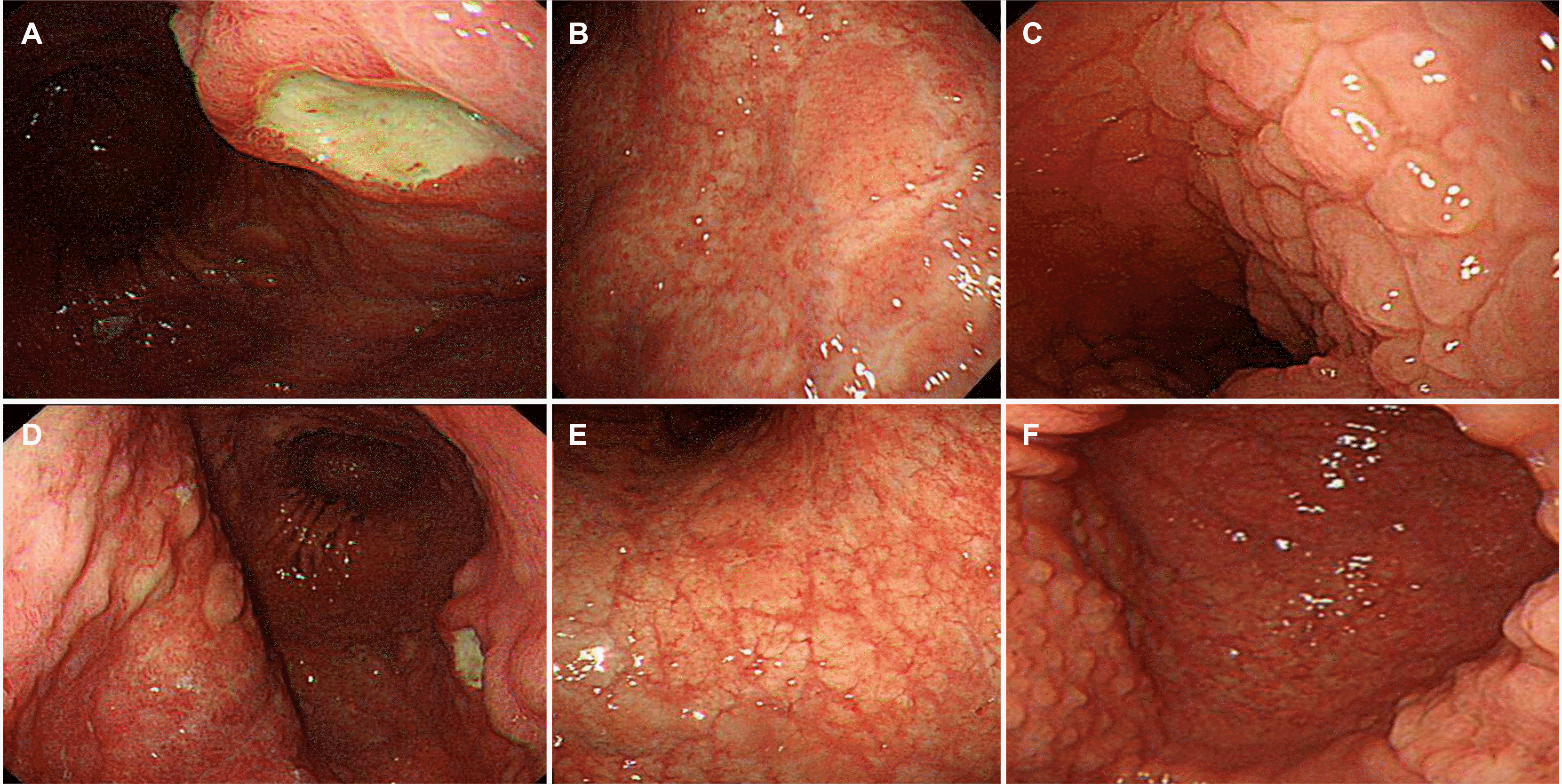
Endoscopy revealed several deep ulcers with an irregular elevated margin in the stomach. The gastric mucosa showed round and flat nodularity, which had a cobblestone appearance with an open-type atrophy. The normal mucosa around the nodule or atrophy lesion looked like a peninsula. The duodenal mucosa also exhibited nodular lesions similar to the gastric mucosa (
Fig. 1). CLO tests and silver staining were negative for a
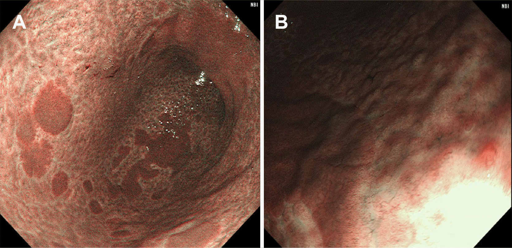
Helicobacter infection. The serum gastrin, pepsinogen I, and pepsinogen II were within the normal ranges. Magnifying endoscopy with narrow-band imaging showed that normal capillary networks were lost in the depressed nodular mucosa. The contrast between nodular mucosal lesions and normal mucosa in the form of a peninsular was more pronounced in narrow band imaging than in white light endoscopic imaging (
Fig. 2). These deep ulcers healed after several weeks of treatment with proton pump inhibitors (PPIs). On the other hand, the diffuse nodular and atrophic changes in the stomach persisted. She received PPI treatment several times for abdominal pain. She was diagnosed with collagenous gastroduodenitis through multiple endoscopy procedures and repeated endoscopic biopsies.
 | Fig. 1Endoscopic images of collagenous gastroduodenitis, showing (A) several deep active ulcers in the stomach, (B) diffuse mucosal nodularity of the gastric body in the shape of cobble stones, (C) normal mucosa around the nodule or atrophy lesion looks like a peninsula, and (D) an open-type atrophy in the stomach. (E) The duodenal mucosa exhibiting nodular lesions similar to those of gastric mucosa. (F) Gastric ulcers were healed after several weeks of treatment with proton pump inhibitors. 
|
 | Fig. 2Magnifying endoscopic findings with narrow band imaging. (A) The contrast between nodular mucosal lesions and normal mucosa in the form of peninsular is more pronounced in narrow band imaging than in white light endoscopic imaging. (B) Loss of normal capillary networks is observed in the depressed nodular mucosa. 
|
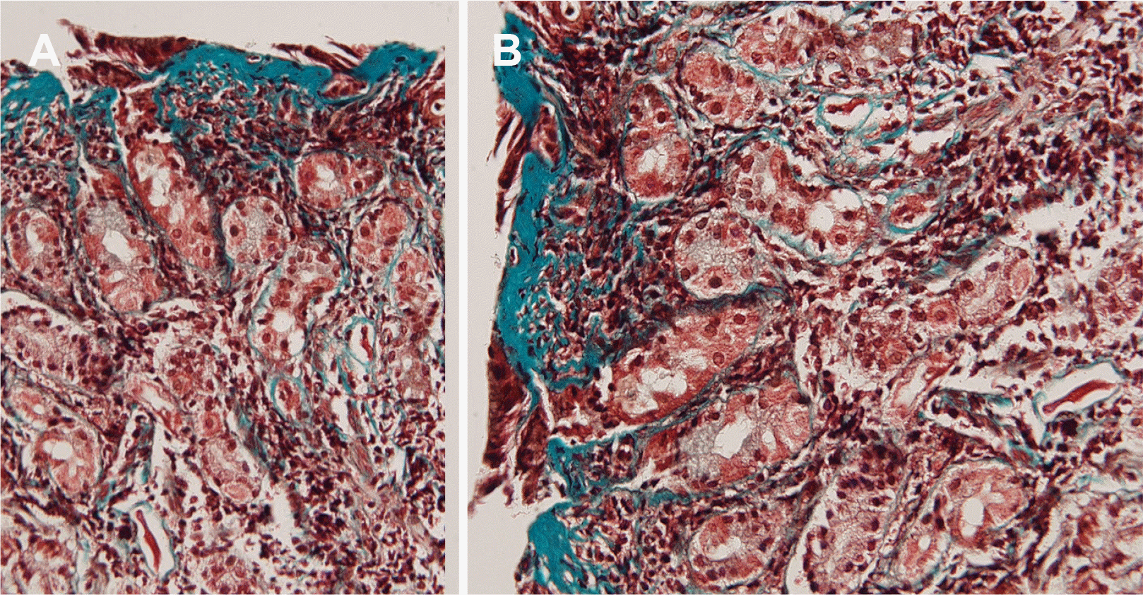
A gastric mucosal biopsy was performed for both gastric ulcer lesions and nodular lesions. A histological examination of the stomach and duodenal bulb showed the infiltration of inflammatory cells in the lamina propria and severely atrophied epithelium. Masson trichrome staining revealed markedly thickened subepithelial collagen bands. These collagen bands were found in the glands of the gastric mucosa and duodenum (
Fig. 3). She has been followed up for more than four years. In the meantime, she has experienced severe epigastric pain intermittently and has taken PPIs. During the follow- up period, endoscopy was performed annually. The gastric ulcer did not recur. On the other hand, nodular mucosal lesions in the stomach and duodenum continued without significant improvement.
 | Fig. 3(A, B) The subepithelial layer is thickened by collagen deposition and stained blue with Masson's trichrome stain (×200). 
|
Go to :

DISCUSSION
CG is a rare form of chronic gastritis defined histologically by a thick subepithelial collagenous band in the lamina propria. Colletti and Trainer
1 first reported a 15-year-old girl suffering from recurrent abdominal pain and gastrointestinal bleeding in 1989. Approximately 60 cases of collagenous gastritis have been reported since 1989.
4 CGD is more uncommon. CG showed a bi-modal age distribution, with the first peak in patients aged <20 years and the second peak primarily in females aged >60 years. In children, it usually occurs before adolescence. It is approximately 1.6-4 times more frequent in females than in males.
5-7 CG is commonly classified into pediatric and adult types. The pediatric type of CG mainly involves the stomach. Its symptoms are abdominal pain and anemia. The adult type of CG involves the small and large bowel. It is associated with collagenous colitis in elderly adults presenting with chronic watery diarrhea. The adult and pediatric types of CG should not be classified as independent diseases but should be considered similar diseases on a continuous spectrum. Recent reports have indicated no clinical or pathological differences between pediatric and adult types of CG. The clinical presentations are related to the region of the gastrointestinal tract involved. Various symptoms of gastrointestinal bleeding (melena, black stools, blood-tinged vomiting, positive for fecal occult blood or ulcer) have been reported in CG patients.
8,9 Multiple case reports have described some association between autoimmune diseases and CG, the most common being celiac disease.
10,11 Most cases of CG had no relationship with infections, such as
H. pylori, autoimmune disorders, or past medications. This case is the second case of pediatric collagenous gastroduodenitis in Asia. The difference between the two patients was that collagen inflammation in the first patient invaded the stomach and duodenum, but also the colon, while this patient did not have colon inflammation.
The nodularity of the gastric mucosa is the most characteristic endoscopic finding of collagenous gastritis. In a review of 60 CG patients, 32 showed endoscopic mucosal nodularity.
4 Mucosal nodules in collagenous gastritis are more irregular in size, spreading broadly throughout the stomach body and antrum, which look like cobblestones.
4 There was a smooth islet-shaped normal area around the nodular lesion in the present case. Atrophic changes in the gastric mucosa were similar to those shown for depressed lesions on endoscopy. Few CGs with gastric ulcers have been reported.
There are few reports on magnifying endoscopy with narrow- band imaging of CG. Kawamura et al.
12 reported that a tubular gastric micromucosal pattern is representative of the infiltration of inflammatory cells and degeneration of the surface epithelium, while an obscure surface structure with irregular, narrowed, and coiled subepithelial capillaries is compatible with moderate to severe mucosal atrophy. The magnifying endoscopy with narrow-band imaging of the present case also showed a similar pattern. The depressed mucosal area had an absent surface pit pattern with a loss of normal capillary vessels.
The cause and pathogenesis of CG are poorly understood. Collagen deposition is believed to be due to underlying injury and chronic inflammation, which could cause abnormal responses. The pathological findings of nodular mucosa showed the deposition of collagen bands just under the mucoepithelial lesion. In addition, she had collagenous duodenitis in part of the bulbs. Collagen depositions were observed in fine fading nodularity/granularity. Her mucosal islands were pathologically normal.
No standard treatment has been identified. Multiple therapies have been used, including PPIs, corticosteroids, sucralfate, histamine-2 receptor antagonists, and 5-aminosalicylates. Some case reports of patients with CG have shown clinical improvement with budesonide, which has been used to treat collagenous deposition in the colon. In a recent large, retrospective, open-label noncontrolled study of CG patients treated with topical targeted budesonide, 89% showed a complete or partial clinical response, and 88% showed a complete or partial histologic response. If the chief complaints of CC cases are diarrhea and weight loss, they could be relatively improved by steroid therapy. Anemic patients are prescribed iron supplements. Medications, such as histamine 2 receptor antagonists and PPIs are also used to protect the damaged mucosa from gastric acid. Although these therapies effectively improve anemia and epigastric pain, they do not contribute to the changes in the endoscopic and pathological findings. Only one patient reported by Hara and Fujisawa showed natural improvement endoscopically and histologically.
13 Steroid treatment was not performed for the present patient. This is because the patient was a growing adolescent. In addition, her collagen gastritis was chronic with continuous inflammation. Moreover, the period and dosage of steroid administration were not determined in previous reports.
This paper reports a rare collagenous CGD of pediatric types presented with epigastric pain and gastric ulcers. It showed the typical endoscopic and histologic findings of CGD. The present case was classified as the nodular type with a normal peninsula mucosa in the corpus to the antrum and duodenum. This case was initially not diagnosed histologically and was later diagnosed as CGD after repeated biopsies. Although endoscopic findings indicated either H. pylori gastritis or lymphofollicular gastritis, the histology in the 1st endoscopy revealed only chronic inflammation and subepithelial collagenous bands. Masson’s trichrome staining was conducted retrospectively later. Because the endoscopic findings of CG or CGD gastritis are characteristic, it is essential to perform repeated biopsies. If CG is suspected endoscopically, even a histological diagnosis is difficult. In summary, this paper presents a case of a pediatric type of CGD showing characteristic mucosal changes with a rare form of multiple gastric ulcers.
Go to :


 PDF
PDF Citation
 Print
Print


 XML Download