

 PDF
PDF Citation
 Print
Print


서 론
방선균증(Actinomycosis)은 일반적으로 방선균(Actinomyces israelii)에 의해 발생하는 감염성 질환으로 두경부, 흉부, 복부 골반 및 중추신경계 등을 포함한 신체 여러 부위에서 발생한다[1]. 또한 방선균증은 대부분 두경부에서 발생하며, 방선균은 구인두나 위장관, 여성 비뇨생식기 등에서 정상균주로 존재하지만 외상이나 비내 수술, 방사선 치료, 불결한 구강위생, 과도한 음주 등의 원인으로 점막손상을 야기하여 이로 인한 기회감염으로 나타나기도 한다[1-3]. 대표적인 임상 증상에는 점액농성 분비물, 두통 및 안면 통증이 있고, 이 외에 방선균의 침범부위에 따른 후각상실, 악취, 비내 출혈 등 다양한 증상을 보일 수 있다[4]. 결절 외 natural killer (NK)/T세포 림프종(extranodal NK/T cell-lymphoma)은 비호지킨 림프종(non-Hodgkin lymphoma)의 아형으로 국내에서 발생하는 전체 림프종의 약 8% 정도를 차지하고 있으나 국소적으로 비강이나 비인두강에서 발생하는 림프종에서는 약 45% 정도를 차지하는 높은 비중을 보인다[5-7].
저자들은 코막힘을 주소로 내원한 환자에서 비인두 내 방선균증이 동반되어 비부비동 NK/T 세포 림프종(extranodal NK/T cell lymphoma, nasal type; NNKTL)이 늦게 진단 된 증례를 경험하였기에 문헌 고찰과 함께 보고하고자 한다.
Go to : 
증 례
60세 여자 환자는 5일 전부터 발생한 양측 코막힘 및 점액 농성의 비루를 주 호소로 외래에 내원하였다. 이 환자는 고혈압 외에 구강에 특이 외상력이나 수술력은 없었다. 외래에서 시행한 비내시경 검사상 비강 내 특이소견은 없었으나 비인두에 점액농성비루와 부종을 동반한 비인두점막 괴사와 구인두의 발적 소견이 관찰되었다(Fig. 1). 급성 부비동염 및 급성 인후염 진단하 일주일간 퀴놀론계 항생제를 사용하였으나 증상이 호전되지 않아 추가적으로 혈액검사, 영상의학 검사 및 비내시경을 이용한 비인두궤양 부분에 조직검사를 진행하였다(Fig. 2A). 혈액 검사 결과 콜레스테롤 수치의 경미한 상승 외에 백혈구 및 C-반응성단백은 정상범위였고, 에이즈, 매독 등을 확인하기 위해 면역혈청검사를 진행하였지만 특이소견은 없었으며, 항호중구세포질항체(anti-neutrophil cytoplasmic antibody, ANCA) 검사에서도 음성으로 확인되었다. 부비동 전산화단층촬영 및 자기공명영상에서 양측으로 광범위한 비인두벽 종대를 동반한 인두 뒤 공간 림프절 종대 소견을 보였다(Fig. 3). 병리조직학적 검사에서는 곰팡이 균사를 가지는 방선균증이 관찰되었으나(Fig. 4), 악성종양 소견은 관찰되지 않았기 때문에 비인두 방선균증으로 진단하에 amoxicillin/clavulanate 항생제 치료를 시작하였다. Amoxicillin/clavulanate 투여 한 달 후, 점액농성비루는 소실되고 비인두점막 괴사의 호전 소견을 보여 기존 항생제 치료를 지속하며 경과 관찰하였다(Fig. 2B). 그러나 한 달 후 환자는 구강 통증을 주 호소로 내원하였고, 비내시경 및 구강 진찰결과 경구개 및 연구개의 궤양소견이 새롭게 발생되어 조직검사 및 부비동 전산화단층촬영, 자기공명영상촬영을 시행하였다(Fig. 2C and D). 두 번째 얻어진 병리검체에서 면역혈청검사를 시행하였고, 그 결과 CD56 약양성 및 CD3, Epstein-Barr virus (EBV) 제자리 부합법 반응(in situ hybridization) 검사에서 양성을 보였고, CD20 검사에서는 음성을 보였다(Fig. 5). 그리고 재시행한 혈액검사에서는 백혈구 3000×103/uL, 혈색소 10.0 g/dL, ANCA 음성이었고, 부비동 전산화단층촬영 및 자기공명영상에서 지속적으로 양측 비인두벽과 인두수축근을 침범하는 광범위한 염증성 침범소견이 확인되었다. 상기 검사결과들을 종합하여 비부비동 NK/T세포 림프종(NNKTL)에 부합된다고 재진단하여 방사선 종양의학과와 혈액종양내과와의 협진을 계획하였다. 그러나 환자는 계획된 방사선 및 항암치료를 시작하기 직전에 호흡곤란과 혈압저하로 응급실을 내원하게 되었다. 혈액검사를 통해 C-반응성단백 및 젖산 수치 상승과 함께 전혈구감소증, 간기능 및 신기능 부전 소견을 보였다. 이는 NNKTL에 의한 이차적 감염 및 기존 감염성 병변의 급성악화로 인한 패혈증 쇼크로 진단되었다. 그리고 내시경 및 목 전산화단층촬영 검사에서 비인두 및 구인두 벽의 광범위한 괴사를 동반하는 심한 부종과 후두부종이 확인되었고, 이로 인한 협착음이 청진되어 즉시 기관절개술을 시행하였다(Fig. 6). 환자의 혈압조절과 염증치료를 위해 집중치료실로 입원하여 광범위 항생제 및 승압제를 지속적으로 투여하던 중 원위부의 손가락과 발가락의 괴사가 발생하여 계획된 항암치료는 시행하지 못하였다. PET-CT 후 비호지킨 림프종 Ann Arbor staging에 따른 Stage II로 최종 진단하여 세기조절방사선치료(54 Gy in 30 fraction)만 단독으로 시행하였으며, 이후 4년이 지난 현재까지 환자에게 재발 및 특이 증상이 없이 경과 관찰 중에 있다(Fig. 7).
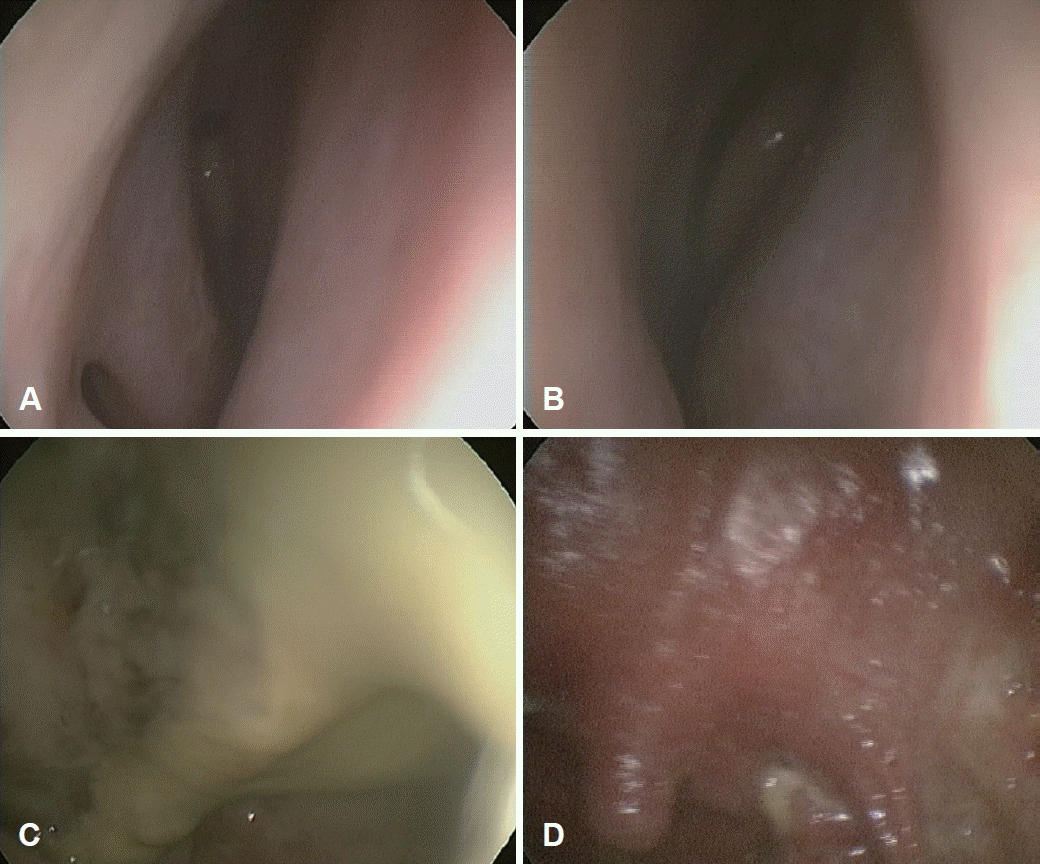 | Fig. 1.Endoscopic findings of nasal cavities, nasopharynx and oropharynx at the patient’s fist visit. A and B: There were no specific findings in both nasal cavities. C: Necrotic swollen mucosa with purulent discharge was found in nasopharynx. D: Oropharynx showed injected mucosa with some exudates.
|
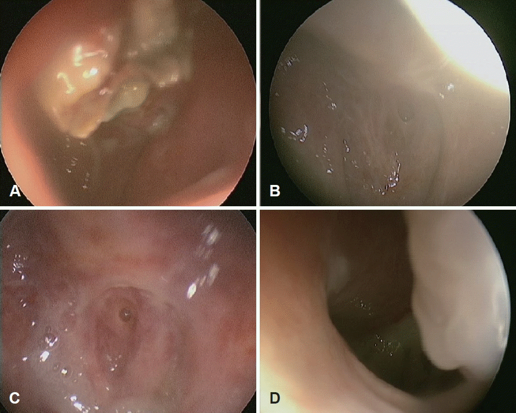 | Fig. 2.Endoscopic findings of nasopharynx at the patient’s serial visits. A: At her second visit, there were some necrotic granulations on the nasopharynx after medication of antibiotics and steroid. B: Inflammation of the nasopharynx was much improved after long term medication of amoxicillin/clavulanate. C: Purulent discharge was found in swollen nasopharynx at her 5th visit. D: Nasopharynx showed swollen mucosa with some exudates before radiation therapy.
|
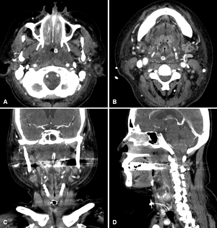
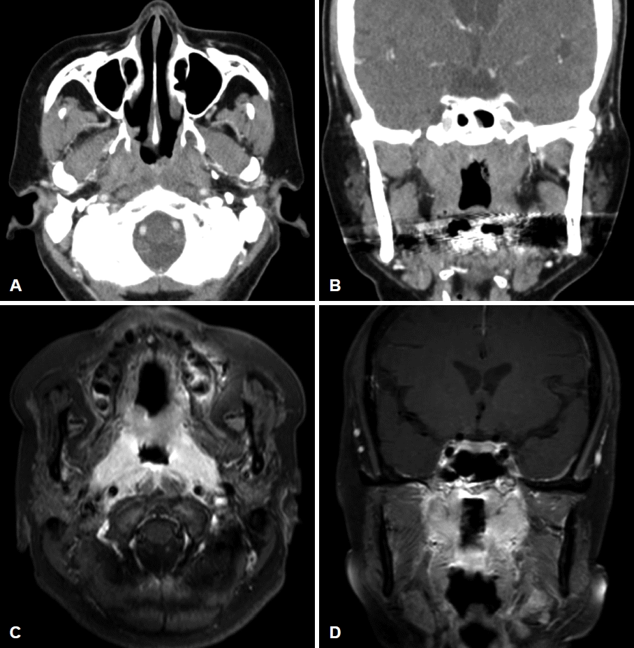 | Fig. 3.CT and MRI images of a 62-year-old female who presented with nasal obstruction and purulent rhinorrhea. A and B: Axial and coronal enhanced paranasal sinus CT demonstrated diffuse infiltrative wall thickening of bilateral nasopharynx and focal invasion of right posterior nasal cavity and both pterygopalatine fossa (PPF). C and D: MRI also demonstrated that diffuse infiltrative enhancing lesions in the nasopharynx involving pharyngeal muscles, preveretbral space, both pterygoid bones, medial part of PPF, and encasement of both distal cervical Internal caroid artery.
|
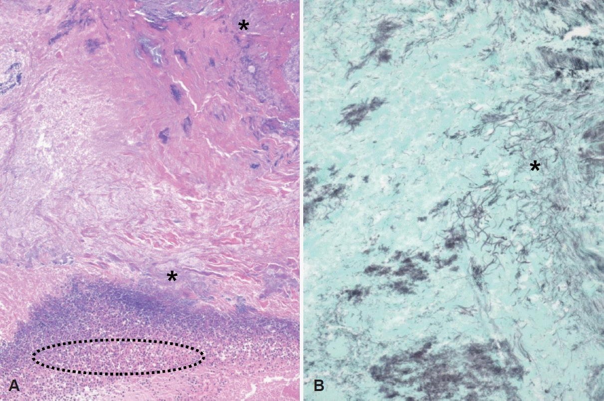 | Fig. 4.Histopathologic findings of actinomycosis. A: Microorganism colonies composed of basophilic radiating filaments (asterisk), which is referred to as sulphur granules, are noted with the neutrophilic infiltration in hematoxylin & eosin staining (dotted circle) (×100). B: Gomori methenamine silver stain highlights thin filamentous bacteria with morphologic features suggestive of Actinomyces species (×400).
|
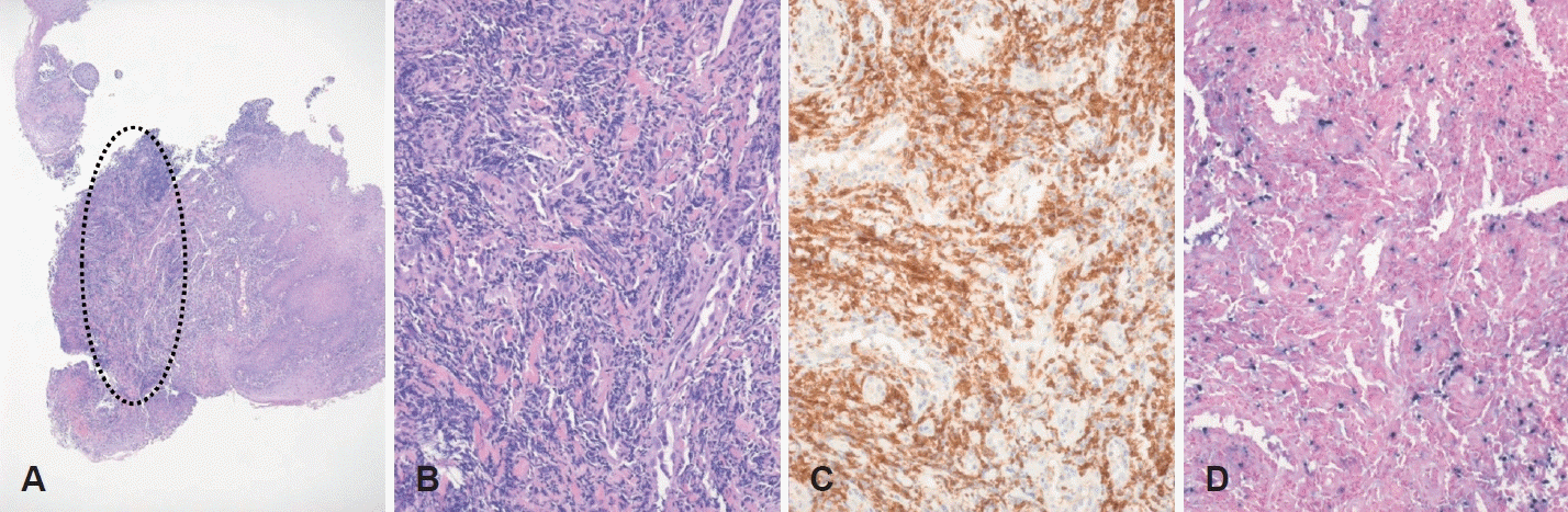 | Fig. 5.Pathological and immunohistochemical findings. A and B: Oropharynx in anterior pillar area shows atypical lymphoid infiltration in submucosal area (A: dotted circle, hematoxylin & eosin [H&E] staining, ×40), consisting of variable pleomorphic cells with small amount of cytoplasm and irregular and folded nuclear contours (B: H&E staining, ×200). C: These atypical lymphoid cells reveal the diffuse immunoreactivity for CD3 (×200). D: In-situ hybridization for Epstein-Barr virus demonstrates reactivity in most tumor cells (H&E staining, ×200).
|
 | Fig. 6.Neck CT scan findings when the patient visited emergency room complained dyspnea. As soon as she arrived at emergency room, she had undergone emergency tracheostomy to secure her airway. A and B: Axial CT scans showed that combined severe edema and necrosis of nasopharynx and orophayngeal wall, and associated diffuse severe inflammatory edematous swelling in the larynx. C and D: Coronal CT scans also showed the diffuse upper airway obstruction and deep neck infection around the pharyngeal mucosal space and cellulitis in the face and upper neck without localized abscess formation.
|
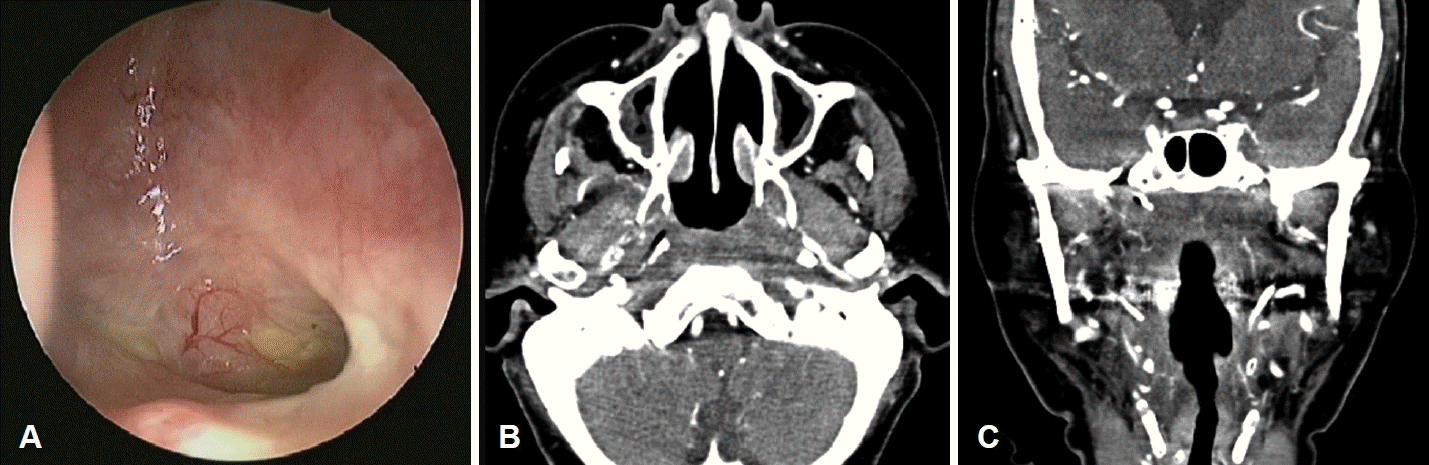 | Fig. 7.Endoscopic findings and neck CT scan findings at her latest visit. A: Some secretions were found in nasopharynx. There was not special abnormality of nasopharyngeal mucosa. B and C: Axial and coronal enhanced paranasal sinus CT demonstrated stable residual less enhancing lesion in the nasopharynx and adjacent prevertebral space. Residual nasopharyngeal mucosa was revealed to be inflammatory lesion without tumor cells by endoscopic biopsy.
|
Go to :
고 찰
방선균의 절반 이상은 두경부에서 발생하지만 방선균은 실모양 형태의 혐기성 그람 양성 간균으로 공기의 통로인 비강 및 비인두 부위 침범은 드물다[2]. 방선균의 진단은 임상 검사와 병리조직학적 검사를 통해 이루어지며 진단 시 4-6주의 고용량의 정맥 penicillin 및 6-12개월 경구 penicillin 치료가 권장된다[1,2]. 주변 조직으로 진행에 따라 침습성과 비침습성으로 분류되고, 비침습성 방선균은 특징적인 전산화단층 촬영의 소견은 없으나 연조직 음영을 동반한 국소적인 석회화 병변이 관찰되거나 림프절 종대가 동반되는 경우도 보고된 바 있다[2,8]. 침습성 방선균증은 만성 화농성 육아종성 감염을 일으키며 전산화단층촬영에서 광범위한 침윤, 괴사 및 뼈 파괴 소견을 보인다[2]. 따라서 침습성 방선균은 치료기간이 길고 광범위하게 침윤하는 특성으로 인해 악성종양, 결핵, 베게너 육아종, 진균감염 등과의 감별진단이 필요하다[2]. 앞서 언급한 것과 같이 방선균증은 임상적인 증상이나 영상의학적 소견, 그리고 수술적 소견으로만 진단하기는 어려운 상황이며 조직 검사를 통해 확진된다[4]. 본 증례의 경우 초진 때 시행한 비내시경 검사에서 비인두 점막 및 구인두의 괴사 소견을 보였고, 조직검사상 방선균이 관찰되었기에 방선균의 비강 침범소견은 드물지만 병리조직검사를 토대로 비인두 방선균증으로 진단하에 amoxicillin/clavulanate 항생제 치료를 시행하였다. 치료 후 병변의 호전 소견은 있었으나 두 달 간의 항생제 치료에도 불구하고 구강 내 새로운 병변이 생기는 등 질병이 다시 진행되는 소견을 보여, 타 질환과의 감별하기 위해 재조직검사 및 면역혈청검사를 시행한 결과 NNKTL으로 재진단되어 방사선치료를 진행하였다.
NNKTL은 비호지킨 림프종의 아형으로 비강에서 림프절 외 침범을 하며 EBV의 잠복감염과 관련 있다[7]. 면역조직화학적으로 CD56, CD2, 세포질 CD3 및 EBV-encoded RNA에 대한 제자리 부합화 검사에서 양성을 보이고 표면 CD3 검사에서는 음성을 보이는 특징이 있다[6]. NNKRL의 병소와 관련하여 한 번의 조직검사는 괴사, 염증, 미생물의 존재 여부만을 확인할 수 있기 때문에 병소의 여러 부위에서 수 차례의 조직검사가 필요할 수 있다[9]. NNKTL의 정중선 비강종괴에 대한 감별진단에는 편평세포 암종, 소타액선 종양, 베게너 육아종증 및 진균감염이 포함되고[10], 전산화단층촬영 검사에서는 종양이 양쪽 비강의 침범과 비강벽을 따라 광범위한 연조직을 침범하는 소견을 보인다[11]. 임상증상으로는 코막힘부터 코피, 비강 내 종괴 등 다양한 증상으로 나타난다. NNKTL이 다른 부위의 원발성을 배제하기 위해 PET-CT 검사도 반드시 필요하다[6,11]. 본 증례에서는 첫번째 조직검사를 통해 방선균만 확진되었고, 방선균증에 대한 amoxicillin/clavulanate 항생제 치료를 시작한 후 이에 대한 효과가 초반에 확연히 나타났기 때문에 다른 병인을 확인하기 위한 재조직검사에 대한 필요성을 간과하게 되었다. 이로 인해 동반된 NNKTL의 진단과 치료가 지연 되어 환자의 상태가 다소 위험해지기도 하였으나 방사선 치료 후 환자는 4년째 재발하지 않는 상태이며, 현재까지 경과 관찰을 진행하고 있다. 따라서 진단된 비인두 방선균증의 치료 시 지속적인 약물투여 중임에도 불구하고 재발되는 염증 및 장기간의 비특이적 증상을 호소하는 경우에는 악성종양 등 타질환과의 감별진단을 반드시 고려해야 하며, 이를 위해 반복적인 조직검사 및 면역조직화학 염색을 적극적으로 시행해야 하겠다.
Go to :
 XML Download