Abstract
A paranasal sinus mucocele is an epithelial-lined, mucus-containing sac that completely fills the sinus and forms an expandable cystic structure. It most commonly affects the frontal and ethmoidal sinuses, and rarely the maxillary and sphenoid sinuses. Orbital displacement or external disfigurement resulting from the expansion of the frontal or ethmoid sinuses is common; however, facial asymmetry caused by maxillary bone remodeling is rare. We describe a case of large maxillary sinus mucocele that destroyed the maxillary sinus bony wall, resulting in notable left cheek swelling and disfigurement, and review the relevant literature.
A paranasal sinus mucocele occurs when the duct allowing the natural drainage of mucus is blocked, resulting in a mucus-filled epithelial sac that expands within the paranasal sinus [1]. Commonly occurring in the frontal and ethmoid sinuses, mucocele cases involving the maxillary sinuses are rare [1,2]. Orbital displacements or cranial nerve symptoms may appear when frontal sinus mucoceles or sphenoid sinus mucoceles occur respectively. However, if a maxillary sinus mucocele becomes large enough to infiltrate bone, it can cause ophthalmic and dental symptoms, or, in rare cases, frank swelling and disfigurement of the mid-face [1,3]. In this case report, an 86-year-old female presenting with a large bulging mass in her left cheek underwent a surgical procedure for removal via the Caldwell-Luc approach. Biopsy of the mass confirmed the diagnosis of a maxillary sinus mucocele through histopathological examination. We report this rare case along with a literature review.
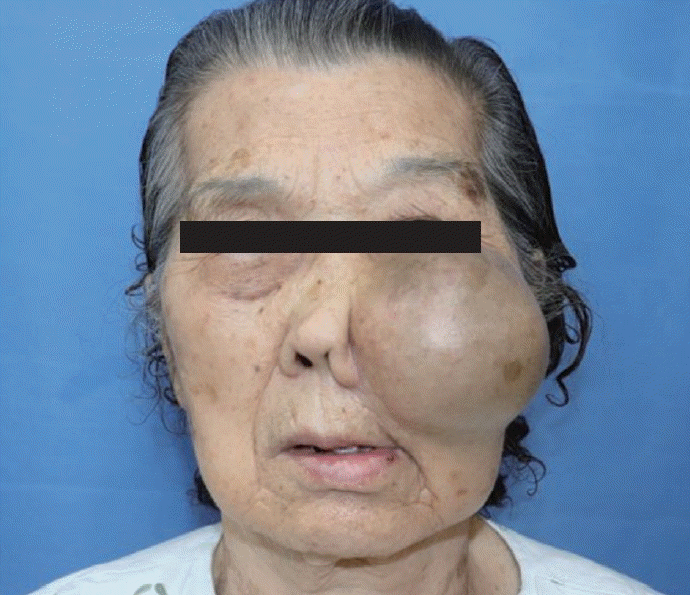
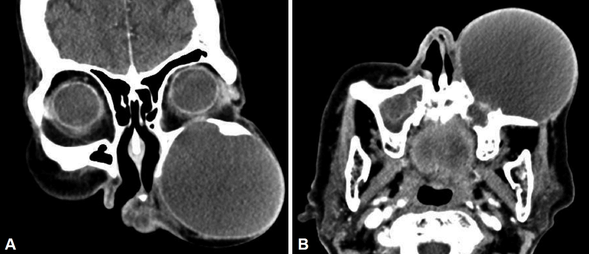
An 86-year-old female visited the Department of Otolaryngology, Head and Neck Surgery at the Veterans Medical Center, presenting with a protruding, non-fixed, well-circumscribed, round 10×10 cm mass on her left anterior midface, which had been gradually growing for 8 months. The patient had past medical history of hypertension but there was no history of sinusitis, allergy, maxillary fracture and surgery including Caldwell-Luc procedure. On gross inspection, the superior boundary of the mass extended to the lower edge of the orbit, while the lateral boundary exceeded the lateral wall of the maxillary bone. The medial margin of the mass surpassed her left nasolabial fold, disfiguring external nose near the left ala (Fig. 1). Within the oral cavity, visible expansion of the mass onto the gingivobuccal sulcus area was also observed. Despite notable external disfigurement, the mass consistency lacked the firmness of malignant tumor. Similarly, the disruption of skin integrity, including necrosis, was also not observed. Additionally, there were no ocular movement abnormalities including diplopia, visual impairment, or facial paralysis. Left endoscopic examination initially demonstrated no visible bony destruction within the lateral wall of the nasal cavity; however, computed tomography of the paranasal sinuses with contrast enhancement did confirm that the lesion (6.8×7.2× 6.1 cm) had also caused bone remodeling and destruction of the anterior and posterior bony walls of the maxillary sinus, ultimately suggesting either a sinus mucocele or malignant neoplastic lesion (Fig. 2). After receiving consent for surgery under general anesthesia, we explored the mass using the Caldwell-Luc approach because mass was vary extensive. A 4 cm incision was made on the gingiva-buccal folds to elevate and dissect the subcutaneous fat and facial muscles. Underneath, well defined cystic wall came into view. Before the cyst removal, aspiration of cyst was performed. Approximately 100 cc of viscous liquefacted mucus containing sand-like melted bone dust was obtained, but not cultured. In order to preserve the cystic wall, the cyst was then meticulously dissected to separate it from the overlying soft tissue as well as or the bony wall of the maxillary sinus. Cyst was then further detached from the upper, lower, and lateral sides of the maxillary sinus (Fig. 3A). During the mass removal, we noticed that the mass had completely eroded through the anterior bone of the maxillary sinus, leaving the area wide open after removal. Similarly, the superior wall of the maxillary sinus was also noted to have been destroyed, demonstrated by the visualization of the inferior periosteum of the left orbit as well as the visible herniation of the inferior aspect of the entire orbit when gentle pressure was applied against the eyeball. On pathological examination, the cells surrounding the cyst and mucinous material were confirmed to be respiratory epithelial cells, and the final diagnosis by permanent biopsy was confirmed to be a sinus mucocele (Fig. 3B and C). The patient had no recurrence as of 2 years after the surgery.
A paranasal sinus mucocele is a cystic disease that was first described by Rollet in 1896 as a benign, dilated cystic lesion completely filled with mucus and surrounded by epithelium [1,3]. It occurs mainly unilaterally and is known to be similar rates of occurrence in both males and females, most common in the 30’s and 40’s. The paranasal sinus mucocele is believed to be induced as a result of the occlusion of the natural foramen of the sinus due to chronic inflammation, previous surgical history, trauma, fibrous dysplasia [4,5]. The obstruction causes accumulation of mucus in the sinus and invading the surrounding bones which can cause remodeling [6].
Mucoceles are divided into primary mucoceles and secondary mucoceles. In primary mucoceles, the mucus discharge is blocked due to idiopathic inflammation, causing blockage of the secretion ducts and the consequent progression of the mucous glands into cysts or following the cystic degeneration of local polyps. Secondary mucoceles frequently occur after trauma or surgery. In both cases, they expand slowly, but often the size alone is enough to cause deformation of the adjacent bones. In the case of ethmoid frontal sinus mucoceles, visual impairment or eye movement limitation may occur [1]. If secondary infections occur, the mucocele may develop into pyoceles.
Sinus mucoceles primarily occur within the frontal and anterior sinuses, while maxillary sinus mucoceles are relatively rare and occur in 2.7%–10% of all mucoceles [7]. In particular, cysts large enough to cause bone destruction are very rare in patients with no history of sinus surgery [6,7]. Although mucoceles are benign, they require treatment and must be distinguished from malignant tumors because of their potential to cause bon destruction to the surrounding sinus walls and structures.
For diagnosis, computed tomography magnetic resonance imaging of the paranasal sinus may be used. On computed tomography, mucoceles without infection demonstrate uniform soft tissue shading that is not enhanced by contrast medium, has a smooth border, and does not show infiltration of surrounding soft tissue. Conversely, malignant tumors are irregular in shape and bony erosion or destruction of the sinuses proceeds after infiltration of the surrounding soft tissues [7,8].
Symptoms are mainly determined by the degree of pressure and erosion caused by the mucocele. Symptoms such as facial swelling, nasal obstruction, eyelid edema, and headache may appear, but there are also asymptomatic cases [5]. When mucoceles erode through the anterior wall, they usually present as painless swelling of the ipsilateral cheek. Nasal obstruction may also occur if the mass extends into the medial wall of the maxillary sinus, eventually compressing the inferior turbinate. With expansion of the superior wall of the maxillary sinus into the floor of the orbit, orbital displacement and visual acuity changes may also occur. If the mucocele extends inferiorly, it may invade the periodontal region and cause loosening of the affected teeth [5].
Treatment of a maxillary sinus mucocele is surgical removal. The traditional treatment is the Caldwell-Luc method, which is an open, external approach that completely removes the inner wall of the cyst [7]. However, with the recent advances in endoscopic surgery, natural endoscopic treatment for normal drainage is preferred [9]. Endoscopic surgery is an effective and rapid method to treat mucous membrane abnormalities with minimal morbidity [7,9]. However, Caldwell-Luc surgery may be a more suitable method for extensive lesions that involve soft tissue invasion of the face or pterygomaxillary fossa, and in cases where the mucocele was incompletely removed in a prior surgery [3].
Although there is no formal definition of giant mucoceles, there have been reports suggesting mucoceles larger than 5 cm to be defined as giant mucoceles [10]. Most of these giant mucoceles were reported to be in the frontal sinus and rarely in the maxillary sinus. This case is the first report of a giant mucocele where the horizontal and vertical diameters of the mucocele both exceed 5 cm, the anterior wall of the maxillary sinus was completely eroded, and notable facial swelling and asymmetry was consequently observed. The authors found that a maxillary sinus mucocele usually grows slowly as a benign disease, but bony destruction is possible after reaching a certain size. In conclusion, this case demonstrated the possibility of giant sinus mucoceles within the maxillary sinus when a patient presents with asymmetric facial swelling, necessitating a thorough examination, palpation, and appropriate imaging.
Notes
Availability of Data and Material
The datasets generated or analyzed during the study are available from the corresponding author on reasonable request.
References
1. Lee TJ, Li SP, Fu CH, Huang CC, Chang PH, Chen YW, et al. Extensive paranasal sinus mucoceles: a 15-year review of 82 cases. Am J Otolaryngol. 2009; 30(4):234–8.

2. Simões JC, Nogueira-Neto FB, Gregório LL, Caparroz FdA, Kosugi EM. Visual loss: a rare complication of maxillary sinus mucocele. Brazilian Journal of Otorhinolaryngology. 2015; 81:451–3.

3. Shahi S, Devkota A, Bhandari TR, Pantha T, Gautam D. Rare giant maxillay mucocele: a rare case report and literature review. Ann Med Surg (Lond). 2019; 43:68–71.

4. Kim DY, Kwon BW, Park DE. Clinical evaluation of paranasal sinus mucocele. J Clin Otolaryngol Head Neck Surg. 2004; 15(1):93–7.

5. Abdel-Aziz M, El-Hoshy H, Azooz K, Naguib N, Hussein A. Maxillary sinus mucocele: predisposing factors, clinical presentations, and treatment. Oral Maxillofac Surg. 2017; 21(1):55–8.

6. Hong SL, Cho KS, Roh HJ. Maxillary sinus retention cysts protruding into the inferior meatus. Clin Exp Otorhinolaryngol. 2014; 7(3):226–8.

7. Mutlu V, Yoruk O, Ozkan O. Destructive giant maxillary sinus mucocele: a case report. Med-Science. 2016; 5(1):306–12.

8. Song HM, Park HW, Chung YS, Jang YJ, Lee BJ. Primary mucoceles of the maxillary sinus. Korean J Otorhinolaryngology-Head and Neck Surg. 2006; 49(1):47–51.
9. Makeieff M, Gardiner Q, Mondain M, Crampette L. Maxillary sinus mucocoeles--10 cases--8 treated endoscopically. Rhinology. 1998; 36(4):192–5.
Fig. 1.
Gross facial appearance of an 86-year-old female. It shows a 10×10 cm sized notable swelling in the left cheek.

Fig. 2.
Radiological imagings. A: A coronal CT scan shows a 6.8×6.1 cm sized cystic lesion. B: A axial CT scan show a 6.8×7.2 cm sized cystic lesion protruding into the cheek.

Fig. 3.
Gross photograph and pathologic findings. A: The surgical specimen shows a 6.2×5 cm cystic surface. B: Microscopic photograph shows mucosa membrane composed of cystic lesions (H&E, ×10). C: Microscopic photograph shows respiratory epithelium where mucous substances are being distributed (H&E, ×40).


 PDF
PDF Citation
 Print
Print


 XML Download