Abstract
Nasopharyngeal tuberculosis can arise from both a primary infection and a secondary spread via the lymphatic or hematogenous system from a primary pulmonary lesion. Primary nasopharyngeal tuberculosis is rare and difficult to detect earlier because of the nonspecific presentations of the disease. As upper airway tuberculosis can be contagious, early initial diagnosis and suspicion of the physicians are needed in clinical practice. Recently, we successfully diagnosed and treated the disease by antitubercular medications of two cases of primary nasopharyngeal tuberculosis. Herein, we report our experience with a literature review.
Tuberculosis (TB) has maintained as one of the world’s oldest and deadliest communicable diseases. It can mostly affect the lung (pulmonary) but also other sites (extrapulmonary) as well. The proportion of extrapulmonary TB has been gradually increasing since 2001 and status of extrapulmonary TB in South Korea accounts for about 11%–17% [1]. However, there is a possibility that extrapulmonary TB is underestimated compared to pulmonary TB because it is relatively difficult to diagnose earlier due to its atypical clinical manifestation and has a low reporting rate [2]. Especially, it was reported that nasopharyngeal tuberculosis (NPTB) takes an average of 2.2 months from onset of the disease to diagnosis [3]. The number of Mycobacterium tuberculosis in the nasopharyngeal lesion is relatively scanty to be detected in the tissue specimen and it resulted in delayed diagnosis in NPTB [4]. Due to its low prevalence, there has been only a few case studies reported. Therefore, we present two cases of primary NPTB that diagnosed and successfully treated by antitubercular medications.
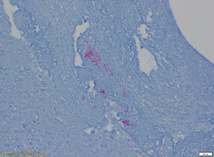
A 56-year-old female presenting with purulent rhinorrhea and postnasal drip of 2-month duration visited the outpatient clinic. Endoscopic examination of the nasopharynx revealed irregular mucosal necrotic ulceration diffusely extending from the opening of Eustachian tube to the soft palate (Fig. 1A). The lesion, somewhat in resemblance of melted cheese, was not detached from the underlying mucosa through repeated suctioning. Computed tomography (CT) demonstrated peripheral rim-enhanced mucosal thickening encompassing the posterior wall of the nasopharynx, the upper soft palate, and the opening of Eustachian tube (Fig. 1B). There were no specific findings in the laboratory blood tests including antineutrophil cytoplasmic antibodies (ANCA) test, chest X-ray, and physical examination of the neck. Deep biopsies were performed at several points of the necrotic tissue including the mid- and right upper-portions of nasopharynx and the left posterior nasal septum in particular. All of them showed chronic caseating granulomatous inflammation and acidfast bacilli (AFB) was detected directly under Ziehl–Nielsen stain (Fig. 2). Sputum AFB smear and culture for M. tuberculosis were all positive. No intrapulmonary lesions were observed on chest CT. As the patient was diagnosed with primary NPTB, she began antitubercular treatment of 6 months. Following the standard antitubercular treatment regimen, the patient received a two-month quadritherapy of rifampicin (RFP), isoniazid (INH), ethambutol (EMB), and pyrazinamide (PZA). After confirming response to medication, treatment was continued for 4 months with double therapy of RFP and INH. After a total of 6 months of antitubercular medication, the treatment was completed with no evidence of recurrence with only mild narrowing of the nasopharynx due to scar formation (Fig. 3).
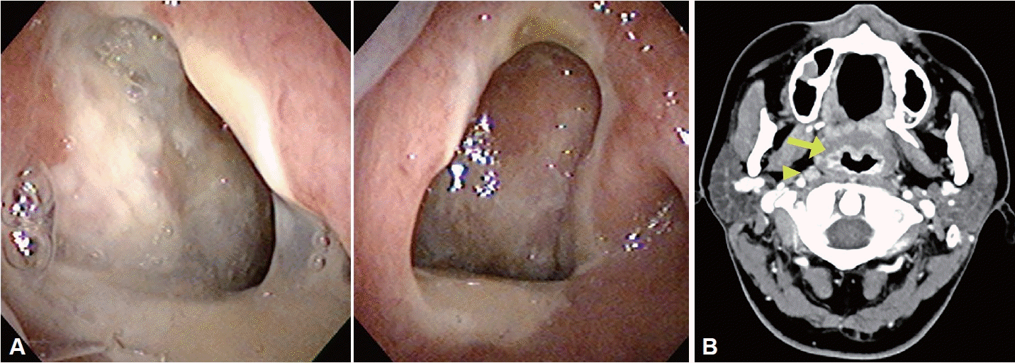
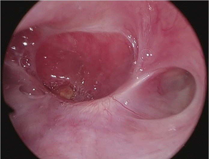
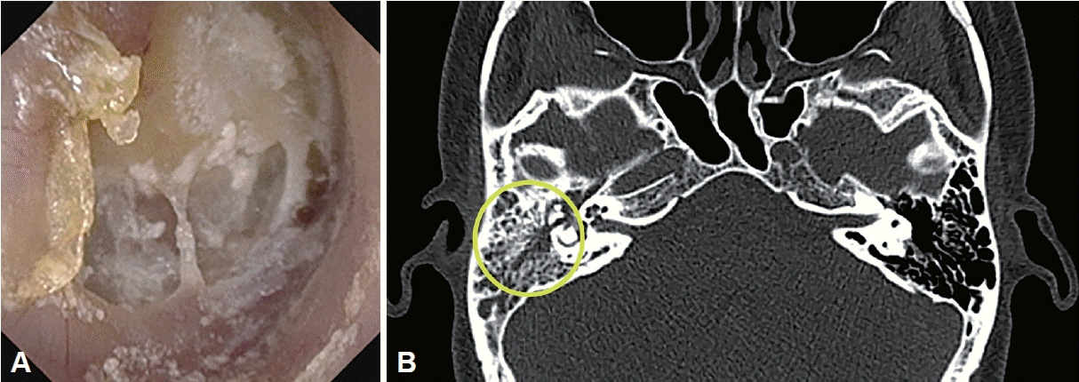
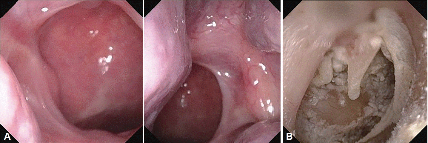
A 34-year-old female who had a one-month history of sore throat, sputum, and mild cough visited the outpatient clinic. On the endoscopic exam, necrotic mucosa was noted on the nasopharynx which was not sucked out (Fig. 4A). Mucosal thickening with increased enhancement at right posterior nasopharyngeal wall and enlarged retropharyngeal lymph node were noted on neck CT (Fig. 4B). It was diagnosed as chronic inflammation with necrosis on biopsy. Five months later, she visited again with ongoing previous symptoms and new onset hearing difficulty with otorrhea on right ear. Moderate conductive hearing loss was revealed on audiometry, and temporal CT demonstrated suspected otitis media with effusion on the right ear (Fig. 5A). Previous nasopharyngeal lesion was remained but relatively improved. Without further follow-up, she visited again 5 months after from the second visit with persistent symptoms. Necrotic tissue was aggravated at right nasopharynx and extended to soft palate superior portion. Perforation of right tympanic membrane with purulent otorrhea was noted (Fig. 5B). Chest X-ray and blood test including ANCA test presented no abnormality. Biopsies were performed again at right superior nasopharynx, right rosenmuller fossa and left posterior septum, which were all diagnosed as TB. Furthermore, AFB staining and culture of otorrhea was positive for M. tuberculosis. The patient started antitubercular treatment regimens by quadritherapy of RFP, INH, EMB, and PZA for 2 months. Due to possibility of auditory neuropathy in EMB, the treatment regimen was changed after 2 months to triple therapy with RFP, INH, and PZA except EMB. After a total of 6 months antitubercular medications, all symptoms except hearing difficulty have improved. Nasopharyngeal mucosa was all improved without any scar nor sequelae (Fig. 6A). Unfortunately, total perforation was noted on the right tympanic membrane which was all melted (Fig. 6B).
Extrapulmonary TB has a relatively low prevalence compared to pulmonary TB, and NPTB is particularly rare. The clinical manifestations of NPTB vary greatly, but most common symptom is cervical lymph node enlargement which was presented in many previous studies. In addition, there are nasal congestion (10%), neck pain, tinnitus, hearing loss, otalgia, and postnasal drip [5]. Therefore, NPTB is a disease that can be met at the first diagnosis by specialists in otology, rhinology, and head and neck surgery. It often takes a considerable amount of time from the initial patient visit to the definitive diagnosis because NPTB mostly presents atypical manifestations and gross lesion can be confused from other nasopharyngeal necrotic diseases like nasopharyngeal cancer, lymphoma, granulomatosis with polyangitis, angiofibroma, fungal infection, sarcoidosis, periarteritis nodosa, leprosy, syphilis, and Castleman’s disease [5,6]. In most cases, it is known that a histological biopsy is standard for diagnosis because it is difficult to differentiate it from other necrotic mucosal diseases using only clinical features, radiological tests, and endoscopic examination [7]. Like a tissue biopsy for diagnosing the malignancy, the clinicians should do the biopsy thoroughly enough to cause bleeding at multiple lesion sites, not the necrotic tissue only. Insufficient or incorrect biopsy results can delay the accurate diagnosis and treatment of the disease and result in the transmission of M. tuberculosis.
The most characteristic finding of the NPTB is the lesion, somewhat in resemblance of melted cheese, not detached from the underlying mucosa through repeated suctioning. Additionally, for diagnosis, it is essential to check the presence or absence of TB in patients or their family history, and close contact with an active TB patient through history taking, and tests such as tissue biopsy, smear, culture, and TB-PCR [8]. We performed CT and deep biopsy under local anesthesia at multiple sites of the lesion for patients with the above-mentioned specific findings on endoscopic examination. A typical pathological report for NPTB is caseating granulomatous inflammation with multinucleated giant cells of Langhan’s type and foreign body giant cells, with or without necrosis [9]. Typical histopathology with caseating granulomatous inflammation mostly must be confirmed with Ziehl–Nielsen staining for AFB. NPTB can be more infectious because TB is spread through the inhalation of droplets and the upper respiratory TB is localized at open lesion. We recently recognized several studies in applying airborne precautions when manipulating open lesions in patients with extrapulmonary TB and in accordance with pulmonary TB, respiratory isolation is recommended from the start of treatment until all negative AFB stain and culture at all 3 sputum specimens at least two interval times were confirmed [7]. Therefore, it is better to refrain from outpatient follow-up in otorhinolaryngology department during the infection period.
Although there was a previous report that surgical excision was performed from the beginning, as in NPTB, surgery is not necessary and only antitubercular medical treatment is sufficient. Surgical treatment should be considered only for NPTB in severe cases of obstruction or multidrug resistant TB unresponsive to medical treatment.
For NPTB, the minimal duration of antitubercular treatment is six months. There were a few previous reports that stated the antitubercular medical treatment started by two months of INH, RFP, EMB, and PZA followed by INH and RFP for 4 to 7 months. However, the preferred regimen of treatment was various according to the clinicians and the course of the disease [10].
In conclusion, NBTB shows various clinical features in the ear, nose, and neck. So even if the patient visits to the hospital for ear symptoms or cervical lymph node problems, if necrosis of the nasopharyngeal mucosa is found on nasal endoscopy, although it is not common, attention should always be paid to the possibility of NPTB. If diagnosis and treatment are delayed, it can spread to others and leave serious sequelae. Accurate diagnosis and prompt treatment through deep and multiple site endoscopic biopsy can reduce sequelae and improve quality of life.
Notes
Availability of Data and Material
The datasets generated or analyzed during the study are available fromthe corresponding author on reasonable request.
References
2. Lee JK, Cho HP, Lee YM, Park JH. Four cases of primary nasopharyngeal tuberculosis presenting as extra pulmonary tuberculosis. J Clin Otolaryngol Head Neck Surg. 2013; 24(2):255–60.

3. Srirompotong S, Yimtae K, Jintakanon D. Nasopharyngeal tuberculosis: manifestations between 1991 and 2000. Otolaryngol Head Neck Surg. 2004; 131(5):762–4.

4. Cho YS, Choi N, Kim HY. A two cases of primary tuberculosis at the nasopharynx. J Rhinol. 2015; 22(2):123–7.

5. Park HS, Boo SH, Son JY. A case of primary nasopharyngeal tuberculosis. J Clin Otolaryngol Head Neck Surg. 2001; 12(1):118–21.

6. Jang DH, Kim HC, Hong YO, Kim JS. A case of nasopharyngeal tuberculosis which was difficult to differentiate from sarcoidosis. Korean J Otorhinolaryngol-Head Neck Surg. 2022; 65(4):231–6.

7. Kawamura I, Kudo T, Tsukahara M, Kurai H. Infection control for extrapulmonary tuberculosis at a tertiary care cancer center. Am J Infect Control. 2014; 42(10):1133–5.

8. Cai PQ, Li YZ, Zeng RF, Xu JH, Xie CM, Wu YP, et al. Nasopharyngeal tuberculosis: CT and MRI findings in thirty-six patients. Eur J Radiol. 2013; 82(9):e448–54.

Fig. 1.
Initial nasopharynx endoscopic and CT findings of case 1. A: Endoscopic finding: melted cheese like necrotic tissue with purulent discharge on right posterior wall of nasopharynx. B: Neck CT: peripheral rim enhancing mucosal thickening of right posterior nasopharynx extended to Eustachian tube (arrow).
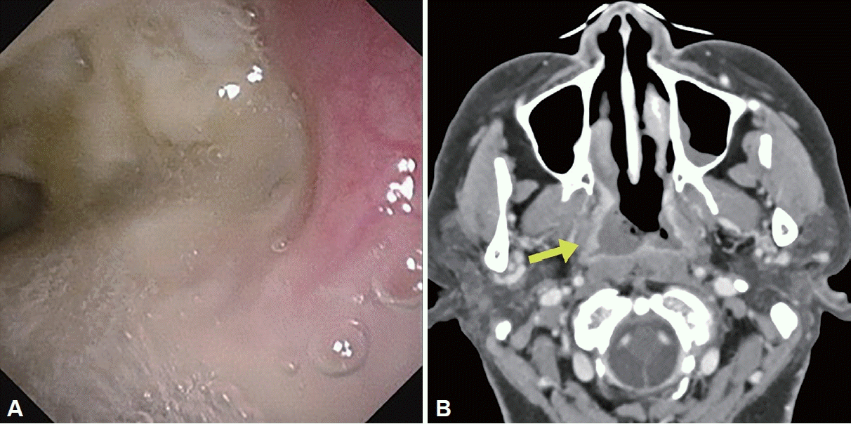
Fig. 4.
Initial nasopharynx endoscopic and CT findings of case 2. A: Endoscopic finding: necrotic tissue like melted cheese with purulent discharge on posterior nasopharynx. B: Neck CT: mucosal thickening and increased enhancement of nasopharynx (arrow) with 0.9 cm lymph node in right retropharyngeal area (arrow head).


 PDF
PDF Citation
 Print
Print


 XML Download