CASE REPORT
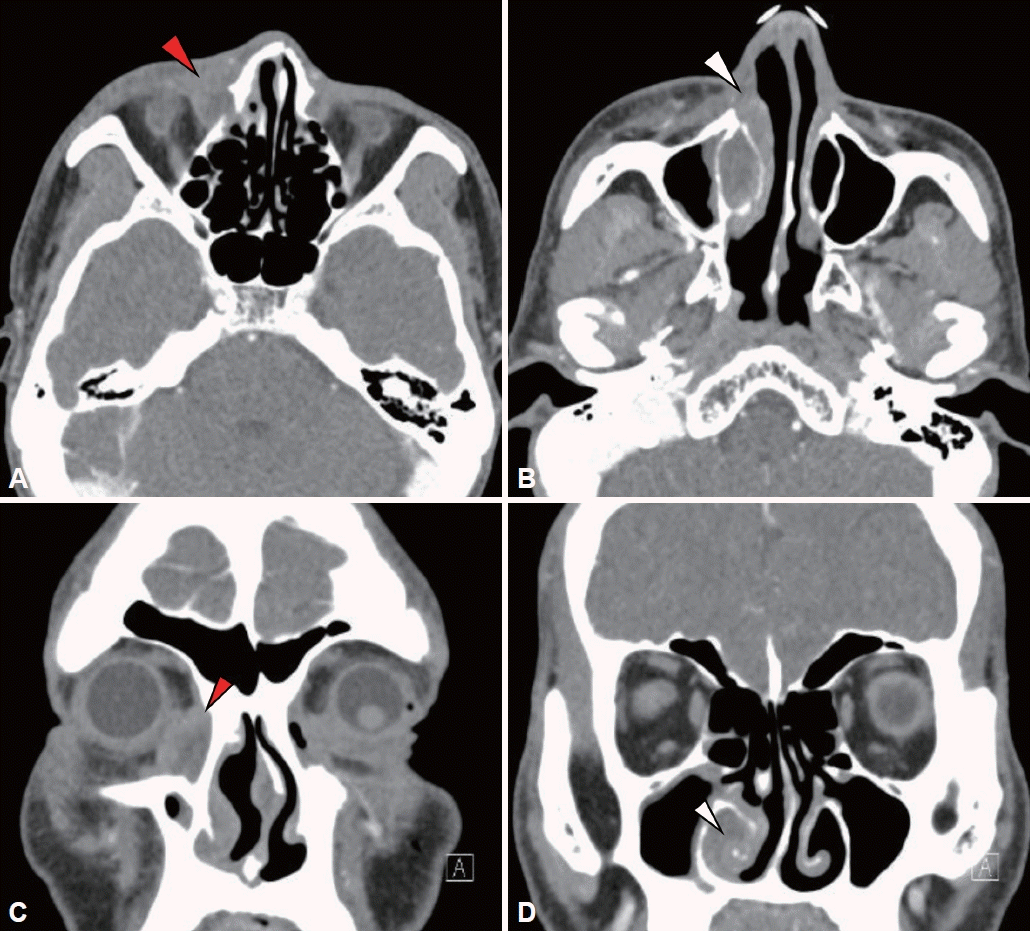
A 58-year-old male patient visited our department of ophthalmology with a chief complaint of right epiphora and right orbital edema which broke out since three months ago. The patient was instilled with antibiotics in the department of ophthalmology for 3 months, but there was no improvement. A lacrimal irrigation test conducted in the department of ophthalmology showed 50% regurgitation of the upper eyelid in the right eye. Since inhomogeneous contrast enhancement and thickening were observed around the right lacrimal duct, and the mucosa of the right inferior turbinate and the inferior nasal meatus showed a sign of thickening in the result of orbital CT taken in the department of ophthalmology, dacryocystitis, hypertrophic rhinitis, and cellulitis were suspected (
Fig. 1). After the patient was hospitalized in the department of ophthalmology and treated with antibiotics (Cefminox and Netilmicin), although orbital edema improved, since epiphora did not improve, a silicone tube implantation operation was performed to insert a silicone tube into the lacrimal duct on the 11th day of hospitalization.
 | Fig. 1.Radiologic findings. Paranasal CT shows inhomogenous enhancement and soft tissue thickening around right nasolacrimal duct (red arrowheads). A: Axial view. B: Coronal view. Paranasal CT shows mucosal thickening of right inferior turbinate and inferior meatus (white arrowheads). C: Axial view. D: Coronal view. 
|
Epiphora did not improve even after ten days of the silicone tube implantation and, since the patient additionally complained of discomfort in the right nose, he was arranged to visit the department of otolaryngology. As a result of the intranasal endoscopy carried out in the department of otolaryngology, edema and synechia of the right inferior turbinate were found and mucous discharge was identified (
Fig. 2A). Since cellulitis of inferior turbinate was suspected to have been exacerbated due to the silicone tube implantation, cephalosporin, a second generation oral antibiotic, was administered for two weeks. Epiphora improved partially after the administration of the antibiotic, but right nasal discharge and nasal congestion exacerbated without improvement. In the intranasal endoscopy carried out by visiting the department of otolaryngology again, the synechia of the right inferior turbinate became more severe than 2 weeks ago, mucopus discharge was identified, and whitish plaque was newly identified along with the overall ulcerative change of the inferior turbinate mucosa (
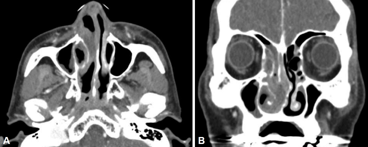
Fig. 2B). In the paranasal sinus CT taken again to differentiate the lesion, the right inferior turbinate, ostiomeatal unit, maxiillary sinus, nasal floor, and the mucosa of the nasal septum became thicker as a whole without contrast enhancement, and thickening of the mucosa was found to have become much more severe than in the CT taken one month ago. The edema around the right lacrimal sac and inside the right lacrimal duct were found to have been rather improved compared to the CT taken one month ago (
Fig. 3). Accordingly, a malignant lesion rather than an inflammatory change of the nasal cavity due to simple dacryocystitis or lacrimal duct implantation was suspected and a biopsy for the lesion of the right inferior turbinate and a bacterial culture test for the sedimentary secretion were performed. At the time of the biopsy, when the crust on the medial side of the right inferior turbinate was removed first, a severely thickened mucosa accompanied by necrosis appeared. The tissue in the size of 5 mm was taken from the necrotic area twice together with the surrounding tissue, and a pathological biopsy was requested with the tissues taken.
 | Fig. 2.Endoscopic findings. A: Shows profuse mucus discharge and swelling of right inferior turbinate adhere to right septal wall mucosa. B: Shows mucopus discharge and adhesion of right inferior turbinate to its adjacent structures. Whitish plaques are newly found on the surface of inferior turbinate. C: Shows no evidence of recurrence. There are no inflammatory change or hypertrophy of nasal mucosa. 
|
 | Fig. 3.Radiologic findings. Paranasal CT (1 month after Fig. 1) shows nonenhanced mucosal thickening of osteomeatal unit, right maxillary sinus, base of nasal cavity and nasal septum. Thickening of mucosa remarkably progressed compared to CT 1 month ago (A: Axial view, B: Coronal view). 
|
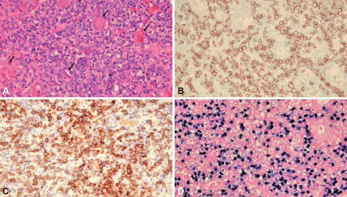
In the bacterial culture test, pseudomonas aeruginosa was detected and, in the pathological biopsy; immunohistochemical staining showed a positive response to CD30, MUM-1, and CD56 which is an NK cell marker; ki-67 staining showed that 60% and more of tumor cells were positive; and in situ hybridization showed positive findings for EBV as a whole (
Fig. 4). Accordingly, it could be diagnosed as NK/T-cell lymphoma. In positive emission tomograpy-CT (PET-CT), hypermetabolic thickenings were found across the right middle meatus, middle turbinate, and inferior turbinate; mild hypermetabolic change was found in the medial canthus of the left orbit; and no metastasis to other organs was found (
Fig. 5A). Nothing peculiar was found in neither the abdomen nor chest CT. The lesion of the left orbit found in the PET-CT was judged to be a result of dacryocystitis. A bone marrow examination was additionally performed and no metastasis to the bone marrow was confirmed. Accordingly, composite anti-cancer radiography was finally planned under the diagnosis of NNKTL.
 | Fig. 4.Pathologic findings. A: The tumor was composed of medium-sized lymphoid cells with irregularly folded nuclei and showed angiodestructive growth pattern (arrows) (hematoxylin eosin stain, ×400 magnification). B: Tumor cells showed membranous positivity for CD56 immunohistochemical stain (CD56, ×400 magnification). C: Tumor cells were also positive for CD30 immunohistochemical stain (CD30, ×400 magnification). D: In situ hybridization for Epstein-Barr virus (EBV)-encoded small RNA (EBER) demonstrated the presence of EBV (EBER, ×400 magnification). 
|
 | Fig. 5.Radiologic findings of positive emission tomograpy-CT. A: Hypermetabolic lymphoma in right middle meatus, middle turbinate and inferior turbinate. B: 2 month after (A) shows diffuse mild hypermetabolic mucosal thickening of right nasal cavity. Focal hypermetabolic thickening of right nasal cavity was disappeared. C: 3 month after (B). Diffuse mild hypermetabolic lesion was disappeared and there are no abnormal hypermetabolic uptake. 
|
After 2 months of the diagnosis, in the third week after completing the radiotherapy, the thickenings of the mucosa inside the right paranasal sinus and nasal cavity were found to have reduced. Both the amount of metabolic uptake and the degree of thickening in the hypermetabolic lesion seen in the left orbit on the PET-CT taken at the same time were found to have reduced compared to the previous images. Although the hypermetabolic lesions which were observed across the right middle meatus, middle turbinate, and inferior turbinate were not found, a mild hypermetabolic lesion was identified in the mucosa inside the nasal cavity (
Fig. 5B). In the 5th month of the diagnosis, combination chemotherapy using etoposide, ifosfamide, dexamethasone, and L-asparaginase was completed. The hypermetabolic lesion which was observed in the left orbit on the PET-CT taken after the anti-cancer treatment was not found, and the mild hypermetabolic lesion which was found inside the mucosa was not seen anymore (
Fig. 5C). The patient was periodically visiting the department of otolaryngology for disinfection of the nasal cavity at that time as an outpatient, and no recurrence of the thickening, inflammation or lymphoma of the mucosa inside the right nasal cavity was seen in the intranasal endoscopy performed in the 3rd month after the completion of the anti-cancer radiotherapy (
Fig. 2C).
Go to :

DISCUSSION
Extranodal NK/T-cell lymphoma is a disease characterized by an ulcerative lesion that rapidly progresses in the nasal cavity and upper respiratory tract [
1], and is known to be related to EBV. In general, NNKTL accounts for 80% of extranodal NK/T-cell lymphoma, and non-nasal types account for 20% [
1,
4-
7]. Although the symptoms of NNKTL patients are nonspecific, it is difficult to distinguish them from rhinosinusitis only with the clinical symptoms as the patients mostly complain about nasal congestion, postnasal drip, nosebleeds, and so on. They may also complain of sore throat, hyposmia, skin lesions, and so on and, according to Hung et al. [
1], 18% of the cases show systemic symptoms such as B symptoms.
NNKTL is characterized by mucosal thickening and ulcerative lesions, and the mucosal thickening of inferior turbinate caused by NNKTL may appear as a clinical manifestation called epiphora as it causes closure of nasolacrimal duct opening. In the present case, although the closure of the nasolacrimal duct opening might have acted as the direct cause of epiphora, dacryocystitis broke out due to the necrosis of tissues caused by NNKTL, which is thought to have exacerbated the epiphora by causing edema of the tissues around the lacrimal duct and lacrimal sac. In addition, the spread of inflammation through the lacrimal sac caused cellulitis to bring about orbital edema, and epiphora and orbital edema showed improvement after the administration of antibiotics. According to Hung et al. [
1], patients who complain of orbital symptoms in relation to NNKTL are rare (4.7%), and among them, almost no cases have been reported in the literature where the patient complains of epiphora. NNKTL showing epiphora may also be confused with a postoperative infection that occurs after DCR as in the present case. It is because the patient may complain epiphor in both cases, and the thickening of the mucosa is accompanied. For this reason, if the patient complains of epiphora, the patient has a past history of DCR, and the thickening of the mucosa is accompanied, a close intranasal endoscopic examination and an imaging evaluation are required. At the time of an endoscopic examination, diffuse large B cell lymphoma, another subtype of non-Hodgkin’s lymphoma, predominantly shows a prominent mass, whereas NNKTL mainly shows mucosal thickening. Also in the study of Hung et al. [
1], only 11 patients (34.4%) among the 32 NNKTL patients who had infiltration in the nasal cavity showed a prominent mass, and the remaining 2/3 of the patients showed no prominent lump tissues but only mucosal thickening. Since mucosal thickening may cause the ambiguous differentiation of NNKTL from other inflammatory diseases, even if no mass is observed, if there is mucosal erosion, severe crusting in the nasal cavity, bleeding to touch, or granulation, NNKTL has to be suspected and additional imaging examination such as CT has to be performed [
1].
CT is a good tool to differentiate NNKTL from other diseases. On CT, NNKTL shows unilateral, non-contrast-enhanced, and homogenous aspects and doesn’t accompany central necrosis. NNKTL predominantly infiltrates into the inferior turbinate, nasal floor, and nasal septum and, only in 50%, infiltrates into the paranasal sinus. According to a study by Hung et al. [
1], only 40% of NNKTL patients’ CT showed mucosal thickening in the nasal cavity only without mucosal thickening in the paranasal sinus. The mucosal thickening in the nasal cavity which has not infiltrated into the paranasal sinus is an important imaging characteristic of NNKTL distinguished from paranasal sinusitis. Also in the present patient case, CT showed mucosal thickening only in the nasal septum, nasal floor, and right inferior turbinate with no infiltration into the paranasal sinus. In some NNKTL cases, mass grow to infiltrate into the paranasal sinus, in which case mucosal thickening can be observed also in the paranasal sinus. In this case, however, the thickening of the nasal floor part (62%) and nasal septum mucosa (56%) is also accompanied at a high frequency, another characteristic of NNKTL CT distinguished from the CT of rhinosinusitis, which rarely shows mucosal thickening in the nasal floor part and nasal septum [
1].
NNKTL is histopathologically diagnosed. When tumor cells are observed and these tumor cells are found to be positive for CD2, CD3, CD3E, CD45, CD56, perforin, Granzyme B, and T-cell restricted intracellular antigen 1 in immunohistochemical staining, the patient can be diagnosed with NNKTL [
4,
8,
9]. According to Harabuchi et al. [
2], it is sometimes difficult to distinguish it from inflammatory tissues since various inflammatory cells such as pleomorphic large or small cells, macrophages, granulocytes, and lymphocytes are also observed in a pathological biopsy.
For NNKTL, anti-cancer radiotherapy is the main treatment method at present. As to the current treatment method for the diseases which fall under phase I/II of the Ann Arbor System, anti-cancer chemotherapy such as DeVIC (dexamethasone, etoposide, ifosfamide, and carboplatin) or MPVICP (methotrexate, peplomycin, etoposide, ifosfamide, carboplatin, and prednisolone) is carried out in combination with radiotherapy [
10]. In the present case, L-asparaginase was added to the DeVIC therapy, and radiotherapy was carried out in combination. In the case of diseases in the metastatic phase such as Ann Arbor System III/IV, SMILE (steroid, methotrexate, ifosfamide, L-asparaginase, and etoposide) therapy combined with radiotherapy may be used [
10]. In the case of a relapse, intractable, or a high risk patient, stem cell transplantation may be considered.
In the case of NNKTL, early diagnosis is very important since it progresses very rapidly to cause bone destruction, which may lead to fatal results [
11], and early anti-cancer treatment has a decisive effect on the prognosis of the disease. The most common clinical manifestations of NNKTL include nasal congestion, postnasal drip, and nosebleeds, and it very rarely shows epiphora too by closing down or infiltrating into the lacrimal duct as in the present patient case. Since NNKTL may show various clinical manifestations, it is difficult to distinguish it from other diseases only with the clinical manifestations. In particular, if a patient complains of a rare symptom like epiphora, it is not easy to suspect NNKTL, a very rare disease. In the present case as well, the authors identified mucosal thickening and inflammatory secretions through intranasal endoscopy at the time of the first visit of the patient to our clinic and suspected acute inflammation of the nasal cavity and paranasal sinus known to break out in 1.5% to 2% of patients after DCR [
12]. The possibility for NNKTL was also taken into consideration since orbital CT showed the thickening of the mucosa of the inferior nasal meatus including the inferior turbinate and nasal floor but, as no lump was observed in the intranasal endoscopy and neither the change in the color of the mucosa nor ulcerative legion was observed, antibiotics were administered first rather than active biopsy because we thought that the possibility for an inflammatory disease which broke out as complications after DCR was higher than NNKTL. However, the authors identified serious synechia and scabs of the inferior turbinate mucosa, exacerbation of edema, ulcerative legions, and leukoplakic through close monitoring without excluding tumorous lesions, and finally diagnosed NNKTL by carrying out CT again and a biopsy.
If the thickening of the mucosa is found along with epiphora in the intranasal endoscopy, NNKTL has to be taken into consideration, although it is rare, and imaging review is required along with close observation of the mucosa. In addition, if ulcerative lesions or necrotic lesions of the mucosa are observed, an active biopsy must be carried out.
Since main mass of NNKTL are blocked out by polypoid mucosa or nasal polyp or is covered by crusts during a biopsy in many instances, close observation of the lesion is required. Since atypical cells are scattered around the necrotic region in the case of NNKTL, if insufficient tissue samples are taken, only chronic inflammation is seen in a biopsy in many cases. Accordingly, many samples have to be taken from all infiltration areas and, if there are crusts, tissues have to be taken beneath the crusts after the crusts are removed. When taking tissue samples, it is recommended to take enough lump tissues, ulcerative lesions, and necrotic lesions of at least 3 mm including the surrounding tissues, and it has to be confirmed that sufficient hemostasis has been achieved after the tissue sampling.
As the authors experienced a rare case of a 58-year-old male patient who persistently complained of epiphora and was diagnosed with NNKTL in a biopsy, we report it along with the literature review result.
Go to :


 PDF
PDF Citation
 Print
Print


 XML Download