This article has been
cited by other articles in ScienceCentral.
Abstract
This case report describes the treatment of an adult female patient with a history of periodontal disease, Class I malocclusion with extrusion, dental spaces, and pathologic tooth migration. The patient was treated with clear aligners, which effectively controlled the strength and direction of orthodontic forces after 3 months of systematic periodontal treatment. The Peer Assessment Rating (PAR) index was calculated from study models before and after treatment. The pretreatment PAR score was 24, and the posttreatment PAR score was 4. The PAR score for this patient changed by 83%. Satisfactory appearance and good function were achieved for this patient.
Go to :

Keywords: Adult treatment, Periodontitis, Aligners, Malocclusion
INTRODUCTION
Periodontal disease is highly prevalent in both developed and developing countries. Smoking, poor oral hygiene, diabetes mellitus, and genetics, among others, are risk factors associated with the development and/or progression of periodontitis.
1 Meanwhile, tooth misalignment increases plaque accumulation and predisposition to periodontal disease. Trauma from occlusion due to tooth position irregularity has also been found to affect periodontal tissues adversely. As the number of adults opting for orthodontic treatment continues to grow, orthodontists are more likely to manage patients with chronic periodontitis.
2 There is no consensus on the positive effects of orthodontic treatment on periodontal health. Nevertheless, current research suggests that orthodontic treatment has no significant negative impact on periodontal tissues when the periodontal infection is properly controlled.
3,4 Combined orthodontic-periodontal treatment is an effective approach for achieving pleasant function and esthetic results in patients with good periodontal health.
5-8 Paolone and Kaitsas
9 claimed that orthodontic movement could increase the amount of periodontal soft tissue and alveolar bone. However, treating adult patients with chronic periodontal disease using fixed appliance therapy is a considerable challenge for orthodontists. Successful treatment of patients with periodontal disease using either labial or lingual fixed appliances requires specific mechanical and biomechanical considerations.
10,11 Compared with conventional fixed appliances, clear aligners are a better choice for patients with periodontal disease because of their ease of oral hygiene maintenance, precise control of force, and mandibular incidence of root resorption.
12-14 This case report demonstrates the successful orthodontic treatment of a 24-year-old woman having inactive periodontitis with clear aligners.
This study was approved by the Ethic Committee of Renmin Hospital of Wuhan University (No. WDRY2021-KS031). Written informed consent was obtained from the patient for the publication of this case report.
Go to :
DIAGNOSIS AND ETIOLOGY
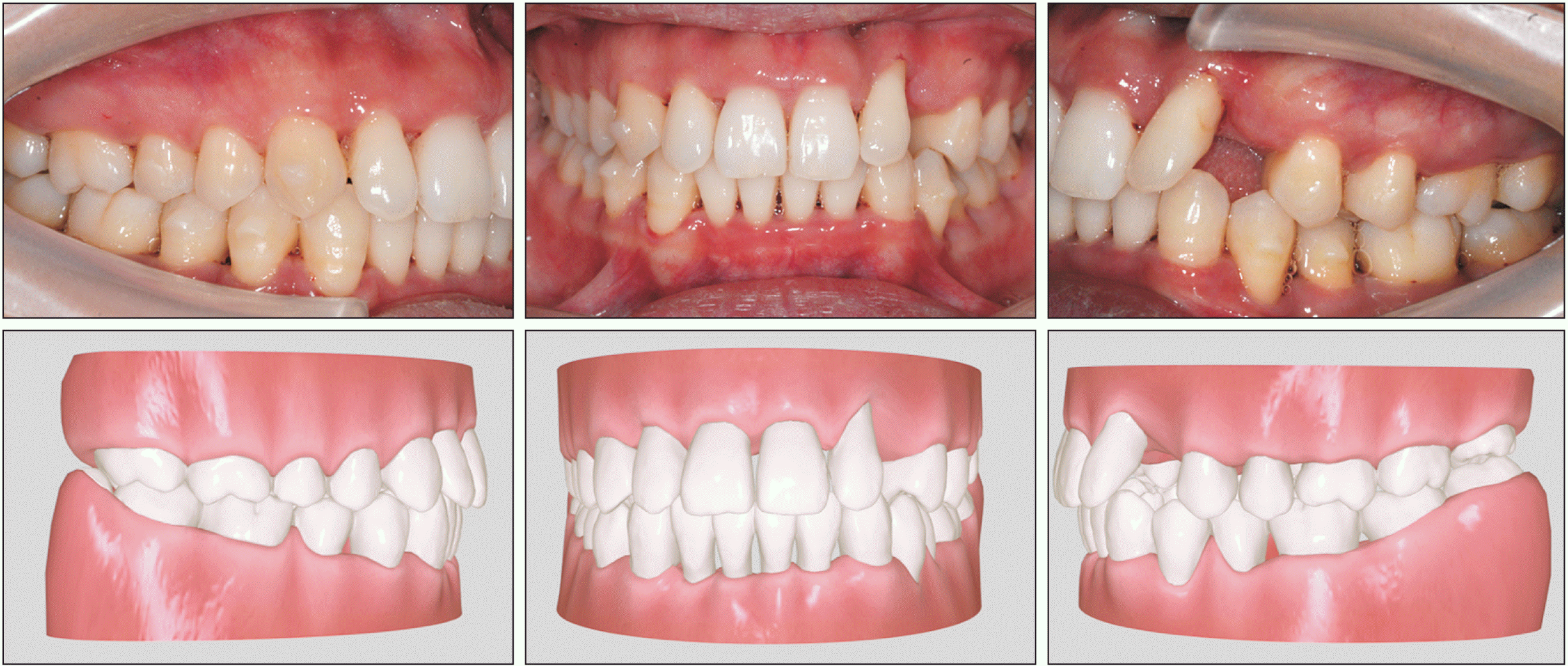
A 24-year-old woman with severe periodontitis and pathologic tooth migration was referred to our orthodontic department. Her chief complaints were malposed maxillary and mandibular anterior teeth. She had no history of systemic disease. However, her mother suffered from misaligned teeth and periodontitis. Notably, poor oral hygiene and traumatic occlusal forces resulting from crooked teeth accelerated the progression of the disease, increasing incisor irregularity. The patient had an almost symmetrical face and a straight facial profile. A Class I canine relationship on the right side and a Class I molar relationship on both sides were observed, with a scissor bite of the maxillary left second premolar and a missing maxillary left canine.
The main problem was located in the anterior region. Her mandibular incisors impinged on the lingual gingiva of the maxillary incisors. The maxillary central incisors and mandibular incisors were extruded with a severe lingual inclination and 8 mm overbite, whereas the maxillary lateral incisors were inclined labially (
Figure 1). There was 4.0 mm of space (the space of the missing tooth was not included) within the maxillary arch and 1.0 mm in the mandibular arch. The anterior Bolton ratio was 78.6%, and the overall Bolton ratio was 93.5%; in which they were measured with the hypothesis that the width of the left maxillary canine was equal to that of the right one. The curve of Spee was approximately 4.5 mm. The patient presented with a generalized gingival recession that was notably severe on the maxillary and lower anterior teeth and in the mandibular premolar region. The recession resulted in sensitivity to hot and cold temperatures, tooth mobility, and esthetic complaints. The initial periodontal conditions were measured using probing depth, plaque, bleeding on probing, and tooth mobility. The patient’s periodontal disease was adequately controlled.
15 No tooth had a probing depth > 4 mm, the full-mouth plaque index was 18%, and the full-mouth percentage of positive bleeding on probing sites was 15% (
Table 1). There was grade I mobility of the maxillary lateral incisors and grade II furcation of the mandibular right first molar.
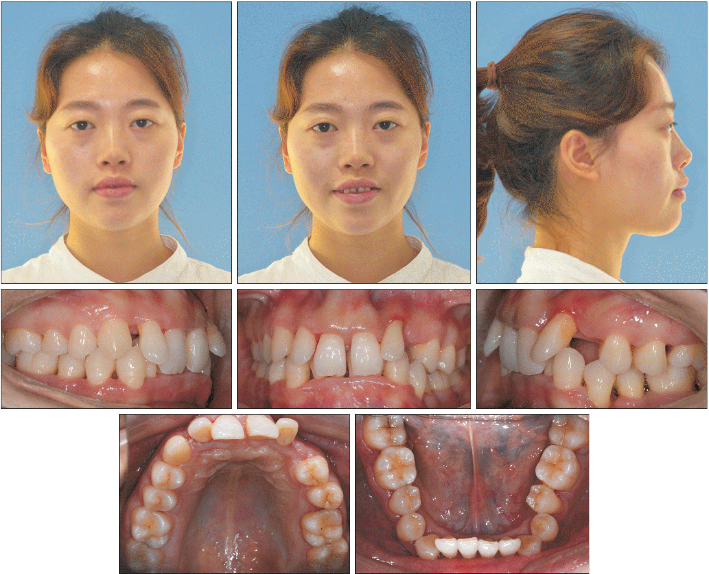 | Figure 1Pretreatment facial and intraoral photographs of an adult with inactive periodontal disease. 
|
Table 1
Results of pretreatment periodontal examination
|
Maxillary teeth |
17 |
16 |
15 |
14 |
13 |
12 |
11 |
21 |
22 |
23 |
24 |
25 |
26 |
27 |
Pocket depth (labial)
(mm) |
111 |
111 |
111 |
111 |
112 |
212 |
212 |
212 |
234 |
|
222 |
323 |
422 |
222 |
|
Bleeding on probing |
|
|
|
|
*
|
|
*
|
|
*** |
|
|
|
|
|
Pocket depth (lingual)
(mm) |
111 |
111 |
111 |
111 |
111 |
212 |
323 |
333 |
223 |
|
222 |
212 |
322 |
222 |
|
Bleeding on probing |
|
|
|
|
|
|
|
|
|
|
|
|
*
|
|
|
Mandibular teeth |
47 |
46 |
45 |
44 |
43 |
42 |
41 |
31 |
32 |
33 |
34 |
35 |
36 |
37 |
Pocket depth (labial)
(mm) |
113 |
343 |
323 |
323 |
323 |
122 |
212 |
222 |
222 |
222 |
244 |
433 |
324 |
322 |
|
Bleeding on probing |
|
*
|
*** |
*** |
*
|
|
|
|
|
*
|
*** |
*
|
|
|
Pocket depth (lingual)
(mm) |
122 |
333 |
333 |
323 |
322 |
322 |
222 |
222 |
222 |
323 |
323 |
223 |
234 |
222 |
|
Bleeding on probing |
|
*** |
*
|
** |
*
|
|
|
|
|
*
|
** |
|
|
|

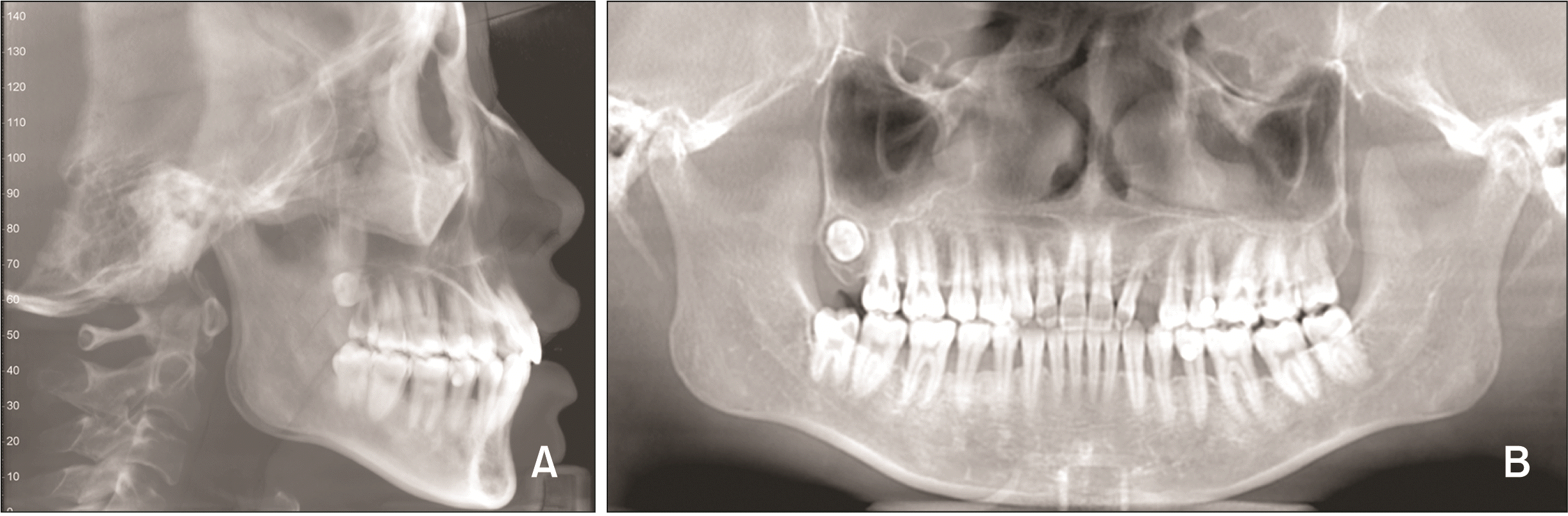
The pretreatment lateral cephalogram is shown in
Figure 2A. Pathological migration of the maxillary left lateral incisor was also observed on the panoramic radiograph (
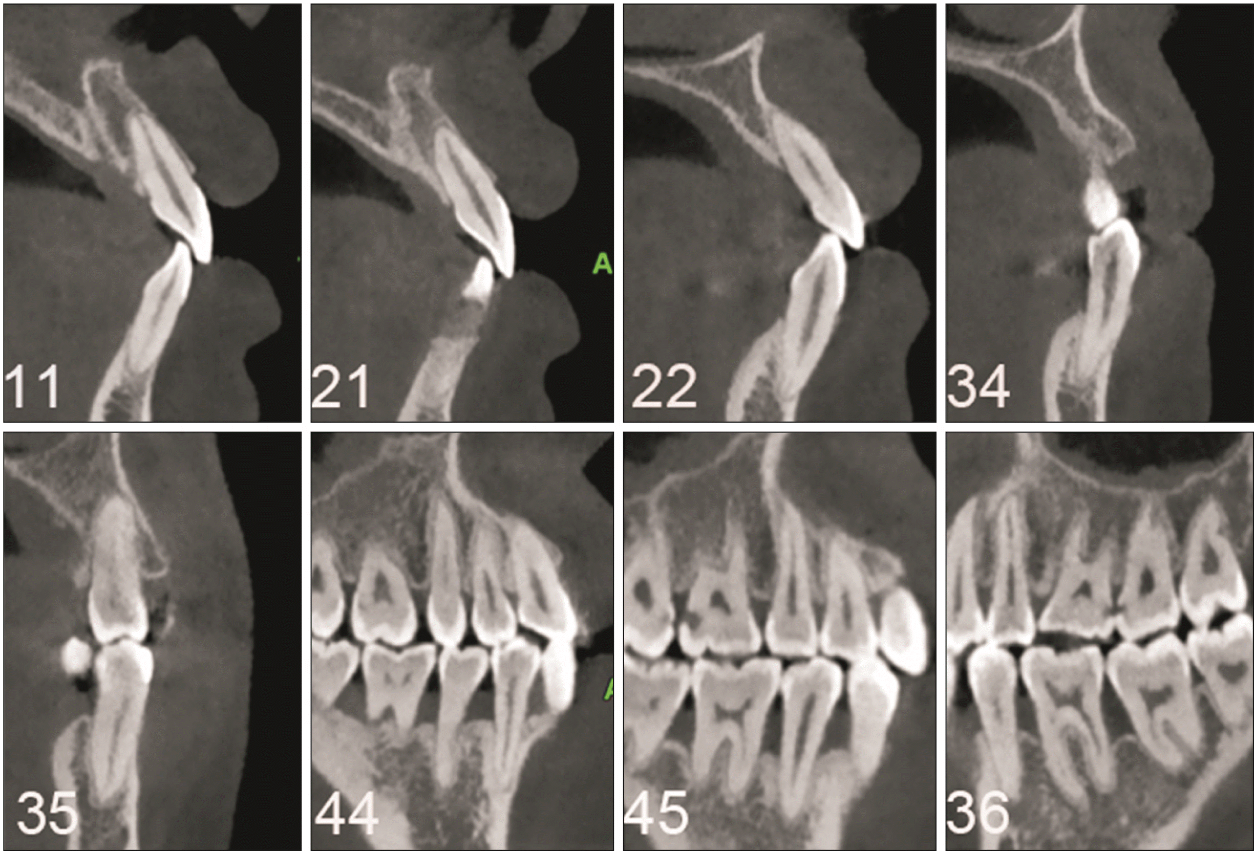
Figure 2B). Multiple sites of alveolar bone resorption were observed, especially in the anterior and premolar areas (
Figure 3). Alveolar bone resorption in the mesial and distal regions of the maxillary left lateral incisor was extensive, involving half to two-thirds of the root. An infracrestal pocket was visible in the distal aspect of the mandibular left first premolar.
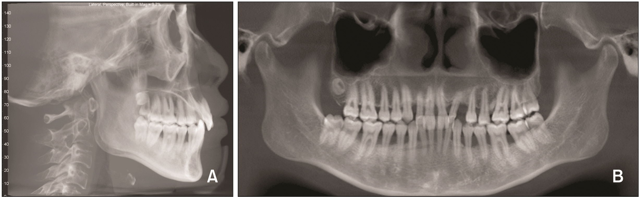 | Figure 2Initial cone-beam computed tomography scan showing cephalometric (A) and panoramic radiographs (B) of an adult with inactive periodontal disease.
|
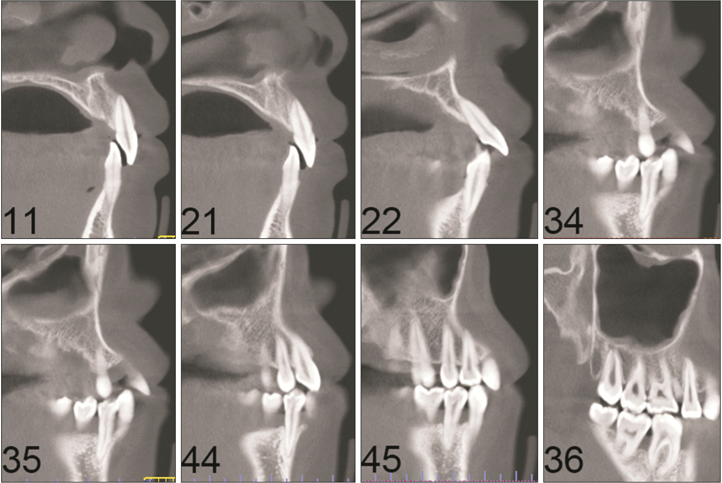 | Figure 3Initial cone-beam computed tomography scan showing dental radiographs of an adult with inactive periodontal disease. The numbers in this figure refer to the FDI notation for permanent teeth.
|
Additionally, the maxillary central incisors and mandibular incisors showed severe lingual inclination (angle between the maxillary incisor and sella–nasion [U1-SN] 81.7°; lower incisor to mandibular plane angle [IMPA] 75.9°,
Table 2). Cephalometric analysis showed Class I skeletal pattern (A point–nasion–B point [ANB] angle of 5.4°,
Table 2) with a slightly high mandibular plane angle (angle between the mandibular plane and sella–nasion [MP-SN] 39.2°,
Table 2). Based on these findings, the patient was diagnosed with skeletal Class I malocclusion with severe periodontal disease.
Table 2
Cephalometric measurements
|
Measurements |
Mean ± SD |
Pretreatment |
Posttreatment |
|
SNA (°) |
82.0 ± 3.5 |
83.4 |
83.1 |
|
SNB (°) |
80.0 ± 3.0 |
78.0 |
77.7 |
|
ANB (°) |
3.0 ± 2.5 |
5.4 |
5.4 |
|
MP-SN (°) |
33.0 ± 6.0 |
39.2 |
40.3 |
|
IMPA (°) |
95.0 ± 7.0 |
75.9 |
87.9 |
|
FMA (°) |
26.0 ± 4.5 |
29.7 |
30.7 |
|
U1-SN (°) |
102.1 ± 5.5 |
81.7 |
93.5 |
|
U1-NA (mm) |
4.3 ± 2.7 |
–0.8 |
1.9 |
|
L1-NB (mm) |
4.0 ± 1.8 |
4 |
7.6 |
|
U1/L1 (°) |
130.0 ± 6.0 |
163.2 |
138.3 |
|
UL-EP (mm) |
–1.5 ± 2.0 |
0.0 |
1.0 |
|
LL-EP (mm) |
–2.0 ± 2.0 |
1.0 |
2.0 |
|
Overjet (mm) |
2.5 ± 2.5 |
5.6 |
3.2 |
|
Overbite (mm) |
2.5 ± 2.0 |
8.6 |
2.4 |
Go to :
TREATMENT OBJECTIVES
The primary treatment goals for this patient with inactive periodontal disease were to:
Move teeth into proper alignment,
Obtain normal overjet and overbite,
Level the curve of Spee,
Maintain the space (7.5 mm) between the maxillary left lateral incisor and maxillary left first premolar for an implant-supported prosthesis, upright the maxillary left lateral incisor, and close residual spaces,
Correct the posterior scissor bite,
Maintainthe patient’s facial profile, and
Establish an esthetic smile by improving the smile arc, leveling the gingival height, and eliminating the lip trap at the maxillary lateral incisors.
Go to :
TREATMENT ALTERNATIVES
Alternative treatment strategies for this patient were restorative or prosthodontic treatment, conventional fixed orthodontic treatment, and orthodontic treatment with clear aligners. The patient was willing to establish appropriate alignment and occlusion of her anterior teeth, which meant that prosthodontic treatment was deemed inappropriate. Excessive tooth removal would be necessary to correct the facially inclined maxillary incisors. Preparing the incisors sufficiently for alignment would not only devitalize the teeth but also leave minimal natural tooth structure to retain the restoration. Orthodontics is typically a better option for correcting apically positioned papillae of anterior teeth due to tooth alignment and interproximal bone loss. Orthodontics before restorative dentistry was recommended to the patient since her misaligned teeth could not be treated with restorative dentistry alone, and this approach could provide an excellent result.
In addition to restorative and prosthodontic options, orthodontic treatment by conventional fixed appliances and clear aligners could be used. Compared with conventional fixed braces, clear aligners are removable and allow patients to practice oral hygiene, which was crucial for this patient, who had periodontal disease. Clear aligners also have an advantage in the segmented movement of teeth to maintain a Class I molar relationship and effectively control anterior intrusion. The displacement-driven system moved the teeth until they lined up with the following staged location of the clear aligners. Such gradual, segmented movement might minimize the excessive proclination of teeth and elicit predictable tooth movement. Clear aligners that required fewer appointments were more suitable for this patient since she lived far away from our office.
Finally, an interdisciplinary treatment plan was adopted, including orthodontic treatment with clear aligners, prosthodontic restoration, and a strictly supervised oral hygiene program. The periodontal pocket depth and bleeding on probing for the periodontal disease were monitored by periodontists throughout the orthodontic treatment period.
Go to :
TREATMENT PROGRESS
Before orthodontic treatment, systemic periodontal therapy was performed, including scaling, root planing, periodontal flap surgery, and oral hygiene instruction. After 6 months of periodontal treatment, the patient was referred to our orthodontic office. Periodontal maintenance was arranged every 3 months during orthodontic treatment.
Clear aligners (Angel Aligner; Angelalgin Technology Inc., Wuxi, China) were used to correct the over erupted and inclined teeth and to close the spaces in the maxillary and mandibular arch. The patient was instructed to wear each aligner for approximately 22 hours every day and move on to the next aligner in the series after 14 days. The treatment progress was divided into two phases: the initial stage (maxillary: 28; mandibular: 28) and the refinement stage (maxillary: 16; mandibular: 11).
A total of 1.9–2.4 mm of anterior incisor intrusion, no extrusion of the posterior teeth, and 14–15 degrees of labial crown movement of lingually inclined incisors were designed in the initial stage of treatment. Optimized attachments and one conventional transverse rectangular attachment were placed on the premolars and the right maxillary canine, providing anchors for the anterior intrusion. Bite ramps built into the lingual surface of the maxillary aligners and elastic deformation of the aligner material generated the force for the intrusion during the first stage (
Figure 4A). Interproximal reduction was employed to reduce the black triangle in the mandibular anterior teeth and provide space for mandibular incisor intrusion. To perform this procedure, fine diamond strips were utilized until the tooth contacts were broken. Then, high-speed burs were used to create 0.5 mm of space in each interproximal contact from the mesial mandibular left second premolar to mesial mandibular right canine (
Figure 4B). Intrusion and lingual or labial movement of the anterior incisors was achieved in 28 steps. The amount of movement was 0.1 mm and less than 1° in one of three dimensions at each step. Maxillary lateral incisors were intruded and moved lingually. Maxillary central incisors and mandibular incisors were intruded and moved labially. The amount of labial movement of the mandibular incisors was no more than that of the maxillary incisors to prevent traumatic occlusion. The left posterior scissor bite was improved by intrusion and lingual inclination of the maxillary second premolar and buccal inclination of the mandibular second premolar with a small amount of intrusion.
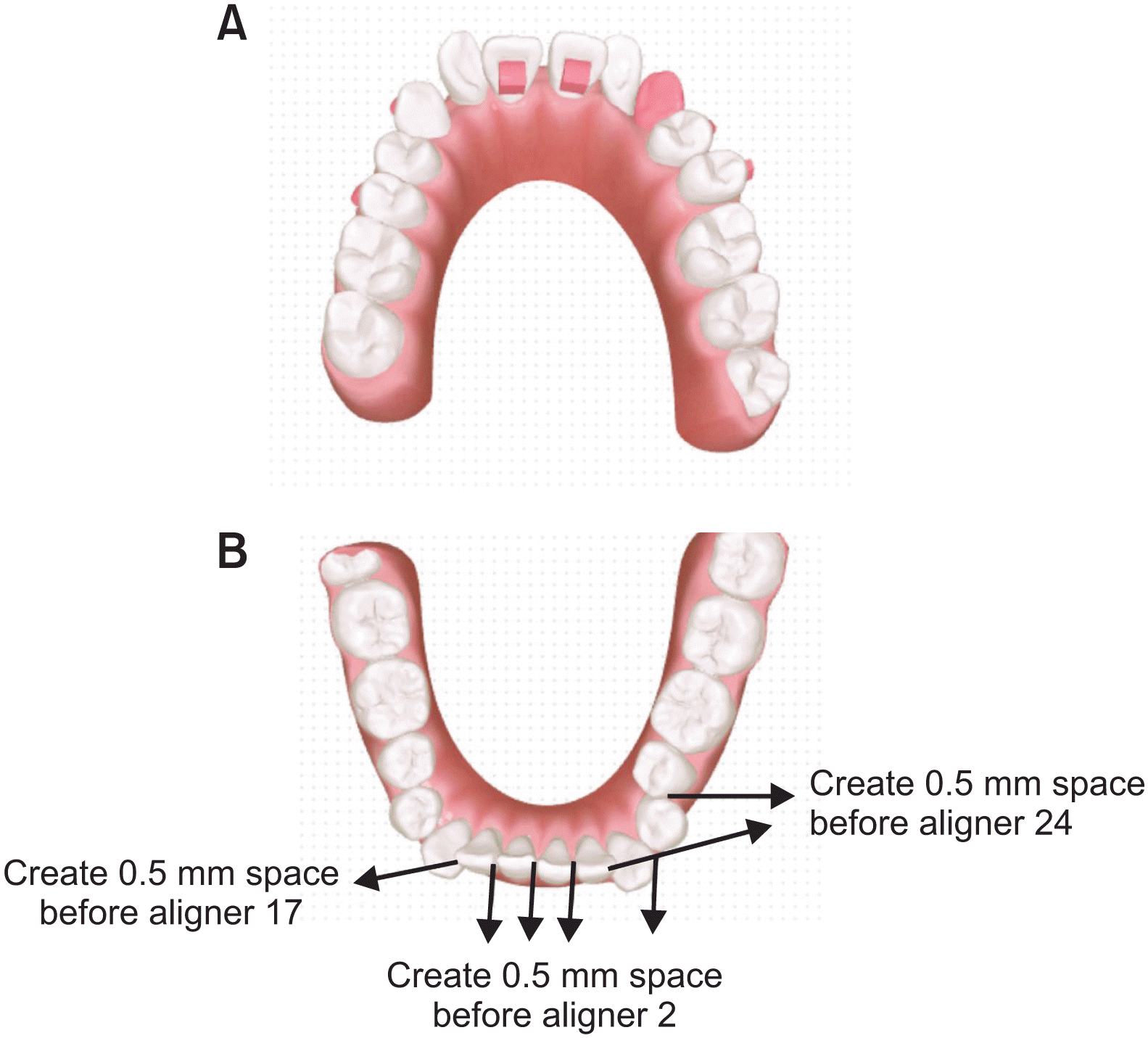 | Figure 4
A, Bite ramps on the lingual surface of the maxillary aligners. B, Interproximal reduction chart.
|
At the end of the initial stage of treatment with aligners 28, we compared the final tooth position with the planned tooth movement (
Figure 5). The results showed that leveling of the bimaxillary arch had almost been completed, the space had been closed, and the deep bite had been corrected. However, the left maxillary lateral incisor was still tilted mesially, and the posterior teeth were not in sound occlusion. Refinement with additional 16 maxillary aligners and 11 mandibular aligners was performed. An additional 22 degrees of mesial root movement of the left maxillary lateral incisor was designed. Rectangular and optimized attachments were bonded to all the premolars and first molars for posterior occlusal settling. Precision cut-outs, where buttons were placed, were designed for cross elastics. Cross elastics from buttons placed on the lingual side of the second mandibular premolar and buccal side of the second maxillary premolar on the left side and anterior bite ramps were used to eliminate the posterior scissor bite with a bite depth greater than 30%.
16 Intermaxillary cross elastics (size 3/16-inch medium, 3.5 ounces) were used for 4 weeks.
 | Figure 5Treatment progress: intraoral photographs 14 months after the start of orthodontic treatment (upper) as compared to patient’s planned tooth positions (lower). The deep bite was corrected, and the spaces between teeth were closed.
|
After 22 months of orthodontic treatment, the mesial inclination of the maxillary left lateral incisor was improved but not corrected. We made additional adjustments for the patient, but she was very pleased with the treatment outcome and refused to continue wearing the adjustment appliances, which is why attachments can be seen in the posttreatment intraoral photographs (
Figure 6). The active treatment lasted 22 months, with 44 maxillary and 39 mandibular aligners. After inactive therapy for 7 months, the patient was referred for prosthodontic treatment. A Maryland bridge, rather than an implant-supported prosthesis, was used for the patient due to the angulation of the maxillary left lateral incisor. The patient was instructed to wear vacuum-formed retainers full-time. Periodontal maintenance appointments were recommended to be arranged every 4 months.
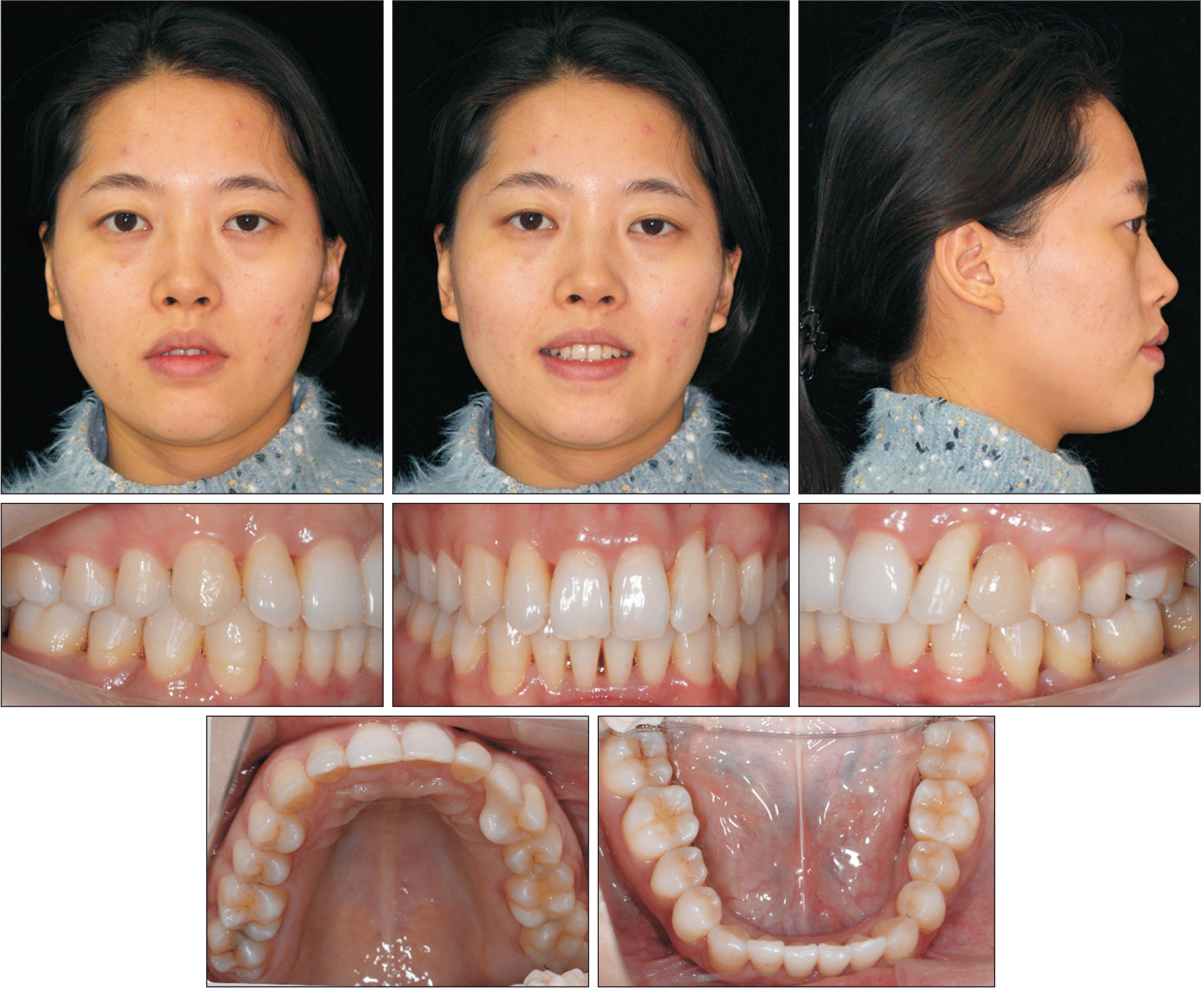
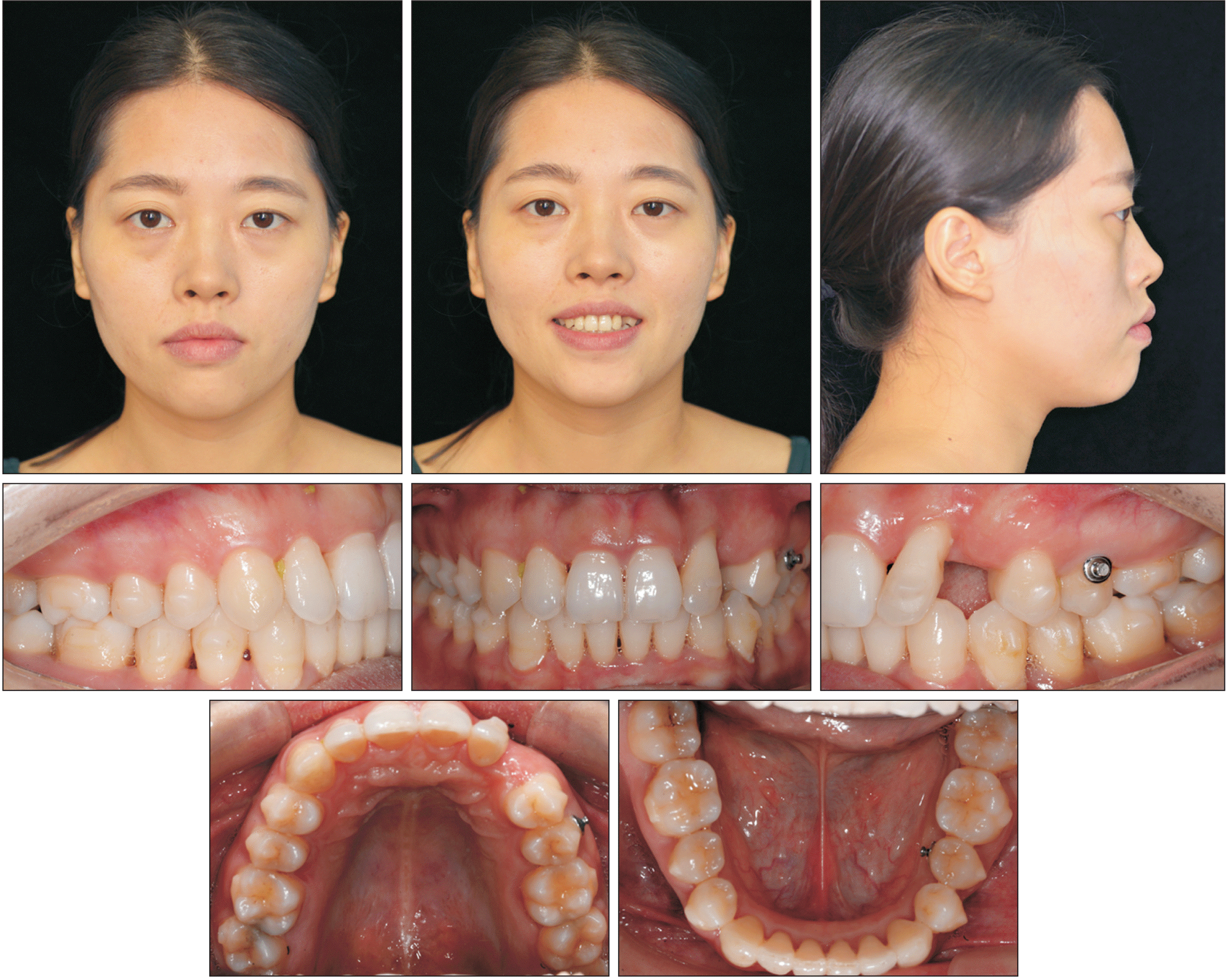 | Figure 6Posttreatment facial and intraoral photographs of an adult with inactive periodontal disease treated with clear aligners. Proper overjet and overbite were achieved, and the lateral profile was maintained.
|
Go to :
RESULTS
Functional and esthetic outcomes were achieved after orthodontic treatment. The harmony between the arc of curvature of the edges of the maxillary incisors and the maxillary border of the mandibular lip resulted in a perfect smile. Normal overjet and overbite were established with better torque and occlusion in the anterior region. The Class I canine and molar relationship was maintained, with a slight midline deviation between the maxillary and mandibular midlines (
Figure 6). In this case, the pretreatment Peer Assessment Rating (PAR) score was 24, and the posttreatment PAR score was 4. There was an 83% reduction (percentage) in the PAR score, indicating improvement.
No significant difference in sagittal skeletal outcomes was observed, as shown in
Figure 7A. The posttreatment cone-beam computed tomography image showed proper space closure and parallel roots, except for the maxillary left lateral incisor (
Figure 7B). Sound periodontal support was maintained in the mandibular left first and second premolars and even improved after orthodontic treatment (
Figure 8,
Table 3). The most notable cephalometric changes were the labial tipping and intrusion of the maxillary central incisors and mandibular incisors. The position of the anterior incisors in the basal bone was improved (U1-SN 93.5°; IMPA 87.9°,
Table 2).
 | Figure 7Posttreatment cone-beam computed tomography scan showing cephalometric (A) and panoramic radiographs (B) of an adult with inactive periodontal disease treated with clear aligners.
|
 | Figure 8Posttreatment cone- beam computed tomography scan showing dental radiographs of an adult with inactive periodontal disease treated with clear aligners. The numbers in this figure refer to the FDI notation for permanent teeth.
|
Table 3
Results of posttreatment periodontal examination
|
Maxillary teeth |
17 |
16 |
15 |
14 |
13 |
12 |
11 |
21 |
22 |
23 |
24 |
25 |
26 |
27 |
Pocket depth (labial)
(mm) |
111 |
111 |
111 |
111 |
111 |
212 |
211 |
112 |
212 |
|
212 |
212 |
222 |
222 |
|
Bleeding on probing |
|
|
|
|
|
|
|
|
|
|
|
|
|
|
Pocket depth (lingual)
(mm) |
111 |
111 |
111 |
111 |
111 |
112 |
211 |
112 |
222 |
|
222 |
212 |
222 |
222 |
|
Bleeding on probing |
|
|
|
|
|
|
|
|
|
|
|
|
|
|
|
Mandibular teeth |
47 |
46 |
45 |
44 |
43 |
42 |
41 |
31 |
32 |
33 |
34 |
35 |
36 |
37 |
Pocket depth (labial)
(mm) |
113 |
212 |
222 |
111 |
222 |
222 |
212 |
222 |
222 |
212 |
233 |
322 |
323 |
322 |
|
Bleeding on probing |
|
|
|
|
|
|
|
|
|
|
*** |
*
|
|
|
Pocket depth (lingual)
(mm) |
122 |
333 |
333 |
323 |
322 |
322 |
222 |
222 |
222 |
323 |
323 |
223 |
234 |
222 |
|
Bleeding on probing |
|
|
|
|
|
|
|
|
|
|
*** |
** |
** |
*
|
Moreover, the buccal bone thickness of the maxillary incisors increased. The posttreatment lateral cephalometric analysis and superimposition displayed no significant skeletal changes (ANB, 5.4°,
Table 2,
Figure 9) and revealed a slight change in the mandibular plane angle (MP-SN, 40.3°,
Table 2,
Figure 9). Mandibular movements were regular, without signs or symptoms of temporomandibular disorders. The active treatment duration was 22 months. The records taken at 7 months posttreatment showed that the results remained stable, and an adhesive fixed partial denture was placed in the maxillary arch (
Figure 10).
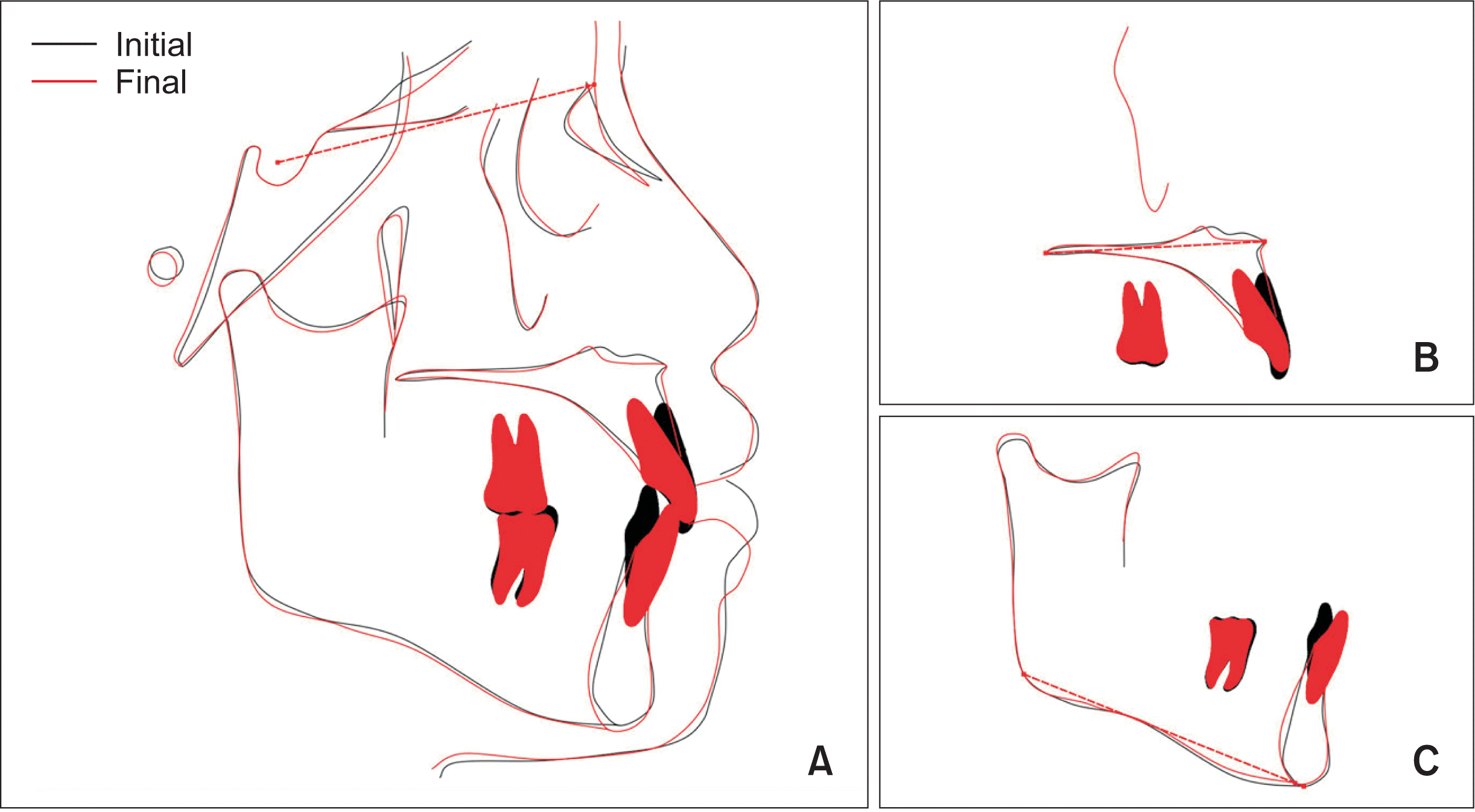 | Figure 9Superimposed cephalometric tracings showing the changes from pretreatment (black) to posttreatment (red) in an adult with inactive periodontal disease treated with clear aligners. A, Superimposed on the sella-nasion plane at sella. B, Superimposed on the palatal plane at the anterior nasal spine. C, Superimposed on the mandibular plane at the menton. The intrusion of the incisors was achieved, and the inclination of the anterior teeth was improved.
|
 | Figure 10Facial and intraoral photographs at 7 months after treatment. Prosthodontic rehabilitation of the left maxillary canine yielded optimal esthetic results.
|
Go to :
DISCUSSION
Orthodontic treatment using clear aligners assisted by periodontal treatment achieved an excellent final result in a conscientious and precise manner, with the preferable occlusal force exerted through the teeth, thus preventing traumatic occlusion and periodontitis.
17 The proclination of the maxillary left lateral incisor and extrusion of both maxillary and mandibular incisors in this patient due to her periodontal disease were challenges for the prosthodontists. Importantly, prosthodontic treatments, including removable partial dentures, fixed bridges, and dental implants, cannot resolve deep overbites and esthetic problems in the anterior region. As other studies have suggested, the orthodontic treatment of adult patients with periodontitis could achieve a more stable, functional occlusal relationship.
18,19 Before orthodontic treatment, periodontal therapy was performed until the periodontal condition was stable. Moreover, the patient’s periodontal status was strictly monitored before, during, and after active orthodontic therapy.
Some researchers have reported no significant differences in plaque or gingival indexes between treatment with fixed appliances and clear aligners in patients with periodontitis.
17 In general, fixed orthodontic appliances can increase plaque accumulation and bacterial colonization, reducing the efficacy of oral hygiene.
20 Clear aligners have increased advantages over regular fixed orthodontic appliances.
21,22 Jiang et al.
14 also indicated in their systematic review that clear aligners, as a type of removable appliance, were more conducive to oral hygiene and periodontal health than fixed appliances. Better periodontal maintenance was one of the reasons for treatment with clear aligners in this case.
Carefully created designs contributed to the patient’s good occlusion and periodontal health. A 0.75-mm-thick appliance was applied to achieve smaller tooth movements of 0.10 mm and 1 degree per step, compared with 0.25 mm and 2 degrees per step in healthy patients without periodontitis. The precise design of the amount of movement induced by clear aligners can more accurately control the size of the applied force. The instantaneous pressure applied when the aligners were inserted and removed was improved by blocking out the undercuts and trimming the edge of the aligners 1–3 mm shorter than those used in healthy patients according to the periodontal status (
Figure 11). These design features also made the aligners easy to wear, considering the diverse angles between the left central and lateral incisors. All the above modifications were used to achieve the suggested light orthodontic forces for adults with a history of periodontal disease.
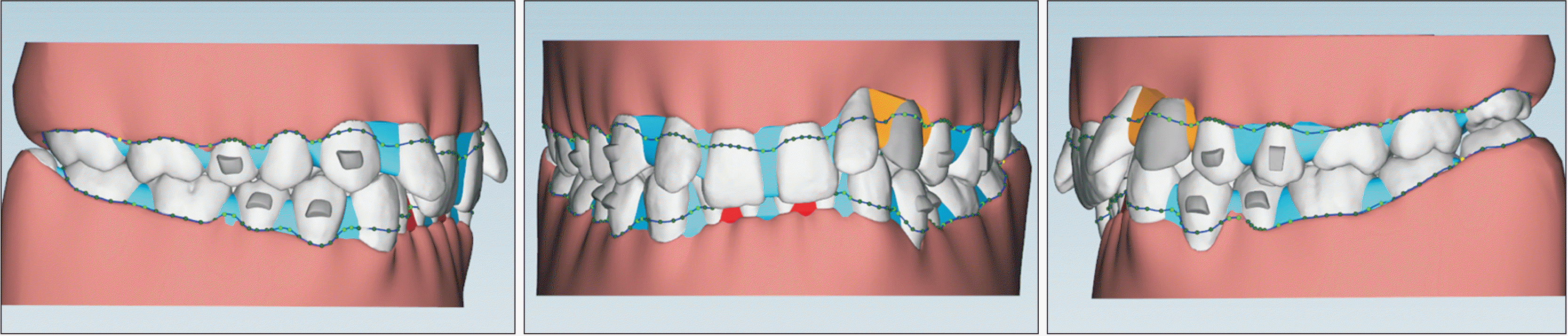 | Figure 11Blocking out the undercuts and trimming the line of the aligners. These designs applied gentle instantaneous force when inserting and removing the aligners to protect the periodontal tissues. The following gaps are considered to be eliminated in this case: (i) triangle-shaped gaps between teeth; (ii) the gaps between the anterior teeth are greater than 1 mm, and (iii) the gaps between the posterior teeth are greater than 2 mm. Blue means undercut areas between the teeth that need to be eliminated. Orange means undercut areas around the virtual pontic for the right maxillary canine in each aligner need to be blocked out. The dashed line is the boundary of the aligners.
|
Planning the design of clear aligners for patients with a deep overbite is still a major challenge for orthodontists. Deep overbites are generally corrected by posterior extrusion and/or anterior intrusion. Due to the profile and lip position in this case, the patient was better suited to being treated by anterior intrusion, which included relative intrusion and some actual intrusion (
Figure 9). The relative intrusion of anterior teeth was gained through the labial inclination of the maxillary central incisors and mandibular incisors. The actual intrusion was not pursued until the anterior incisors were appropriately positioned within the alveolar bone. Incisal flaring is often observed as a side effect when intruding teeth by using fixed appliances. It is also troublesome to control the extent of labial movement of the maxillary central incisors using a fixed orthodontic appliance, preventing the proclination of the incisors. For this patient, the excessive labial inclination of the maxillary incisors was harmful to the profile. However, we improved the torque of the maxillary central incisors step by step and evaluated the relationship between the axial inclination of the maxillary incisors and the lip position in a timely manner by using clear aligners.
Periodontal health was also considered in the attachment design. A small amount of tooth movement in the posterior segments was allowed because posterior teeth were used as anchors to correct the severe deep bite during the first treatment phase. Since the larger teeth of periodontal patients allow aligners to fit firmly, only three beveled horizontal rectangular attachments and a rectangular attachment were placed on the premolars and the right maxillary canine. This design also made it easier to insert and remove the aligners, thus further reducing the forces experienced by the patient upon insertion and removal. After the anterior teeth were correctly oriented, attachments for rotation correction and root uprighting were set on the posterior teeth for occlusal adjustment during the refinement process.
Ultimately, excellent control of the torque of incisors facilitates the stabilization of anterior tooth roots in the central region of the alveolar bone, eliminating the abnormal direction of the bite force and helping to maintain good periodontal status. The results demonstrated that the orthodontic therapy did not result in any irreversible periodontal destruction. This interdisciplinary approach led to good functional and esthetic outcomes, providing the optimal treatment plan in a complex clinical case, and these results agree with those of a study by Lee et al.
23 Follow-up at 7 months posttreatment confirmed the excellent choice of treatment design and the stability of the occlusion.
However, there are still some deficiencies in this case. A slight deviation of the mandibular midline remained. The results might have been better if the patient had completed the modification phase. Additionally, the maxillary lateral incisor showed slight mesial inclination. We speculated that the angulation was not corrected due to the attachment design. During the initial stage of treatment, no attachments were added to the maxillary left lateral incisor to avoid applying too much force to the teeth while inserting and removing the aligners. During treatment refinement, an optimized attachment was used. However, for teeth that need excellent control of root angulation, the optimized attachments are not as effective as root control attachments. Root control attachments include a gingival attachment to allow the aligner to produce a force in the direction of root movement, and the second attachment can be placed occlusally, providing a second-order moment. Therefore, root control attachments are recommended for achieving good root angulation.
Go to :
CONCLUSIONS
The mechanism responsible for overbite opening in this patient was primary proclination of the inclined incisors and intrusion of anterior incisors. Continuous and repeated professional periodontal maintenance, good oral hygiene, and motivation are vital elements for the successful orthodontic management of periodontal patients. Orthodontic treatment with clear aligners for periodontal patients may provide a substantial mutual benefit in periodontal therapy and prosthodontic treatment. Clear aligners might be better recommended for patients with adequately controlled chronic periodontitis.
Go to :
SUPPLEMENTAL VIDEO
Go to :


 PDF
PDF Citation
 Print
Print


 XML Download