

 PDF
PDF Citation
 Print
Print


INTRODUCTION
Moebius syndrome (MBS) is characterized by congenital, unilateral or bilateral, non-progressive facial weakness and limited abduction of the eyes.1 Von Graefe first described bilateral facial paresis in 1880.2 In 1888, Moebius classified various congenital cranial nerve palsies and singled out the combined 6th and 7th cranial nerve weakness.2 It is a rare neurologic disorder estimated to occur in one in every 50,000 live births and is reported to have no sex differences.1
The etiology and pathogenesis of the syndrome are unclear, but two explanations have been proposed, a primary genetic cause and a primary ischemic cause.3 Teratogenicity is suggested to be an important etiologic factor in both explanations.3
This syndrome is diagnosed when there is a unilateral or bilateral non-progressive pattern of facial paralysis and limited ocular abduction. Because it is diagnosed based on clinical symptoms, the CLUFT (cranial nerve, lower limb, upper limb, face, and thorax) criteria are used for the evaluation.4 Patients with MBS are usually diagnosed in the early neonatal period because patients do not suck as strongly and drool due to incomplete lip sealing and hypotonic mimetic and lip muscles. Patients have also been observed to have incomplete eyelid closure during sleep, ptosis, and defects in abductive and adductive ocular movements.5,6 As children start to learn to speak, they may have some difficulties making sounds that involve using the lips and tongue, and making facial expressions.6 In approximately one-third of MBS patients, limb malformation is seen, the most common of which is club foot; hand anomalies also occur and may be bilateral or unilateral.5,7 There are other similar disorders that exhibit the symptoms of MBS. The oromandibular limb hypogenesis syndromes (OLHS) are considered a group of rare conditions characterized by congenital malformations involving the tongue, mandible, and limbs. Six defined syndromes are gathered under the umbrella term OLHS, including MBS, hypoglossia-hypodactylia syndrome, Hanhart syndrome, glossopalatine ankylosis syndrome, limb deficiency-splenogonadal fusion syndrome, and Charlie M. syndrome.5,8
Reports on the clinical, genetic, neurophysiological characteristics, and surgical treatment of MBS have been published by numerous authors after Von Graefeʼs first report, published in 1880.9 However, there is no clear protocol for treatment, which is performed according to the symptoms. Children with MBS have a complex of signs and dysfunction affecting motor skills, feeding, communication, and cognition.9 Therefore, patients’ diagnosis and treatment require a multidisciplinary engagement from a team of specialists, including pediatricians, neurologists, plastic surgeons, maxillofacial surgeons, orthodontists, ophthalmologists, and speech pathologists.6,10 However, there are very few publications on the orthodontic treatment protocol for dentoskeletal anomalies and long-term stability in patients with MBS.11
Therefore, we report the orthodontic treatment and long-term stability of a patient diagnosed with MBS at the age of 2 years and who displayed the typical symptoms.
DIAGNOSIS AND ETIOLOGY
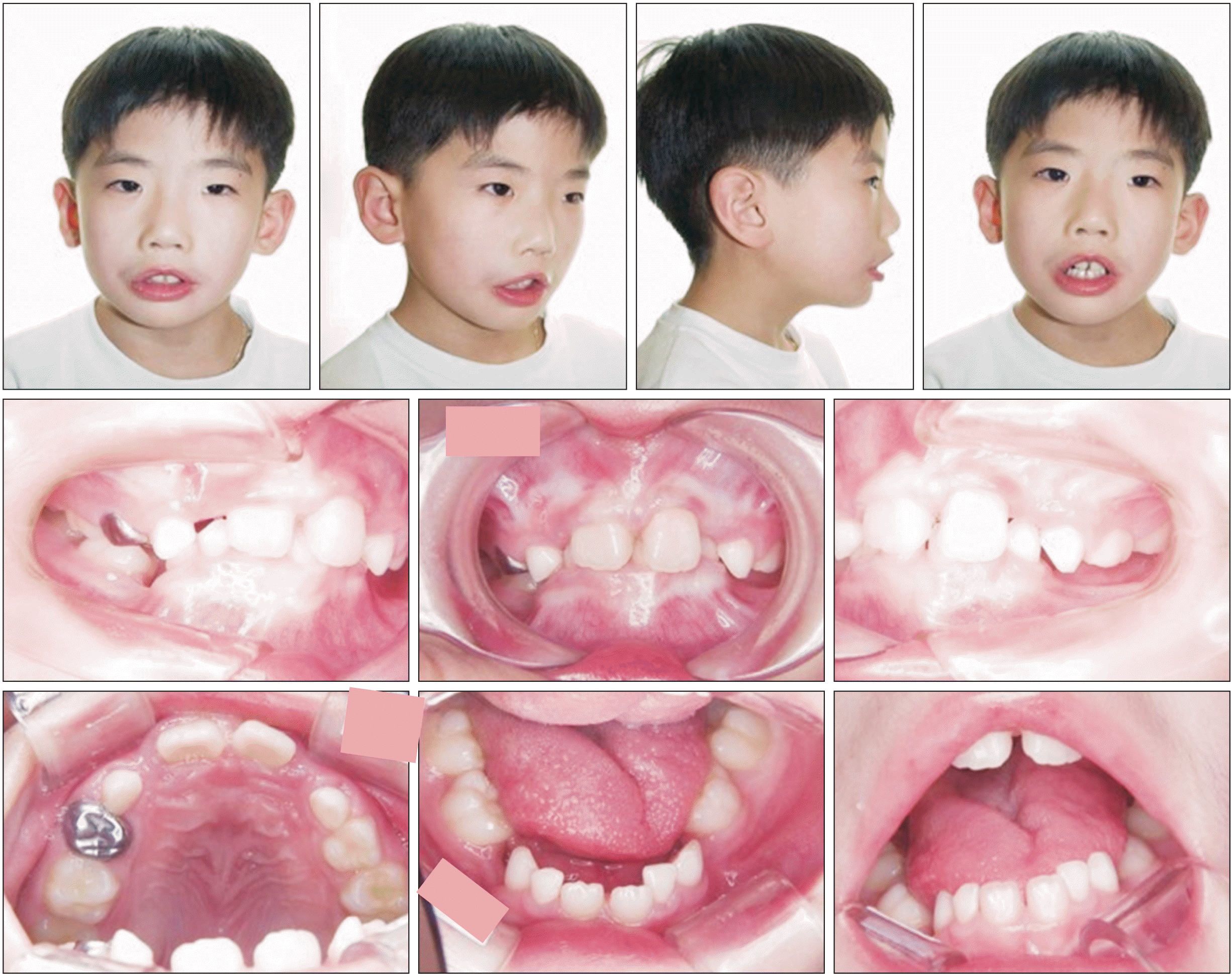
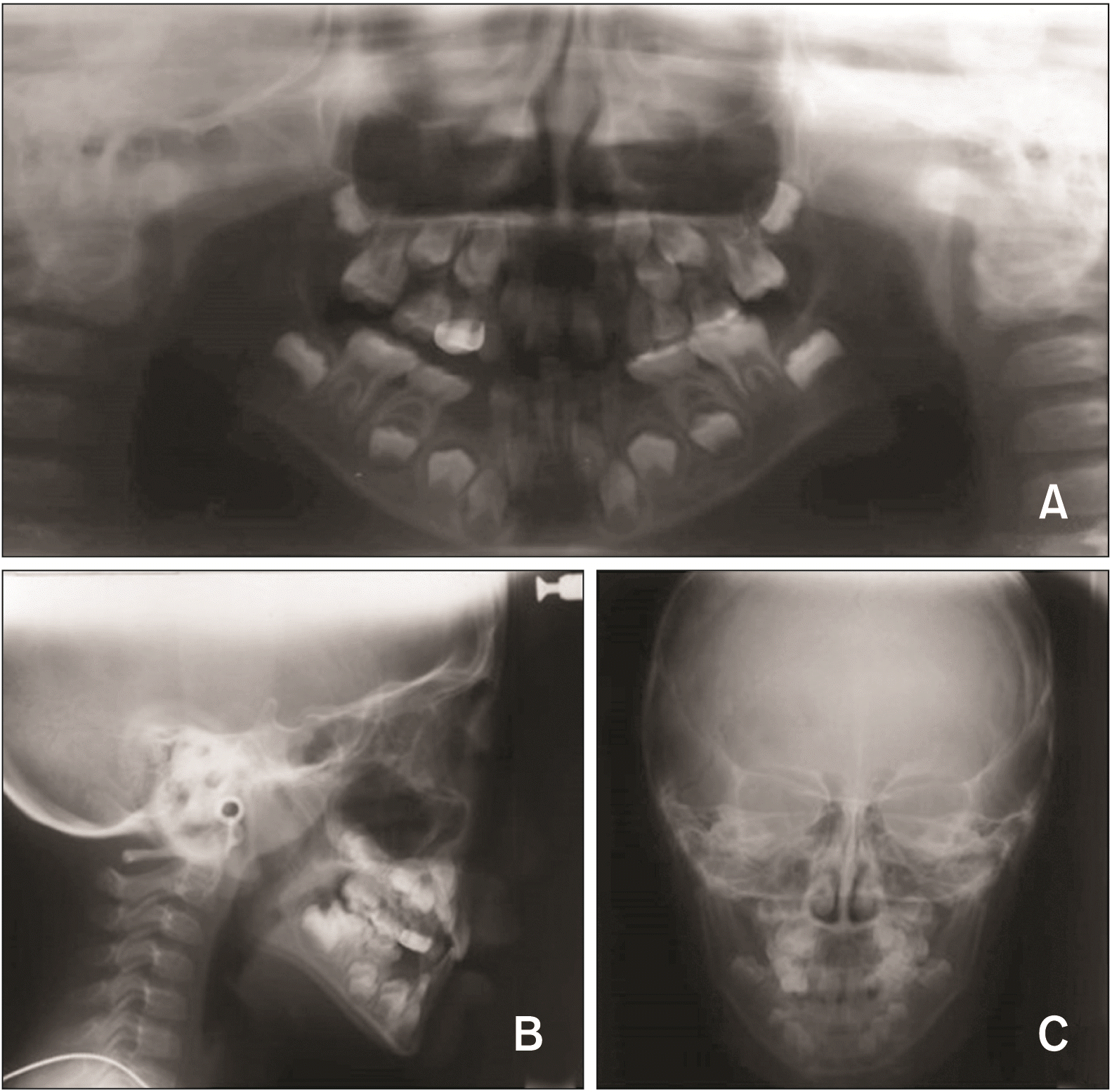
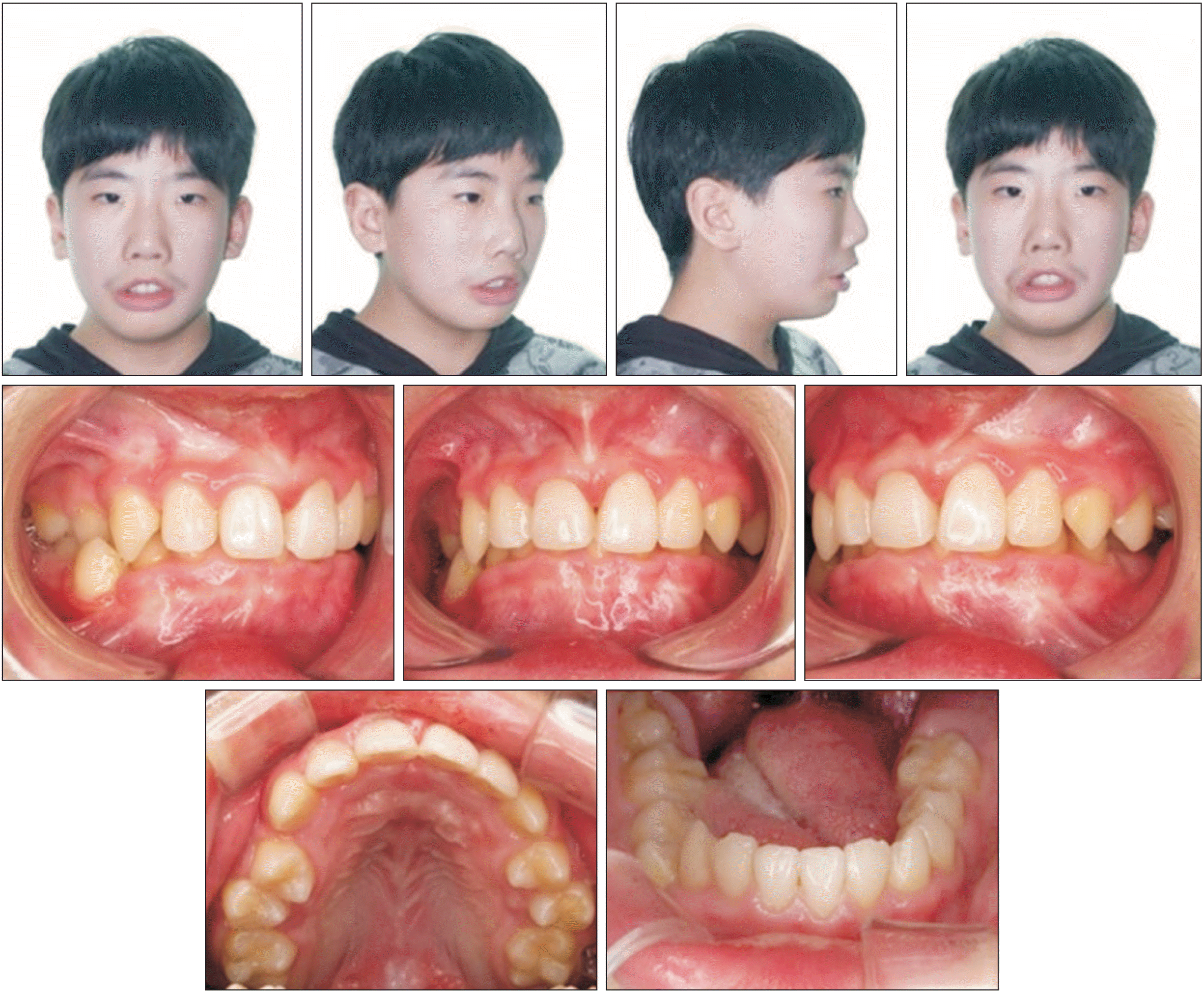
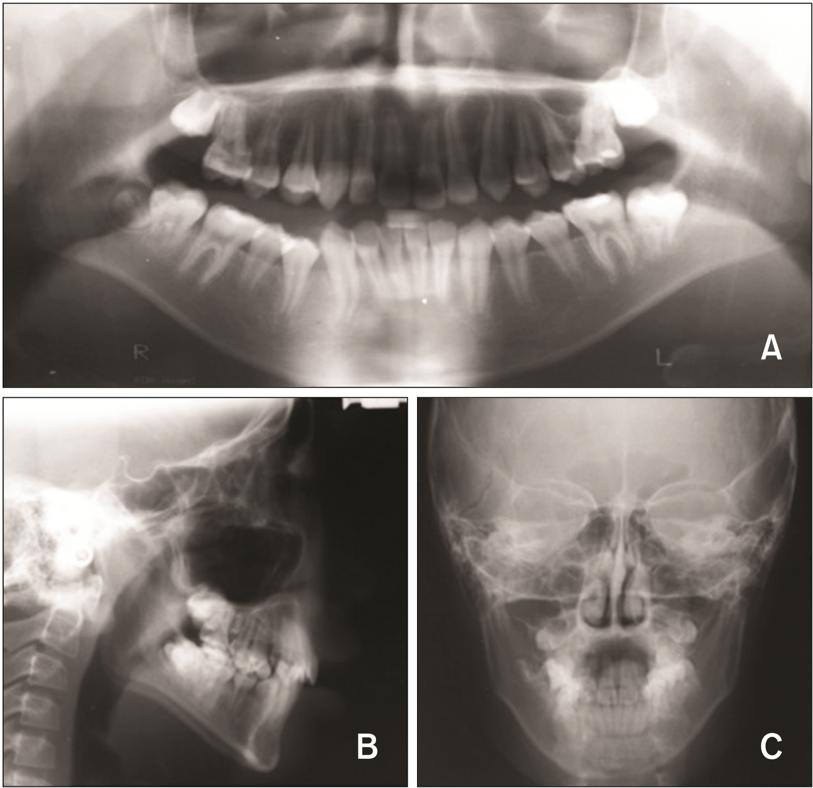
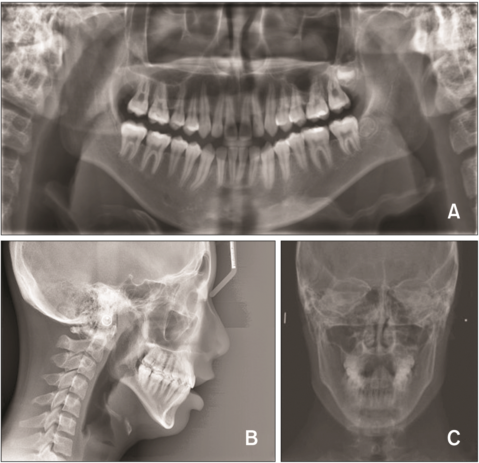
A 7-year-old male patient, who was diagnosed with MBS at the age of 2 years at Hanyang University Hospital, Seoul, Korea, presented for orthodontic treatment at Gachon University Gil Medical Center, Incheon, Korea. He had previously undergone various surgeries at other institutions, including clubfoot surgery at the age of 2 years and strabismus surgery at the age of 5 years. He displayed the typical clinical symptoms of MBS that included lower limb deformity, inability to implement eye movements, inadequate facial expressions, and limitation and deformity of tongue motion and mouth opening limitation. The intraoral examination showed anterior deep bite, left scissor bite, mandibular narrow arch shape, and generalized lack of eruption space. A panoramic radiograph showed this lack of eruption space and no congenital missing teeth. Cephalometric analysis showed a skeletal Class II pattern (A point-nasion-B point angle [ANB]: 4.88°), hyperdivergent pattern (mandibular plane angle: 40.92°), and lingual inclination of mandibular incisor (lower central incisor to mandibular plane angle [IMPA]: 75.29°). From the clinical and radiographic findings, this patient was diagnosed with skeletal Class II malocclusion with deep bite and left-side scissor bite (Table 1, Figures 1 and 2).
TREATMENT
A two-stage treatment was planned to resolve the skeleto-dental problems.
Phase I
Objectives and treatment plan
The following treatment objectives were planned: (1) left scissor bite correction, (2) space management of mandibular left dentition, (3) permanent tooth eruption path guide, and (4) establishment of stable molar occlusion and long-term stability.
Treatment alternatives
For patients with skeletal Class II malocclusion in the initial mixed dentition, it is necessary to consider observing the growth up to the early permanent dentition and then to implement 1 stage orthodontic treatment with growth control. However, in this case, 2 stage orthodontic treatment was considered. Primary treatment was required to correct the left scissor bite and resolve and prevent the deterioration of the developmental abnormality of the mandible, due to the tongue deformity and motion limitation. After permanent tooth eruption, the patient could receive secondary orthodontic treatment to improve the oral and facial functions and the esthetics. This treatment plan was explained to the patient and guardian, and it was decided to implement the two-stage treatment with the consent of the guardian.
Treatment progress
Treatment for the left scissor bite correction was initiated using a removable appliance. A hook was made on the left side of the mandibular plate, and a button was bonded to the lingual cusps of the left mandibular primary second molar and permanent first molar, and buccal traction was started using elastic (1/8-inch [in], 3.5 oz). The patientʼs compliance was good, and the appliance was maintained in place for 16 to 18 hours every day.
Treatment outcomes and follow-up results
After 5 months of treatment, the left scissor bite had improved, and the retainer was subsequently delivered (Figures 3 and 4). The patient visited the hospital every 3 months and received follow-up assessments. Although stable molar occlusion was maintained, the deep overbite continued with a narrow mandibular width. Because of the dysfunctional self-cleansing, restricted tongue movement, incomplete lip sealing, and mouth opening limitation, treatment of caries and prophylactic procedures such as fluoride application were regularly performed.
Phase II
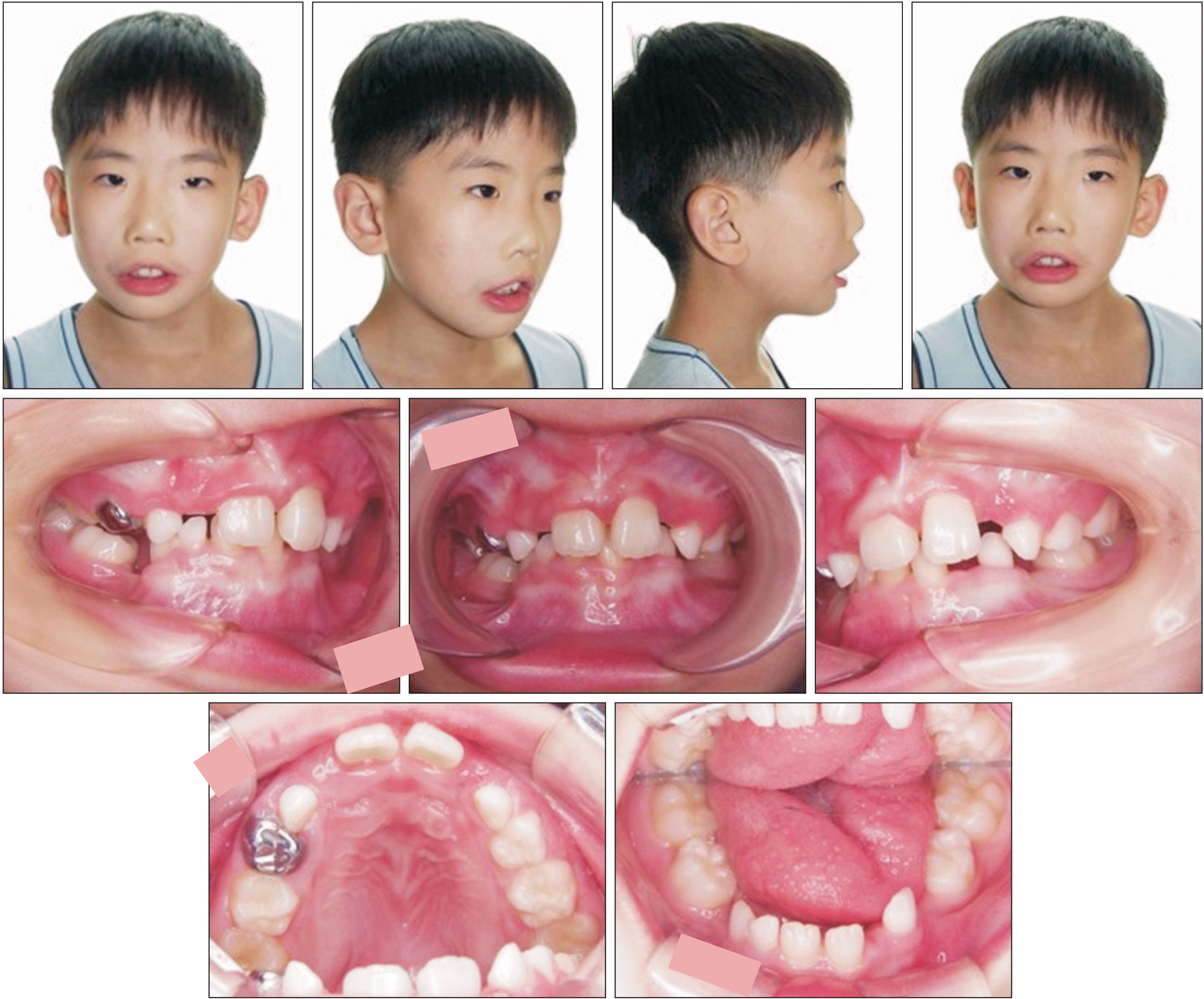
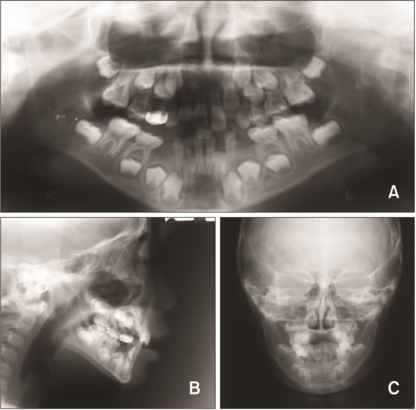
After 4.5 years of phase I treatment, a diagnostic examination in preparation for the next stage was performed when the patient was 12 years old. The intraoral examination found a midline discrepancy, anterior deep bite (overbite: 5 mm), left premolar scissor bite, right first premolar crossbite, mandibular narrow arch shape, tongue deformity and motion limitation, and lack of mandibular eruption space (arch length discrepancy: 3.40 mm). The cephalometric analysis found a skeletal Class II pattern (ANB: 5.57°), hyperdivergent pattern (mandibular angle: 40.19°), and lingual inclination of the mandibular incisor (IMPA: 77.65°). The patient was diagnosed with skeletal Class II malocclusion with a deep bite (Table 1, Figures 5 and 6).
Objectives and treatment plan
The following treatment objectives were planned: (1) deep bite correction, (2) expansion of the mandibular arch width, and (3) establishment of normal occlusion.
Treatment alternatives
In deep bite patients, a treatment plan for orthognathic surgery or incisor intrusion, molar extrusion may be required to address the functional and aesthetic needs of the patient while considering the pattern of skeletal growth. In this case, we considered that the deep bite was caused by the excessive growth of the maxilla and the inadequate growth of the mandible. Thus, deep bite correction is necessary to first resolve the maxillary and mandibular anterior interference and induce the growth of the mandible. When the anterior interference was resolved, the position of the mandibular condyle was moved more to the center, so that the anterior and lateral growth of the mandible could be induced.12
Surgery is considered an option to correct deep bite, cross bite, scissor bite, and skeletal discrepancy, when growth is complete. However, in this case, the skeletal discrepancy was not severe, so we recommended observing the growth pattern of the mandible. It was explained that surgical correction would be possible if the severe dentoskeletal abnormalities continued. The patient and guardian opted for fixed orthodontic treatment as a more reliable treatment option and accepted the fact that orthognathic surgery could be considered after the end of the patient’s growth.13
Treatment progress
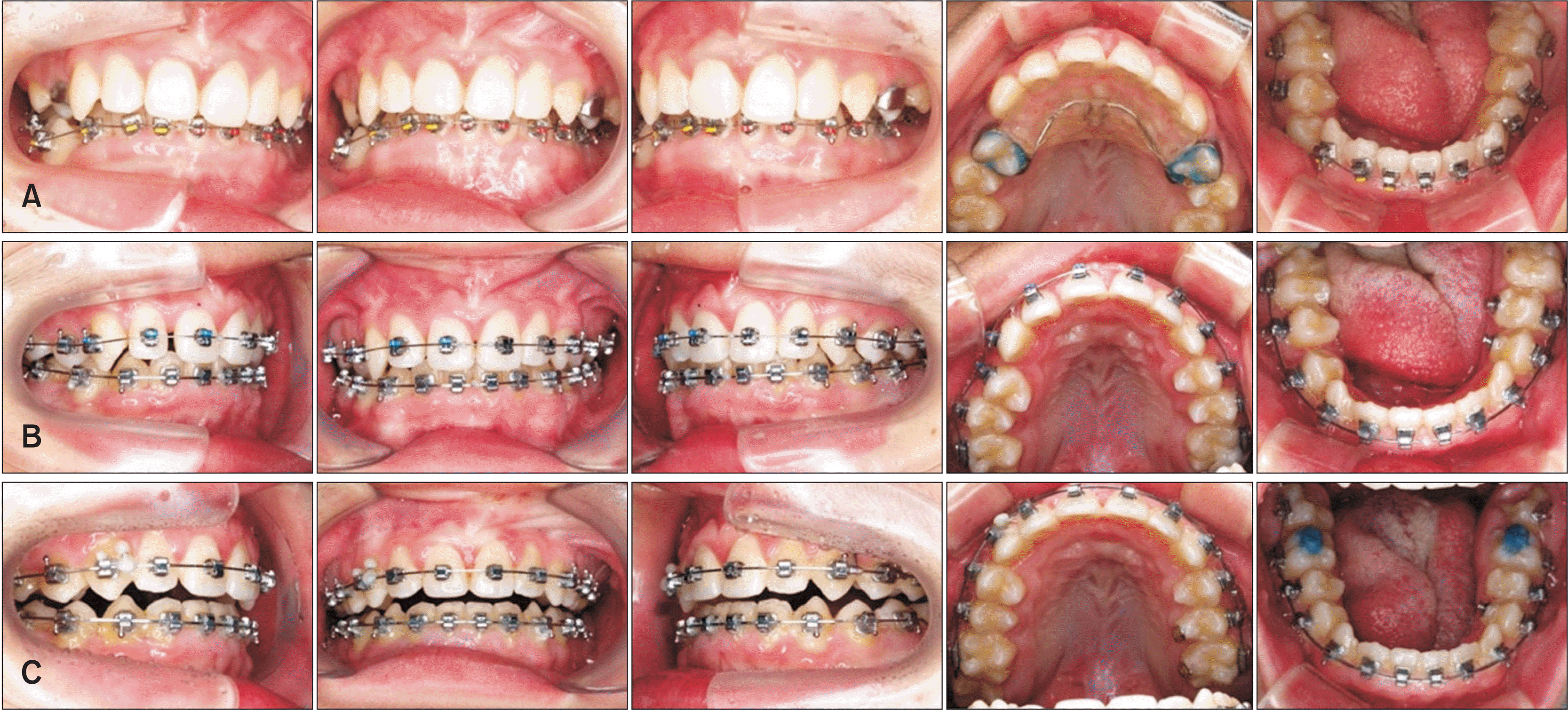
Treatment was initiated by setting the anterior bite plate for deep bite correction with lower molar extrusion and lower incisor intrusion. After that, the lower teeth were bonded with the 0.018 slot, Roth prescription self-ligation SPEED bracket (SPEED System Orthodontics, a division of Strite Industries, Cambridge, ON, Canada) (Figure 7A). Lower arch leveling was performed with a 0.012-in nickel-titanium (NiTi) wire, 0.014-in NiTi wire, 0.016 × 0.016-in reverse-NiTi wire, and 0.016 × 0.022-in NiTi wire. The wide width arch form was used during the initial leveling to extend the width between the canines and the premolars. During leveling, up and down cross elastics (5/16-in, 3.5 oz) were applied between the left upper premolars buccal button and lower premolars lingual button for left premolar scissor bite correction.
After 7 months of treatment, the upper anterior bite plate was removed; the upper teeth were bonded with a SPEED bracket, and leveling was performed (Figure 7B). With the upper and lower 0.016 × 0.022-in stainless steel arch wire, after 10 months of treatment, occlusal correction was performed using various types of up and down elastics. After 18 months of treatment, to arrange the lingual inclination of both the lower second molars, bite raising was performed on both lower first molars; the bracket was then bonded, and leveling was done (Figure 7C). After 30 months of treatment, the alignment of the dental arch and stable molar occlusion was achieved. The appliances were removed, and the removable type retainer was delivered (Figure 8).
Treatment outcomes & follow-up results
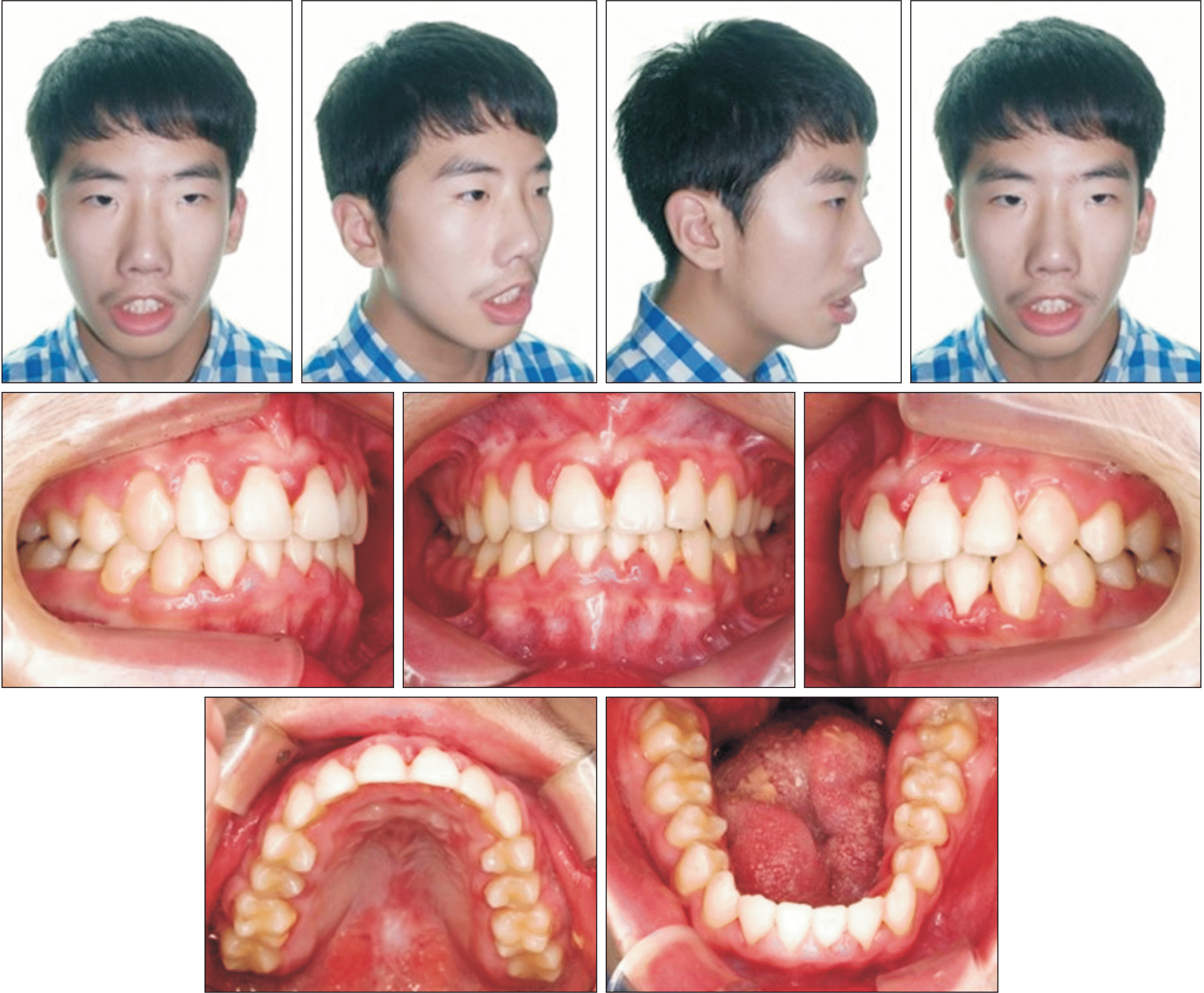
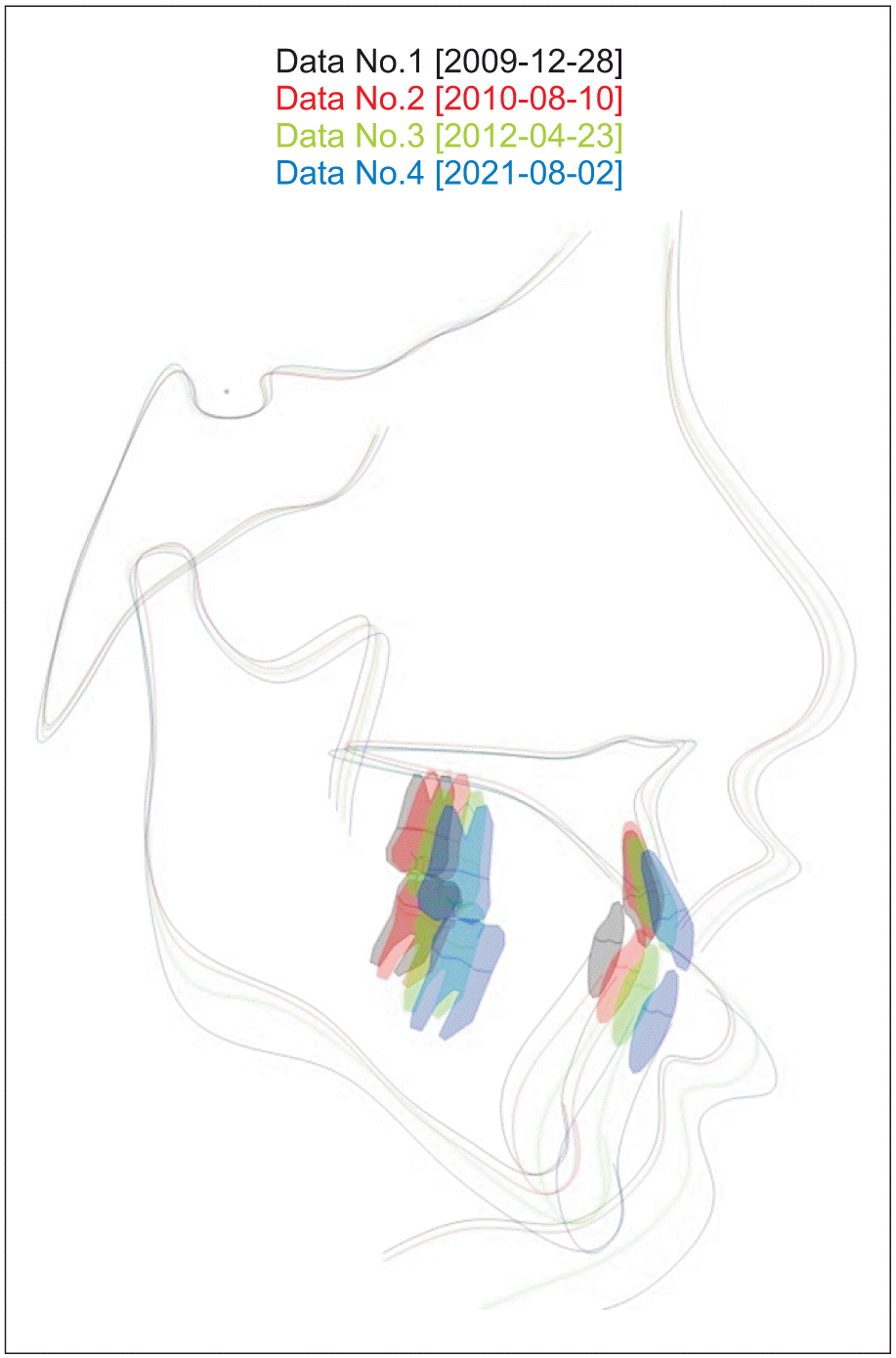
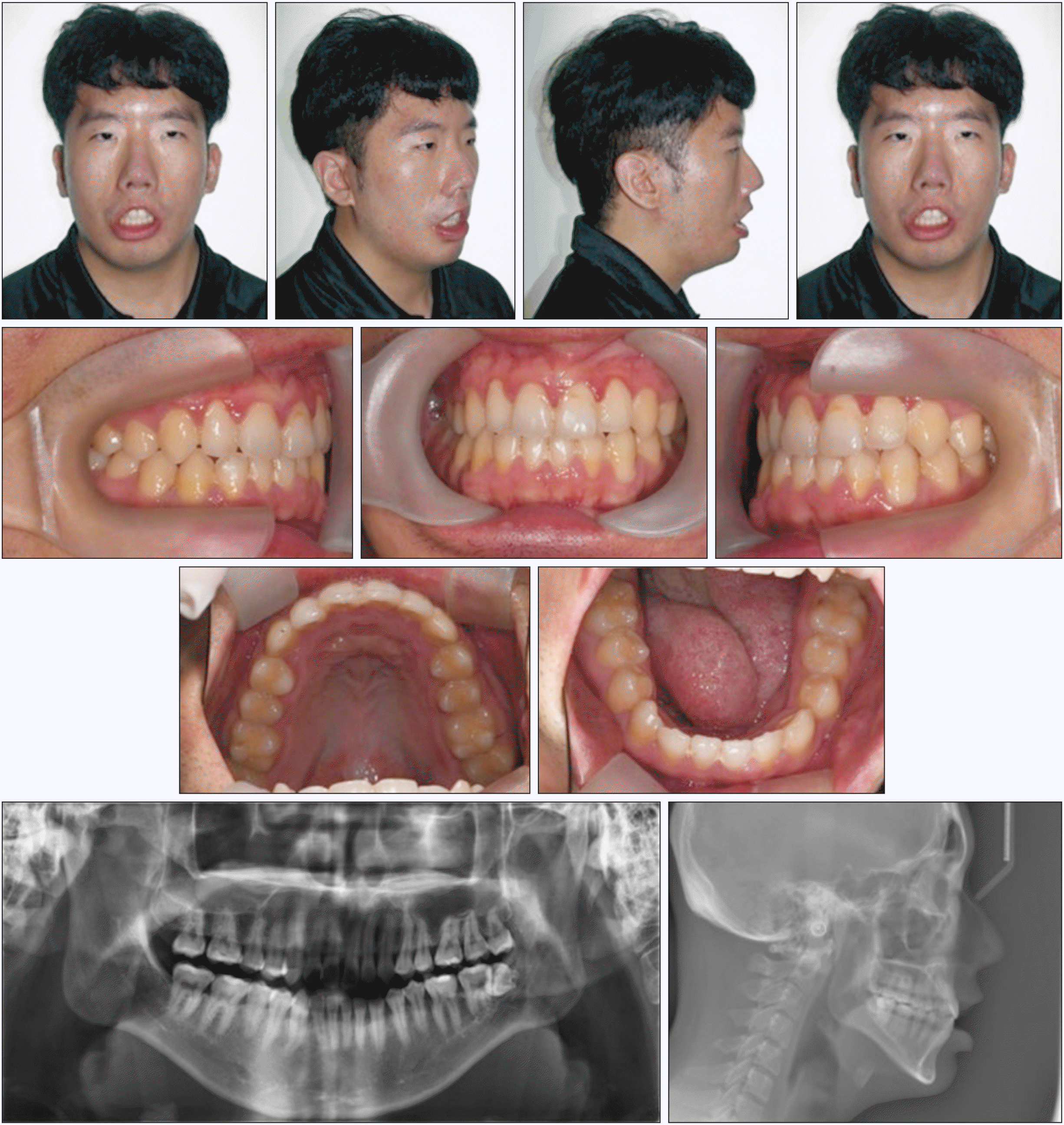
The post-treatment intraoral photos revealed a Class I canine and molar relationship and normal occlusion with appropriate overjet/overbite (Figure 8). The lateral cephalogram revealed that the values of the upper central incisor to sella-nasion (SN) and IMPA angles had increased, which produced more labioversion of the incisors in relation to each other (Figures 9 and 10). The value of sella-nasion-B point angle (SNB) increased, indicating anterior growth of the mandible relative to the cranial base (Table 1, Figure 10). The ratio between the posterior facial height and anterior facial height (PFH/AFH) decreased at the end of the treatment; however, PFH/AFH increased after 9.4 years of retention to 65.75% and reached a value close to the norm (67.24 ± 3.69%) (Table 1). There were no significant changes in the patient’s facial profile as it was not accompanied by surgical rehabilitation, such as orthognathic surgery or smile surgery.
We instructed the patient to use a retainer all day except during meals as the patient had a high possibility of mandibular collapse, due to his tongue deformity and hypotonic lip muscle. The patientʼs cooperation was good, the retainer was continuously used, and the follow-up checks could be performed. Slight tooth movements, such as edge-to-edge bite of the right lateral incisor, were observed; however, function and occlusion remained stable, with a Class I canine and molar relationship. A normal overjet/overbite was maintained, with good PFH/AFH after 9.4 years of retainer use (Table 1, Figure 11).
DISCUSSION
Studies reporting MBS in Korea are rare, and papers on orthognathic surgery with orthodontic treatment for MBS have been reported,13,14 but there are few studies reporting treatment inducing mandibular growth through orthodontic treatment. Further, there have been several papers reporting the general dental management and orthodontic treatment of MBS patients,10,15,16 but there are very few studies reporting the long-term retention and stability after orthodontic treatment. Therefore, we reported on the long-term stability after orthodontic treatment without orthognathic surgery of this patient with MBS.
MBS patients are diagnosed based on general clinical symptoms and are usually diagnosed in the early neonatal period. Orofacial dysfunction is a marked symptom of MBS patients. Orofacial dysfunction in patients with MBS includes microstomia, micrognathia, hypotonic mimetic and lip muscles, tongue deformity, deep overbite, maxillary hyperplasia, high arched palate, and mandibular hyperplasia or features indicating mandibular hypoplasia.5 The most frequent dentofacial malformation in patients with MBS is micrognathia.13 The maxilla usually presents with anterior and downward growth in subjects with normal facial development, but in patients with MBS, excessive anterior growth of the maxilla is induced due to decreased muscle activity of the upper lip, whereas downward growth is arrested.16 Lack of lip seal is closely related to the high prevalence of the Class II pattern with micrognathia and excessive maxillary development.13
In this case, the patient was also diagnosed with Class II malocclusion with a large overjet/overbite, tongue deformity and motion limitation, and lip closure incompetency. Treatment was initiated using a removable appliance for correction of the scissor bite during a 5-month period after permanent tooth eruption, and fixed appliance treatment was performed for correction of the arch width discrepancy and deep overbite.
In cases of deep bite, early clinical intervention is required for the treatment because it can interfere with anterior and lateral movements of the mandible, worsen the growth of the mandible, and cause temporomandibular joint disorders.17,18 In growing patients with deep bite, when the anterior interference was resolved, the position of the mandibular condyle was moved more to the center, so that the anterior movement of the mandible could be induced.12 In this case, the anterior bite plate was used to resolve the anterior interference caused by the excessive growth of the maxilla and the inadequate growth of the mandible, due to the low activity of the facial muscles. After the 7-months setting of the anterior bite plate, the molar occlusion was moved more anterior; this short-term spontaneous molar relationship change could be interpreted as a change in the mandibular position due to the forward movement of the mandibular condyle. When comparing the growth pattern of the mandible on cephalogram, the value of AFH increased from 122.84 mm to 126.05 mm but the value of PFH underwent a relatively smaller change, from 77.13 mm to 78.67 mm. The value of the Y-axis to SN increased from 75.05° to 76.15°, and the mandibular plane angle increased from 40.19° to 41.69°, which was expected to have increased the anterior facial height, and the clockwise rotation pattern of the mandible. After the anterior interference was resolved, molar occlusion was achieved. Thus, the bite plate was removed and the mandibular growth pattern was followed up (Table 1, Figure 10). In the mandible, a self-ligation system and wide-width arch form wire were used during treatment to expand the width between the canines and the premolars.19 The dental arch form was subsequently improved. By adjusting the dental arch form, the maxillary and mandibular width discrepancy improved. At the first visit, the mandibular incisor was severely lingually inclined at an IMPA of 77.65°, but by allowing labial inclination, space was obtained, and the large overjet could be corrected.
After 30 months of phase II treatment, the value of SNB increased from 77.82° to 80.36°, the value of pogonion to the N perpendicular line increased from –10.70 mm to –7.43 mm, and the value of PFH/AFH decreased from 62.79% to 61.56%. It was possible to confirm the anterior growth pattern of the mandible along with the clockwise rotation pattern of the mandible. According to Woods,20 the forward movement of the mandible (forward movement of the B point and pogonion) was observed to be more prominent compared to the control group, when the deep bite was improved in skeletal Class II patients. However, after 9.4 years of retention in this patient, the SNB value increased from 80.36° to 81.99°, and the PFH/AFH increased from 61.56% to 65.75% with the counterclockwise rotation pattern of the mandible. As the anterior interference was resolved, it allowed mandibular anterior growth, and the intrinsic horizontal growth of the mandible was generated,20 which was expected to have a counterclockwise rotation pattern (Table 1, Figure 10).
This reported patient has a high possibility of mandibular collapse, due to his tongue deformity and hypotonic lip muscle. Thus, the retainer was continuously used, and it was confirmed that 9.4 years after treatment, the treatment results were stably maintained until recently, when growth was completed.
Orofacial dysfunction has marked symptoms in MBS patients, and treatment of MBS patients is technically difficult, particularly when taking impressions and fitting fixed appliances. Technical problems occur due to the severe limitation of the mandibular opening and the restricted tongue muscle. The impaired functioning of the tongue and mimetric muscle restricted the use of orthodontic appliances and handpieces. Oral care is also difficult due to these characteristics, which cause frequent caries and periodontal disease. In this regard, caries management, periodontal management, and preventive management, such as periodic fluoride application, should also be provided.15,16
CONCLUSIONS
MBS is a rare neurologic disease, and patients have several dento-skeletal abnormalities and orthodontic problems. In this case, treatment was initiated using a removable appliance for the left scissor bite correction. Stable molar occlusion was achieved; however, the large overbite continued with a narrow mandibular width. After permanent tooth eruption, fixed appliance treatment was performed for correction of the arch width discrepancy and deep overbite. The alignment of the dental arch and the stable molar occlusion was achieved, and the treatment outcome was stable at 9.4 years after the end of the treatment. In patients with MBS, it is important to achieve long-term stability through early and appropriate diagnosis, and to implement a multidisciplinary treatment approach with periodic follow-up assessments.
SUPPLEMENTAL VIDEO
A video presentation of this article is available at https://youtu.be/e6An2pB5psU or www.e-kjo.org.
 XML Download