

 PDF
PDF Citation
 Print
Print


INTRODUCTION
Proper planning of the incisal position is important to achieve optimal esthetics and function1 and clinicians should plan the incisal position based on the individual characteristics of patients, including their periodontal biotype2 and malocclusion.3,4 The importance of considering the incisal inclination from the treatment planning was first described by Tweed,5 and ideal incisal inclinations were proposed by Andrews6 for the straight-wire appliance with preadjusted brackets.7 Subsequently, various inclination prescriptions have been suggested for incisors by Roth,8 Alexander,9 McLaughlin - Bennet - Trevisi (MBT),10 Ricketts,11 and others. Nevertheless, a single prescription may not fulfill the esthetic requirements of different patients7 and specific treatment biomechanics may influence the choice of the inclination prescription. For this reason, Damon brackets include multiple standardized inclination prescriptions for maxillary (high, standard, low) and mandibular incisors (standard, low).12,13 Despite the importance of choosing an appropriate bracket prescription in straight-wire appliance system,14 the clinically achieved incisal inclination is often inconsistent with such prescriptions.15 Force application far from the center of resistance,14 wire-play,16 and the mechanical properties of materials17 may also influence the final dental inclination. Some studies investigated the clinical outcomes of the bracket prescription by measuring the root inclination with respect to the occlusal plane on cone-beam computed tomography (CBCT).18 Others measured the inclination of the long axis of the tooth with respect to facial planes on cephalometric radiographs.19 However, the method used to measure the angular information of the slot and the incisal inclination should be consistent to achieve a meaningful comparison.20 In particular, incisal inclination (“torque”) should be measured on dental models as the angle formed by the line perpendicular to the occlusal plane and the line that is tangent to the bracket site.6 Furthermore, to the best of our knowledge, the clinical outcomes related to the use of passive self-ligating brackets with different prescriptions have not been previously reported in the published literature.
Objectives
The changes in incisal inclination generated by the use of a certain bracket may significantly differ from the angular information of the bracket slot and the primary aim of the present study was to assess the effects of different inclination prescriptions for central and lateral incisors on the final dental inclination of these elements.
MATERIALS AND METHODS
Participants and treatment
The sample size was based on the minimum requirement of 25 patients for performing a linear regression analysis.21 Given the retrospective nature of the study, 28 adolescents (15 females and 13 males; mean age, 12.9 ± 1.3 years) consecutively treated at the Dental School of the University of Brescia (Brescia, Italy) were included. Patients with facial asymmetries, syndromes, a history of orthognathic or orthodontic treatment, and impacted or missing teeth were excluded. Only non-extraction cases, with dental Class II, skeletal Class II tendency (mean ANB angle = 3.8° ± 1.3°; range, 2.1°–6.6°), and mild crowding were included. All patients were treated by the same orthodontist (G.M., with ten years of experience in performing the adopted technique22). A fixed multibracket appliance with passive self-ligating (Damon©Q; Ormco, Glendora, CA, USA) preadjusted (0.022" × 0.028" slot) brackets was used. Incisal inclination prescriptions were high (+22°, n = 7, mean crowding = 1 mm), standard (+15°, n = 17, mean crowding = 0 mm), and low (+2°, n = 4, mean crowding = 2 mm) for maxillary central incisors; high (+13°, n = 4, mean crowding = 0 mm), standard (+6°, n = 16, mean crowding = 0 mm), and low (–5°, n = 8, mean crowding = 1 mm) for maxillary lateral incisors; standard (–3°, n = 14, mean crowding = 0 mm) and low (–11°, n = 14, mean crowding = 0 mm) for mandibular central incisors; and standard (–3°, n = 15, mean crowding = 1 mm) and low (–11°, n = 13, mean crowding = 1 mm) for mandibular lateral incisors. Archwires (Damon©; Ormco) were sequentially used: 0.014" copper-nickel-titanium (CuNiTi) during initial alignment (about 6 to 8 months, with light Class II elastics); 0.014" × 0.025" CuNiTi and 0.018" × 0.025" CuNiTi during final alignment and dental arch development (about 6 to 8 months, with medium Class II elastics); 0.019" × 0.025" titanium molybdenum alloy (TMA), 0.016" × 0.025" stainless steel (SS), and 0.016" SS or 0.018" SS during final refinement and occlusal settling (about 4 to 6 months, with heavy Class II elastics); and finishing, which included esthetic bends mainly in the upper anterior segment, with 0.019" × 0.025" SS or 0.019" × 0.025" TMA (about 4 to 6 months). The mean treatment duration was 23.9 ± 3.9 months. The study was approved by the Ethics Committee of the University of Brescia (registration number: NP3899 of clinical study ISW01).
Data acquisition
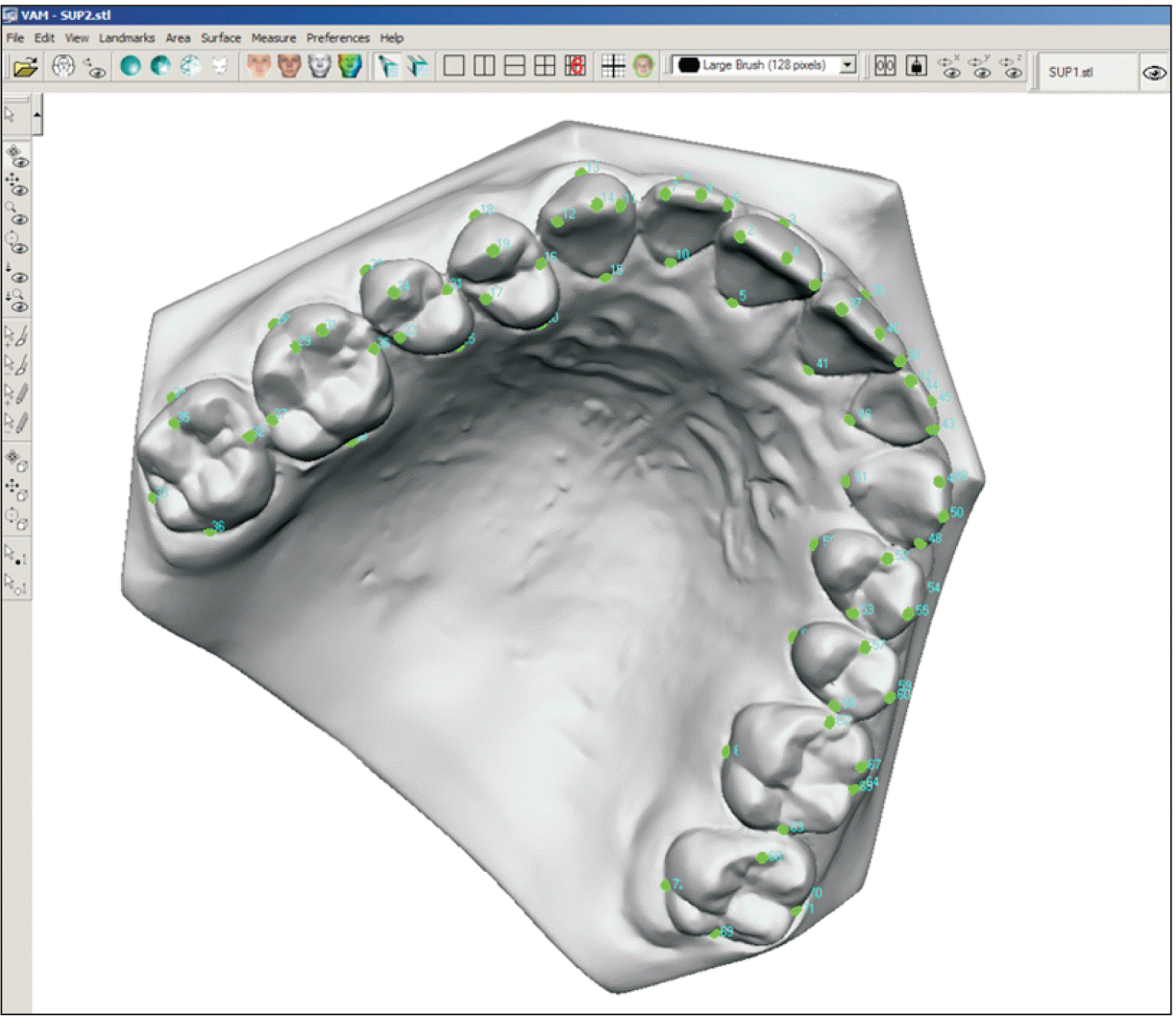
Pre- and post-treatment maxillary and mandibular digital casts were analyzed with VAM (Vectra Analysis Module) software (Vectra©; Canfield Scientific, Parsippany, NJ, USA) according to the method described by Huanca Ghislanzoni et al.20 (Figure 1). Teeth from incisors to second molars were included, and five points were recorded for each tooth. The most mesial and most distal points of the occlusal surface were identified. The facial axis of the crown (FACC) was determined,6 and the gingival limit of the buccal FACC, the occlusal limit of the buccal FACC, and the gingival limit of the lingual FACC were identified. Points were digitalized using space coordinates (x, y, z), and a reference plane was established as the best-fit plane among all lingual points. The coordinates were converted to the new reference system, and linear distances and angles were calculated by using custom-made trigonometric macros (Microsoft Excel©; Microsoft, Redmond, WA, USA). A 90° angle with respect to the occlusal plane was used as the null value (0°), considering labial inclinations positive and lingual inclinations negative. The dental crowding, intercanine distance, and intermolar distance between first molars were calculated at the beginning and end of treatment. The same evaluator repeated the entire process (from the identification of points to all measurements) on 10 models after a wash-out period of approximately one month.
Statistical analysis
The intra-assessor agreement was calculated with the intraclass correlation coefficient,23 which was excellent (> 0.8) for all measurements. The controlled variable was the selected inclination prescription (Tsel), while the outcome variables were the initial dental inclination (Tin), the final dental inclination (Tfin), the obtained inclination change (ΔT), the differential inclination applied (Tsel-Tin), and the differential inclination obtained (Tsel-Tfin). The Shapiro–Wilk test showed that data were not normally distributed. Paired-sample Wilcoxon test was used to assess changes between pre- and post-treatment. Mann–Whitney U test was used to compare low and standard inclination prescription of the mandibular incisors. Kruskal–Wallis one-way analysis of variance (with the Mann–Whitney U test as the post-hoc test and adjusted significance α = 0.05/3 = 0.017) was used to compare low, standard, and high inclination prescription of the maxillary incisors.
A multiple linear regression model was developed with ΔT as the outcome variable (investigating the factors contributing to the final inclination change), and another one was developed with Tsel as the outcome variable (investigating the factors affecting the prescription choice). Predictors for the first model included Tsel, Tsel-Tin, intercanine distance change, and intermolar distance change. Predictors for the second model were Tin, initial dental crowding, initial intercanine distance, and initial intermolar distance. Data analysis was carried out with statistical software (SPSS© ver. 22; IBM, Armonk, NY, USA) at significance level α = 0.05, and the threshold for clinical relevance was > 1 mm or > 1º.
RESULTS
Changes in maxillary incisor inclination
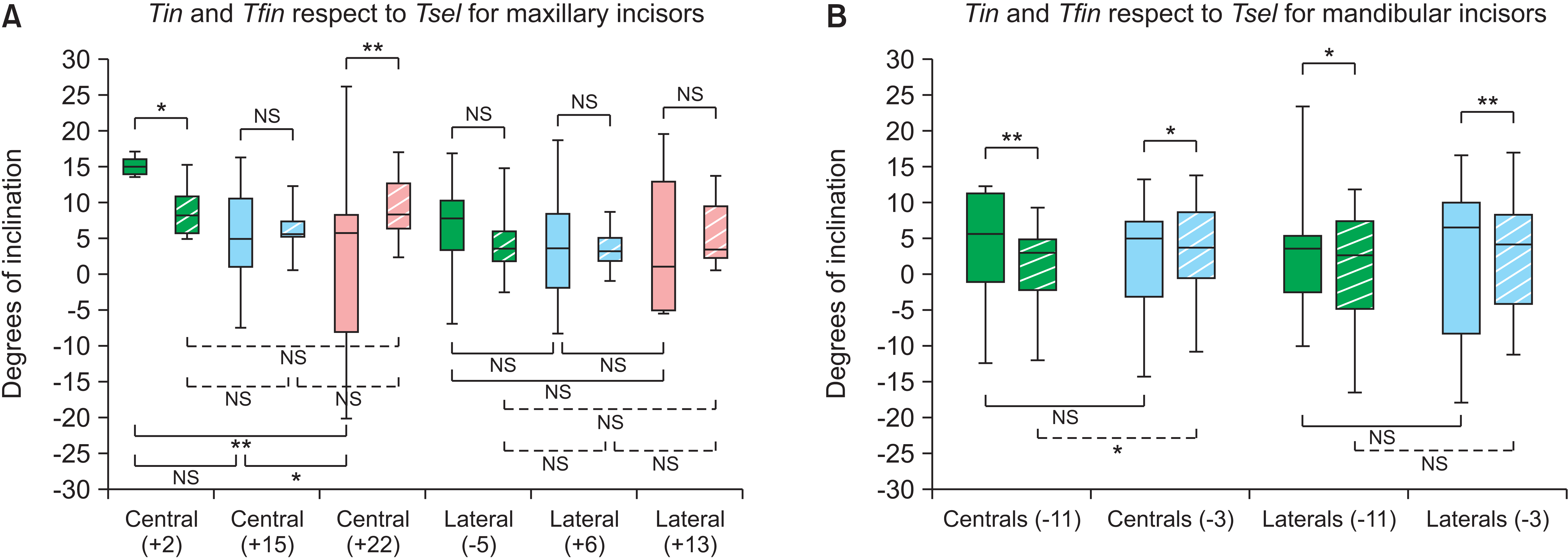
For central incisors, when low prescription was used (+2°, intended to incline them lingually), the correction was followed by the teeth (p = 0.046). When standard prescription was used (+15°, intended to incline them labially), the initial dental inclination was maintained. When high prescription was used (+22°, intended to incline them labially), the correction was followed by the teeth (p = 0.005).
Changes in mandibular incisor inclination
For central incisors, when the low prescription was used (–11°, intended to incline them lingually), the correction was followed by the teeth (p = 0.005). When the standard prescription was used (–3°, intended to incline the teeth lingually), incisors inclined labially instead (p = 0.045).
For lateral incisors, when the low prescription was used (–11°, intended to incline them lingually), the correction was followed by the teeth (p = 0.010). When the standard prescription was used (–3°, intended to incline them lingually), they inclined labially instead (p = 0.005) (Figures 2 and 4, Table 2).
Comparisons among different inclination prescriptions for maxillary incisors
For central incisors, the applied differential inclination was different between low and standard (p < 0.001), low and high (p = 0.001), and standard and high (p < 0.001) prescriptions. Similarly, the obtained differential inclination was different between the low and standard (p < 0.001), low and high (p = 0.001), and standard and high (p = 0.011) prescriptions. However, the final inclination was similar among different prescriptions.
For lateral incisors, the applied differential inclination was different only between the low and standard (p < 0.001) and low and high (p = 0.001) prescriptions. Similarly, the obtained differential inclination was different between the low and standard (p < 0.001) and low and high (p < 0.001) prescriptions. However, the final inclination was similar among different prescriptions (Figure 2, Table 1).
Comparison between different inclination prescriptions for mandibular incisors
For central incisors, the differences were present between low and standard prescriptions for each parameter except for the initial incisal inclination.
Model showing the factors related to the clinical inclination change (ΔT)
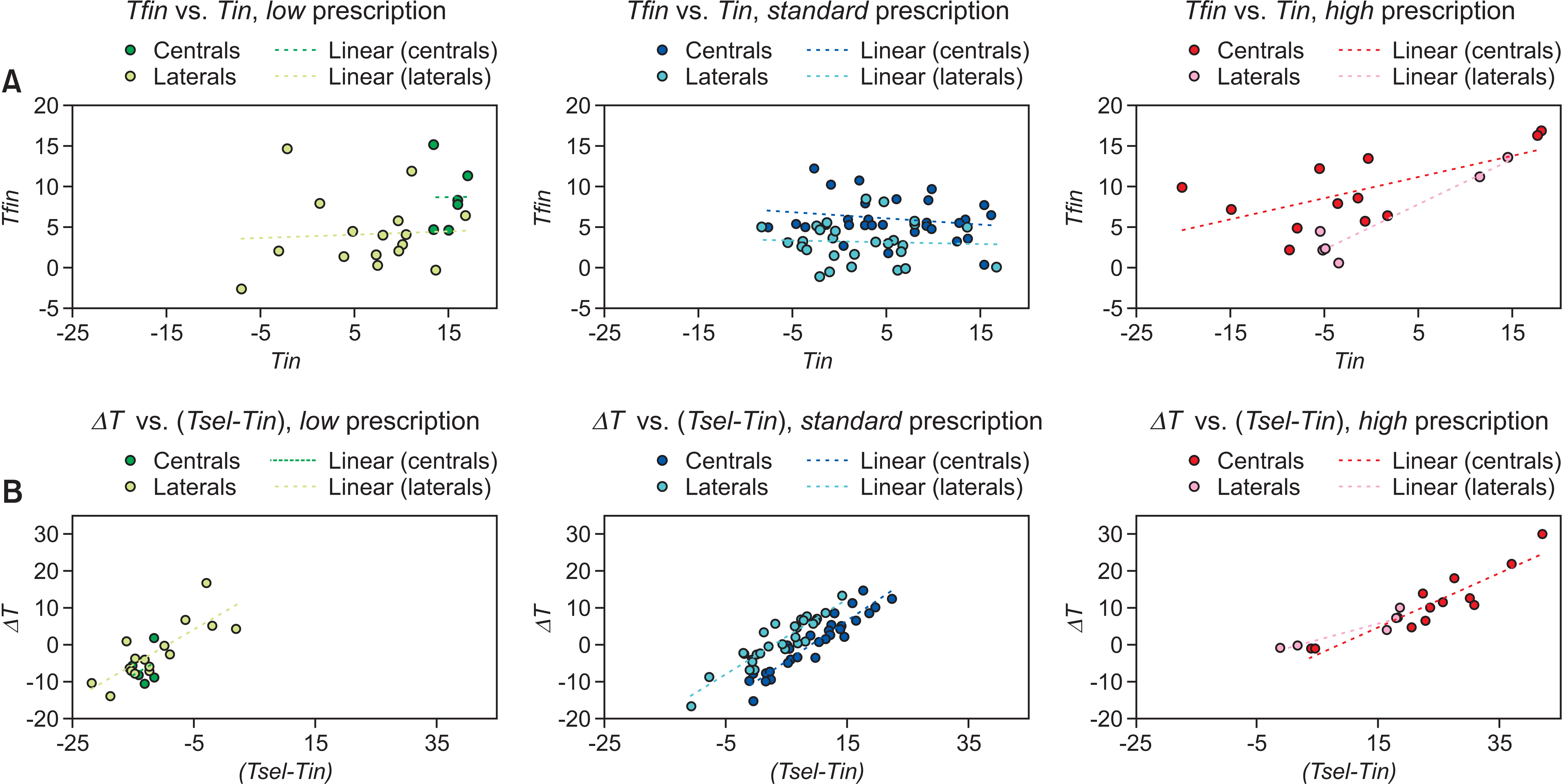
For maxillary incisors, the model for central incisors showed that an increase in the applied differential inclination (β = 1.255, p < 0.001) was related to increased ΔT, while an increase in the selected inclination prescription (β = –0.476, p < 0.001) or intermolar distance (β = –0.203, p < 0.001) was related to reduced ΔT. The model for lateral incisors showed similar results, in addition to the fact that the change in the intercanine distance was relevant (β = –0.180, p = 0.022), rather than the intermolar distance.
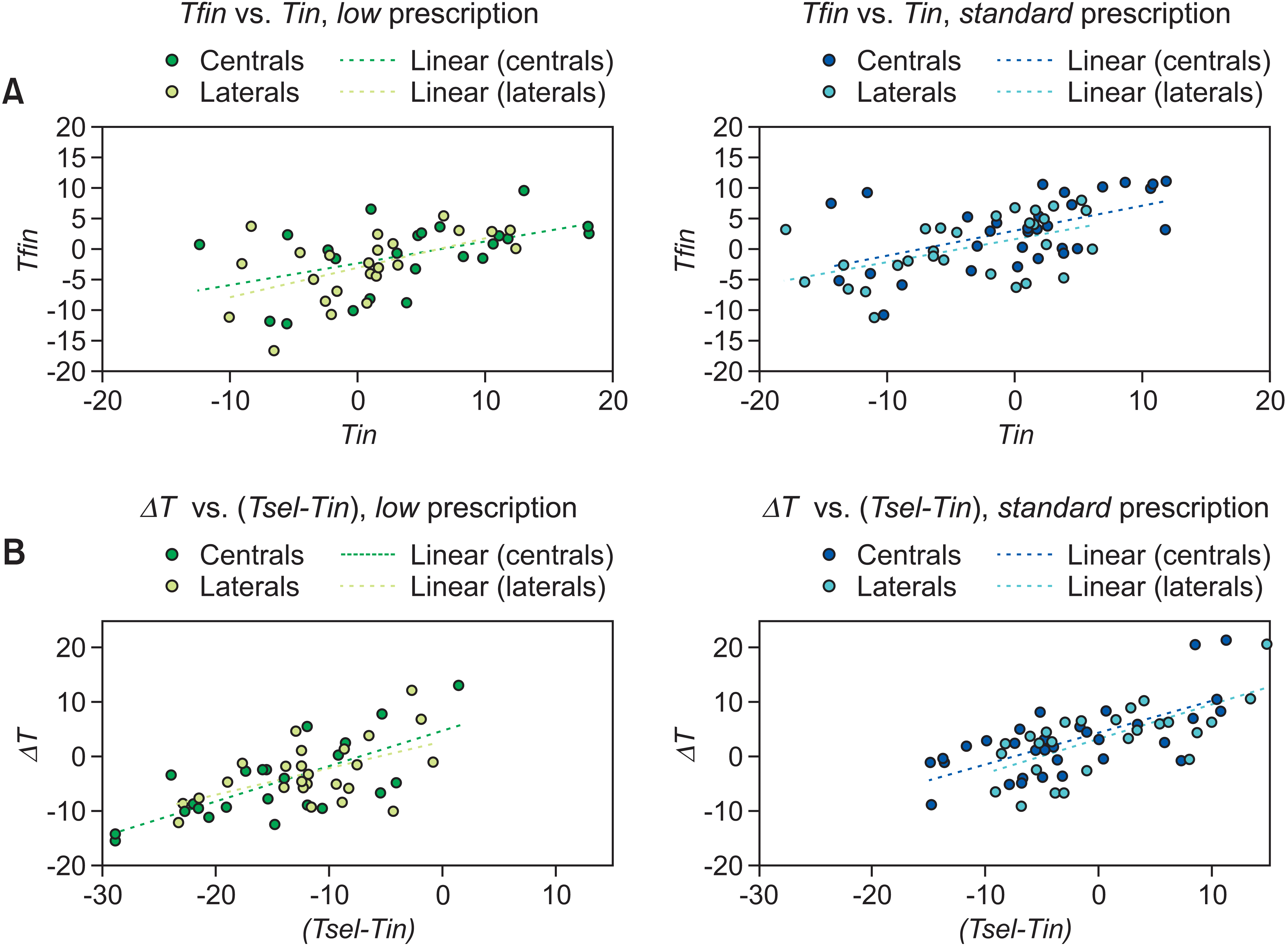
For mandibular incisors, the model showed that an increase in the applied differential inclination was related to increased ΔT for both central (β = 0.767, p < 0.001) and lateral (β = 0.735, p < 0.001) incisors (Table 3).
Model showing the factors related to the selection of the inclination prescription (Tsel)
For maxillary incisors, the model for central incisors showed that the higher the initial inclination (β = –0.484, p < 0.001) and the greater the initial intercanine distance (β = –0.247, p = 0.047), the lower was the Tsel. The model for lateral incisors showed similar findings for the relevance of the initial intercanine distance (β = –0.409, p = 0.028), but the initial inclination was not significant.
For mandibular incisors, the model for central incisors showed that only the initial inclination was relevant (β = –0.292, p = 0.036). The model for lateral incisors showed that both the initial inclination (β = –0.361, p = 0.007) and the intercanine distance (β = 0.290, p = 0.028) influenced the Tsel (Table 3).
DISCUSSION
The choice of inclination prescription is based either on the intention to change the initial incisal inclination or to counteract its undesirable change.19 If the initial labio-lingual incisal position is appropriate, but the inclination is not, a force couple to selectively change the inclination is required. Conversely, if the initial inclination is correct, but the labio-lingual position is not, a force couple is required to counteract undesirable inclination changes during tooth translation. Lastly, both incisal inclination and labio-lingual position may require correction, which is a complex scenario involving multiple approaches based on the concordance/discordance between the direction of the angular and translational corrections. In addition, the prescription choice is also affected by the wire-play16 that needs to be added/subtracted according to the direction of the planned movement.14 Mechanical considerations related to variable forces exerted by different wires17 and biological variables such as craniofacial type7 add further complexity to the choice of the bracket prescription. Thus, prescription selection is a clinical decision based on a combined evaluation of these factors, and assessing its appropriateness was beyond the scope of this study.11
For discussion purposes, in the present work, the initial dental inclination was defined as “lingual” or “labial” based on a reference of +7° for central maxillary incisors, +4° for lateral maxillary incisors, and –6° for central and lateral mandibular incisors.7 The present findings showed that inclination prescription was higher when the initial tooth inclination was low. In particular, for maxillary central incisors, the low prescription was chosen for the most labially inclined teeth (about +15°); the standard prescription for teeth with normal inclination (about +6°); and the high prescription for the most lingually inclined teeth (about –2°). Accordingly, the initial inclination was the most relevant parameter for the choice of the bracket inclination prescription for all teeth except for the maxillary lateral incisors. This may be justified by the fact that although clinical assessments can easily identify the initial inclination of central incisors, lateral incisors are more challenging since they are not clearly visible on lateral cephalograms.16 The second most significant predictor for the selection of the inclination prescription was the initial intercanine distance, since the transverse dental arch development is arguably one of the most important parameters to be considered for proper incisal positioning.24 In fact, if intercanine distance was small, low prescription was used for mandibular incisors (for reducing incisal proclination) while high prescription was applied for maxillary incisors (for achieving adequate interincisal relationship), since proclination of the lower incisors is a common consequence during tooth alignment even in cases with an increased intercanine distance.25
Interestingly, the final inclination of the maxillary central incisors was similar among different inclination prescriptions. In fact, all upper central incisors converged toward an ideal inclination of +7° (ranging between +6° and +9°), despite the marked initial discrepancies (ranging between –2° and +15°). Notably, for maxillary central incisors, the standard prescription (+15°) was used despite an almost ideal initial inclination of +7°, so that the lingual inclination caused by Class II elastics could be counteracted. Maxillary lateral incisors also converged toward their respective optimal inclination of +4°, and eventually showed a similar inclination at the end of treatment (ranging between +3° and +5°). These findings show that in the context of passive self-ligating brackets with an estimated wire-play of approximately ±7° between a 0.019" × 0.025" finishing wire and a 0.022" slot,26 patients with similar types of malocclusions may need specific inclination prescriptions based on the initial dental inclination, even when the final position to be achieved is the same. However, in both cases, high prescription led to overcorrection, providing reasons to limit its use only for teeth with severe lingual inclination.
Conversely, mandibular central incisors showed different final inclinations based on the inclination prescription adopted. In particular, the low prescription limited incisal proclination, but the standard prescription did not, as reported by previous authors.18 In fact, the initially proclined mandibular incisors, which received the low prescription, showed partial correction of proclination. However, mandibular incisors that were not severely proclined at the beginning of treatment, which received standard prescription, were excessively proclined after treatment. Thus, low rather than standard prescription should be considered if proclination of the mandibular incisors should be limited.18
Although the inclination value of the slot is seldomly achieved by the incisors,15 the prescription selection had a predictable influence on the direction of the correction (lingual vs. labial). In general, there was an agreement on the differential inclination imposed, which should reflect the rationale for selection of the bracket prescription,19 and the inclination change achieved. On the other hand, a simple comparison of the difference between pre- and post-treatment inclinations may have limited clinical relevance,19 since it overlooks the nature of the couple applied to the tooth. The present findings stress the importance of assessment of incisal inclination on dental models - rather than using lateral cephalometry or CBCT imaging19 - since the discrepancy between the slot and tooth inclination (which determines the moment of the force) can be estimated only by using the same occlusal reference for both parameters. Moreover, it was not obvious that compared to standard, low prescription for mandibular incisors led to lower final inclinations. In fact, the minimum amount of differential inclination that should be applied to generate a clinically significant effect is debatable, since the difference between Roth and MBT incisal prescriptions was reported to be irrelevant.27 Nevertheless, the consistency between the amount of imposed inclination and the amount of clinical change is a more complex topic. For example, for maxillary central incisors, the consistency was minimal for standard prescriptions aiming at a +9° inclination change, and it was good for low and high prescriptions aiming at –13° and +24° inclination changes, respectively. Such behavior was in contrast with the fact that a smaller inclination correction should be easier to achieve. However, the wire-play may have affected the accuracy of the system,28 leading to small corrections being less controllable. In addition, mandibular central incisors with the standard prescription showed even negative consistency between the clinical results and the imposed angular change. In particular, for mandibular incisors, their labioversion may be explained by the use of Class II elastics, or by variations in dental crowding and curve of Spee,29 highlighting the importance of considering all biomechanical variables in the choice of the inclination prescription.
Limitations
The method used in the present study was validated by a comparison with the prescription values declared by Andrews,6 and showed overlapping results.20 However, measurement of dental inclination changes did not allow discrimination between “tipping” and “torque” movements. In addition, even though central and lateral mandibular incisors had the same values of low and standard prescriptions (–11° and –3°, respectively), these teeth were analyzed separately for internal validation of the method, and they showed similar findings. Due to different ligation systems, self-ligating brackets may exert different engagement force on the archwire compared to conventional brackets,30,31. Nevertheless, conventional ligatures may not apply standardized forces and may act as a confounding factor.27 Although prospective randomized trials would remove the allocation bias such trials may present ethical concerns since randomization may assign high inclination prescriptions to patients with already protruded incisors and vice versa, potentially worsening their malocclusion. Lastly, the observed clinical changes may have been influenced by growth and should not be attributed solely to the treatment.
CONCLUSIONS
The chosen inclination prescription caused clinical change in dental inclination that was coherent with the direction of the correction (labial vs. lingual), and this was applicable to every teeth that was analyzed except mandibular central incisors.
The imposed inclination change was the most relevant predictor of the clinically achieved change. However, the final inclination had limited consistency with the value of the selected prescription.
The initial incisal inclination was the leading factor determining the choice of the bracket prescription.
A comprehensive consideration of all biomechanical variables affecting the force system is important in choosing the bracket prescription.
 XML Download