Journal List > Korean J Orthod > v.52(6) > 1516080230

See the reply "READER’S FORUM" in Volume 53 on page 137.
Abdelwassie, Kaddah, El-Dakroury, El-Boghdady, El-Ghafour, and Seifeldin: Effectiveness of low-level laser therapy in facilitating maxillary expansion using bone-borne hyrax expander: A randomized clinical trial

Abstract

Objective

The objective of this randomized clinical trial was to study the skeletal and dental effects of low-level laser therapy (LLLT) along with a miniscrew-assisted expander (Hyrax) after six months of retention.

Methods

After sequence generation, concealed allocation, and implementation, 24 female patients were randomly divided (11) into two-groups bone-borne rapid palatal expansion (BBE) without LLLT (n = 12) and BBE with LLLT (n = 12). Eligibility criteria included female patients aged 10–13 years old with bilateral posterior crossbites. Intraoral and extraoral photographs, cone-beam computed tomography images, and digital study models were obtained before expansion and six months after retention. The 7 mm Hyrax appliance was anchored to four palatal mini-screws, which were activated twice daily for 15 days, then locked and kept in place as a retainer. LLLT was performed in the laser group during expansion and retention, according to the guidelines provided.

Results

The records of 24 patients were analyzed. According to the post-retention measurements, both groups showed a significant increase in nasal and maxillary widths and total facial height. In the laser group, the Sella-Nasion-Point A and Point A-Nasion-Point B angles and the interpremolar apical distance were significantly increased.

Conclusions

Within the limitations of this study, the results suggest that the parameters and protocol of LLLT do not clinically affect the efficiency of BBE in prepubertal and pubertal patients.

INTRODUCTION

Maxillary transverse deficiency is a common orthodontic complication.1 Many maxillary deficiency cases are related to posterior crossbite and crowding of the maxillary arch, which compromise the function of mastication and esthetics.2,3 In 1860, Angell4 was the first to describe maxillary expansion. This procedure gained popularity by Haas5 in 1961. The aim of rapid maxillary expansion (RME) was to produce heavy forces by expanders to obtain maximum skeletal response by opening the mid-palatal suture (MPS) with minimum orthodontic movement.6,7
Nonsurgical treatment of maxillary transverse deficiency before or during puberty has never been a challenge. The main difficulty in dealing with transverse discrepancy is associated with the limited range of tooth movement in transverse dimension, described by Proffit et al.8 as the “transverse envelope of discrepancy.”
The major goal of RME is to increase the width of the maxilla through skeletal expansion of sutures. Bone-borne maxillary expanders promote bi-cortical engagement of the four mini-screws into the cortical bone of the palate.9 It avoids the side effects of tooth-borne expanders, such as alveolar bone resorption, which leads to tooth movement in the same direction.10 Tooth-borne expanders concentrate the force at the dentoalveolar area, which might be iatrogenic to the surrounding periodontal tissue and limit the skeletal effect of the appliance.11
The treatment that affects the biostimulation potency of the laser, which is not accompanied by a local temperature increase in tissues by more than 1°C, is called low-level laser therapy (LLLT). LLLT is noninvasive, nonthermal, painless, and requires a relatively short application time. The procedure is common in orthodontic clinics for reasons such as reducing pain, accelerating tooth movement, and stimulating bone regeneration in the MPS after palatal expansion.12-15
The aim of the current study was to measure the skeletal and dental effects of a miniscrew-assisted expander (Hyrax), six months after expansion, along with LLLT in orthodontic patients with maxillary constriction.

MATERIALS AND METHODS

Trial design

This prospective randomized clinical-controlled trial followed the Consolidated Standards of Reporting Trials guidelines16 for reporting randomized clinical trials (RCTs), allowing a detailed description of the study interventions and assessment methods. This RCT consisted of parallel groups, with a 1:1 allocation ratio.

Participants, eligibility criteria, and settings

The study was conducted at the clinic of the Department of Orthodontics, Faculty of Dentistry, where the subjects were selected and the trial was performed. The study protocol was registered at the Evidence-Based Center and approved by the Research Ethics Committee of the Faculty of Dentistry, Cairo University (IRB no: 16-9-15). All patients were informed of the study, and informed written consent was obtained. The inclusion criteria for this study were female patients with an age range of 10–13 years old with an average growth pattern and an open MPS, which was verified using an occlusal radiograph, and a bilateral posterior crossbite due to apical maxillary deficiency. Good oral hygiene, a healthy periodontal condition, and medically free were the other inclusion criteria. Patients with surgical or other treatments that might affect RME, congenital malformations, previous orthodontic therapy, systematic disease, or active periodontal disease were excluded from this study.

Intervention

Records

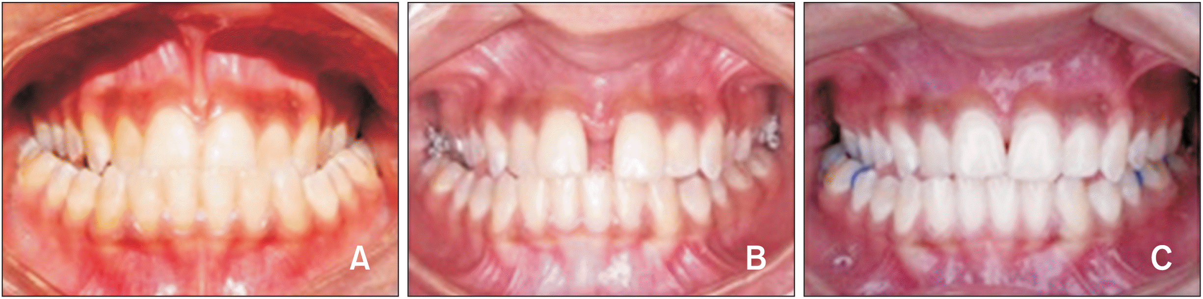
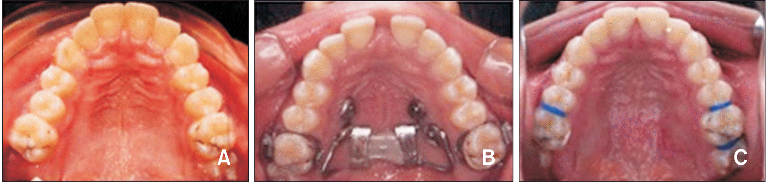
Intraoral and extraoral photographs and cone-beam computed tomography (CBCT) images were obtained before treatment (T0) and after 6 months of the retention phase (T1) (Figures 1 and 2).
CBCT images were taken before expansion for proper diagnosis, to determine mini-screw implementation sites, and as the baseline for frequent measurements. Another CBCT image was taken six months after expansion to evaluate dentoskeletal changes after retention.
CBCT images were acquired using a next-generation i-CAT scanner (Henry Schein Dental, Melville, NY, USA). The machine was supplied with an amorphous silicon flat panel sensor with a cesium iodide scintillator, 0.5 mm focal spot size, 14 Bit grey scale resolution, and operating at the following protocol for all the scans of the study: 120 kVp, 37.07 mAs, 0.3 mm voxel size, 17.8 seconds scanning time, and medium diameter field of vision. The Digital Imaging and Communications in Medicine format of the images was processed using InVivo 5.01 Anatomage software (DEXIS, Quakertown, PA, USA).
The landmarks, lines, and planes that were used for skeletal and dental evaluation were traced on the CBCT images, as described in Tables 1 and 2.

Bone-borne Hyrax expander

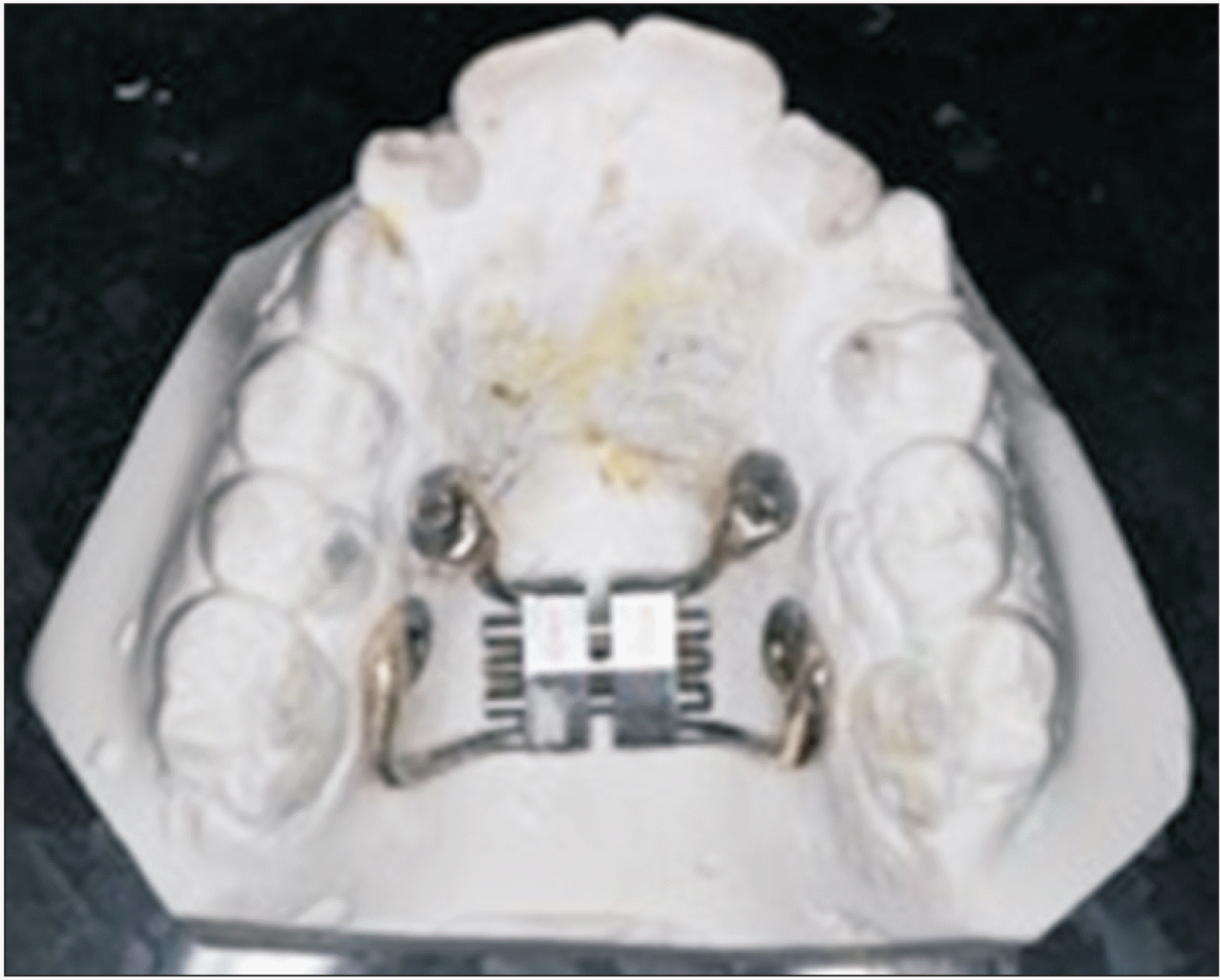
The bone-borne Hyrax expander used in this study was supported by four mini-screws (dimensions 1.5 mm diameter and the length 10 mm with a trans-mucous section of 2 mm) that were placed two on each side, between the first and second premolars, and between the second premolars and first molars (Figure 3).
CBCT images were used to plan the insertion location of the mini-screws in an area of at least 3.5 mm of inter-radicular space to receive the 1.5 mm diameter mini-screw. A rubber base impression was obtained after mini-screw insertion for the Hyrax appliance fabrication. The appliance was soldered into custom-made stainless steel rings that were used to support the appliance on the mini-screw head. Upon delivery, the initial activation of the appliance was four turns.
For safety measures, after applying the appliance to the patient’s palate, it was ligated to the cleats on the lingual surface of the banded upper first molars. Patients were instructed to activate the expansion screw twice daily for the next 15 days of the active expansion phase by verbal explanation and demonstration on the first day of expansion. The patients in the intervention group visited the clinic every day to ensure that they were activating the appliance properly and for LLLT application as per the LLLT protocol of Cepera et al.13 During follow-up, the hyrax was checked to ensure that it was activated twice, as instructed.
After 15 days of expansion, the Hyrax appliance was locked using light-cured composite and was kept in place for the 6 months of retention. During this phase, the patients were asked to visit every three weeks to follow up on their oral hygiene and appliance stability.

LLLT application

In the laser group, low-level laser treatment was performed using Epic 10 Console (Biolase, Foothill Ranch, CA, USA) with an active medium indium gallium arsenide semiconductor diode via the tooth whitening tip (rectangular 35 mm × 8 mm = 2.8 cm2) according to the manufacturer’s instructions using the following parameters: wavelength, 780 nm; power density, 40 mW; energy density, 10 J/cm2; energy per point, 32 J; continuous wave, time, 8 seconds. The energy density used in this study was similar to that used by Cepera et al.13
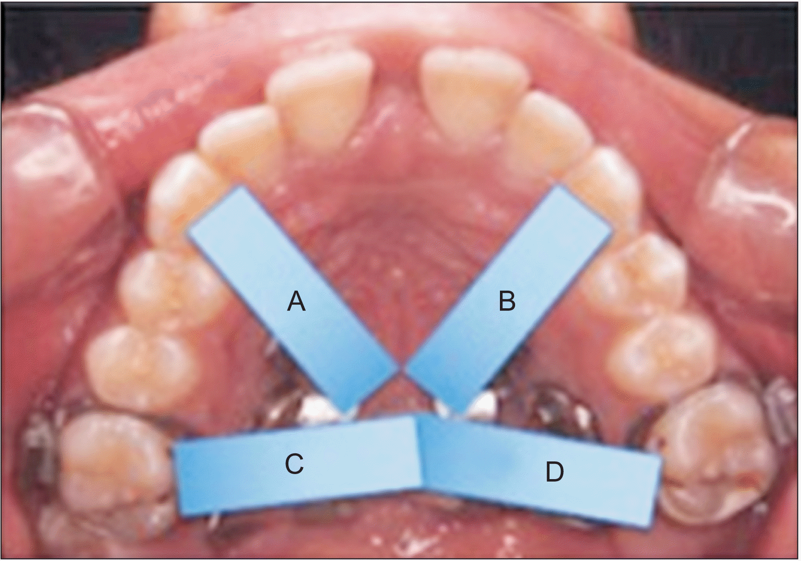
A dental diode laser was used along with a fully engineered deep tissue hand piece to control important parameters, such as spot size and power intensity. The hand piece was protected using a plastic wrap according to biosafety standards. During laser application, the operator and patient wore filter glasses for a wavelength of 780 nm. Application was performed in points distributed in four application areas around the MPS (Figure 4):
  • Two anterior areas from jackscrew to the canines.

  • Two posterior areas from the jackscrew to the first molars.

LLLT was applied in the following stages, for a total of 11 doses:
  • Day 1 to 5 (first 5 days of expansion).

  • Day 16 to 18 (day 1, 2, and 3 of the retention phase).

  • Day 25 (7 days after the previous dose).

  • Day 32 (7 days after the previous dose).

  • Day 39 (7 days after the previous dose).

The laser group patients received LLLT in the first five days of expansion and the first three days of retention, followed by three separate doses every seven days.

Outcomes

The primary outcome of this RCT was to determine the effect of LLLT on bone-borne rapid palatal expansion (BBE) after six months of retention. This was done by comparing the measurements on the CBCT images at T0 and T1. A total of 48 CBCT images were obtained at the end of this study (24 pre-expansion and 24 post-expansion), upon which the analysis was processed using InVivo 5.01 Anatomage software. A custom analysis of dental and skeletal landmarks, lines, and planes (Table 1) was performed to measure the effect of LLLT on maxillary expansion with and without LLLT after 6 months of retention (Table 2). The CBCT image measurements were transferred into an Excel sheet containing the measurements in rows and the subjects’ names in columns.

Sample size calculation

A study of a continuous response variable from an independent variable was conducted. In a previous study,13 the responses within each subject group were normally distributed, with a standard deviation of 1.6. If the true difference in the means of Groups I and II is 2.2, we will need to study nine Group I subjects and nine Group II subjects to be able to reject the null hypothesis such that the population means of the two groups are equal with a probability (power) of 0.8. The type I error probability associated with this test of null hypothesis was 0.05. Considering the dropout rate, a sample size of 12 per group was appropriate.
In this RCT, the sample comprised of 24 female patients who were randomly divided into two groups: BBE without LLLT (n = 12), and BBE with LLLT (n = 12).

Randomization

Participants were randomly assigned to either the control group (non-laser group) or the experimental group (laser group) at a 1:1 allocation. Sequence generation was performed using computer-generated random numbers in Microsoft Office Excel 2007 (Microsoft, Redmond, WA, USA). Allocation concealment was performed by writing random numbers obtained from the sequence generation on opaque papers, folded four times, and kept in sealed, opaque envelopes in a box with the secretary of the department. After the patient was examined for eligibility, they were sent to the secretary of the department and allowed to choose one of the envelopes to detect the assigned group.

Blinding

The first assessor performing this study assessed the CBCT measurements blindly. The second assessor fed the data into the computer in separate data sheets so that they could analyze the data without having access to the information about the allocation.

Statistical analysis

Data were coded and entered using the statistical package SPSS version 25 (IBM Corp., Armonk, NY, USA). Data were summarized using the mean, standard deviation, median, minimum, and maximum quantitative data. Pre- and post-intervention comparisons in each group were performed using the paired t-test. Comparisons between the two groups regarding quantitative variables were performed using the non-parametric Mann–Whitney test.17 Statistical significance was set at p < 0.05.

RESULTS

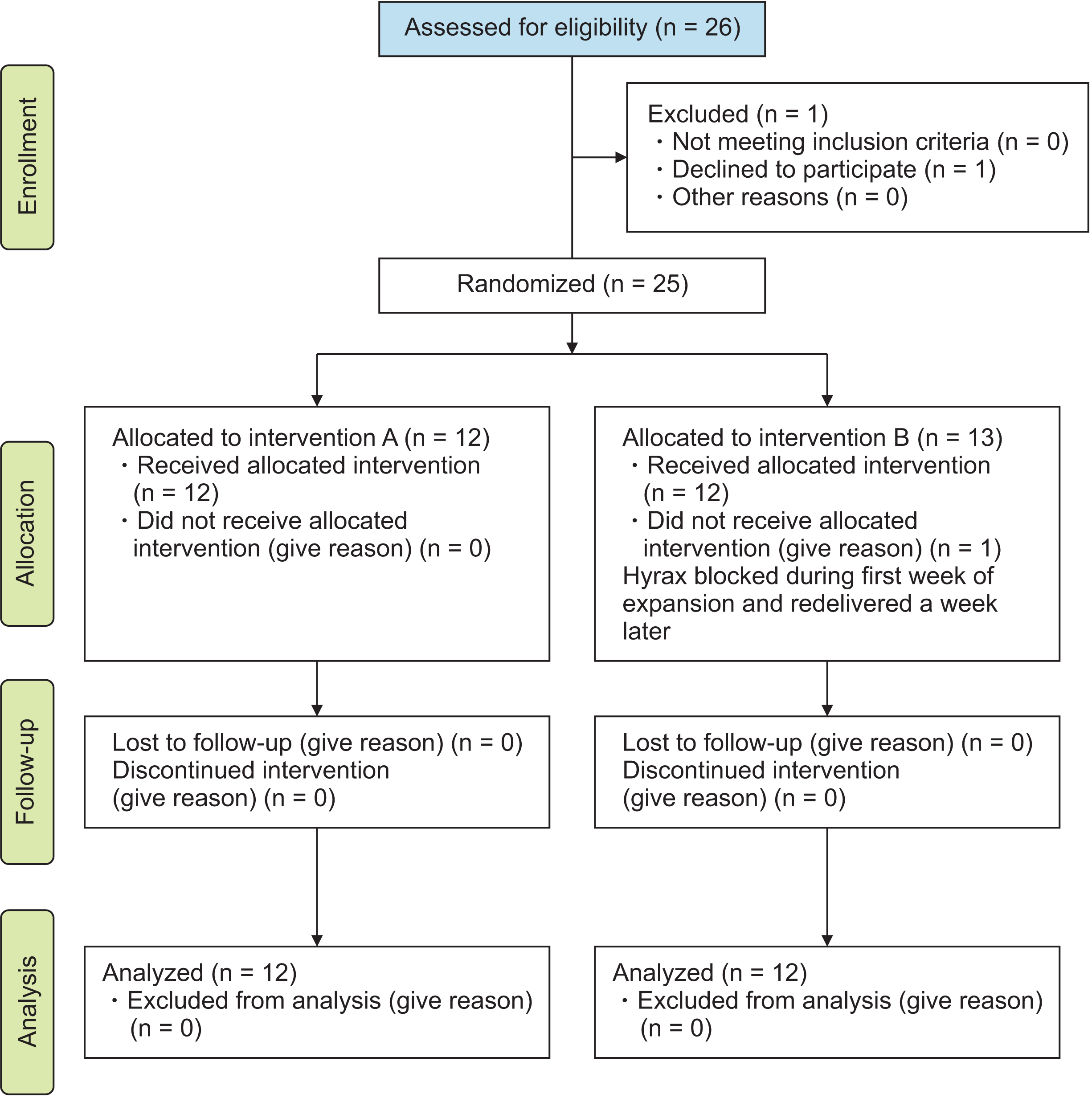
Participant flow, dropouts, and numbers analyzed (Figure 5)

In the non-laser group, 12 patients underwent bone-borne palatal expansion without LLLT. Fourteen participants were initially assigned to the laser group. One patient was excluded because she was not able to visit the clinic regularly for LLLT. Another patient was lost because the Hyrax screw was blocked during the first week of treatment and was delivered a week later to continue the expansion phase after replacing the Hyrax appliance. The records of all 12 participants in the laser group were analyzed.

Recruitment

The first patient was treated on October 13th, 2016, while the last patient was treated on April 6th, 2018. All patients included in this study were followed up for 6 months.

Data baseline

Each group consisted of 12 female patients with bilateral skeletal posterior crossbite.

Outcomes and estimation

General clinical findings

Patients in both groups experienced a significant amount of palatal expansion. A diastema was observed in the midline between the central incisors on the fourth day of expansion. The diastema was approximately 2–4 mm in both groups on the 15th day of expansion. The midline diastema closed during the first 14 days of the retention phase, with central incisor tipping. Crowding relief in the upper arch was also evident by the end of the retention phase after diastema closure due to widening of the upper arch (Figures 1 and 2).

Post-retention CBCT measurements

CBCT measurements were performed after 6 months to evaluate the effect of LLLT on dental and skeletal retention within each group and between both groups (Tables 3 and 4).
According to the post-retention measurements of the transverse skeletal changes in the maxilla and circummaxillary structures in the non-laser group, the nasal and maxillary widths were significantly increased, and no statistically significant changes were observed in the other transverse skeletal measurements. In the laser group, the mean and standard deviation values of the nasal, maxillary, and left maxillary-mandibular widths were significantly increased, with no statistically significant change in the rest of the transverse skeletal measurements. There was no statistically significant difference in the transverse skeletal linear measurements between the two groups.
Regarding the post-retention vertical skeletal linear measurements, a statistically significant increase in the total facial height was found in both groups. However, in the non-laser group, the increase was due to a statistically significant increase in the lower facial height. In contrast, in the laser group, the increase was due to a statistically significant increase in middle facial height.
In the post-retention vertical skeletal angular measurements, no statistically significant difference was observed in the non-laser group. In the laser group, the angle between the mandibular line and the maxillary plane and Frankfort horizontal plane was significantly decreased. The angle between the mandibular line and Frankfurt horizontal plane was significantly different between the two groups.
There was no statistically significant change in the anteroposterior skeletal angular measurements after the retention phase in the non-laser group. In the laser group, the Sella-Nasion-Point A (SNA) and Point A-Nasion- Point B (ANB) angles were significantly increased after expansion, with no statistically significant change in the Sella-Nasion-Point B (SNB) angle. There was no statistically significant difference in the anteroposterior skeletal angular measurements between the two groups.
In the post-retention transverse dento-alveolar linear changes (inter-dental widths), all measurements in the non-laser group were significantly increased, except for the apical inter-premolar and apical inter-incisor widths. In the laser group, all dentoalveolar linear measurements significantly increased. The interpremolar apical distance was significantly increased in the laser group, and no statistically significant difference was observed in the other transverse dentoalveolar linear measurements between the two groups.
According to the transverse dental angular changes (buccolingual dental inclination) and vertical dental changes of the upper first permanent molar, there was no statistically significant change in any of the measurements after expansion treatment within each group or when comparing the differences between the groups.

Harm

Participants who were not committed to oral hygiene instructions in both groups experienced moderate to severe palatal mucosal inflammation. Three patients experienced palatal overgrowth of the screws and part of the appliance arm during the expansion phase. Inflammation receded during the retention phase after strictly following the oral hygiene instructions and finally diminished a few days after the appliance and mini-screws were removed.

DISCUSSION

Several reports and trials11,18,19 have shown the clinical success of BBE; however, this is one of the first RCT to assess the effects of LLLT on BBE. LLLT is a noninvasive, nonthermal, and painless procedure that requires a relatively short application time. Owing to its biostimulatory effect, it is believed that LLLT accelerates the opening and bone regeneration and healing of the MPS in RME procedures by stimulating collagen synthesis, a basic component of the osteoid matrix.20-22 To test whether this positive effect of LLLT on bone regeneration extended to influence the quality of expansion, rendering it more skeletal in nature, application of LLLT to a bone-anchored hyrax was presented. Similar to the Beiderman23 hygienic appliance, the expansion appliance used in this study lacked acrylic palatal coverage, preventing any inference with laser application along the MPS. This is in addition to the ease of maintaining oral hygiene and being better tolerated by patients.
Since a greater response to RME has been reported in younger subjects,19,24,25 pre-pubertal and circumpubertal patients were recruited for this study to improve the prognosis of expansion. Female patients were chosen to exclude any gender variability, in addition to their cooperation and high motivation for treatment.26 CBCT was used to allow 3-dimensional evaluation of the skeletal and dental effects of LLLT on expansion. Pretreatment and post-expansion CBCT images after 6 months of retention were collected to minimize radiation exposure.27
Following the laser protocol used by Cepera et al.,13 the results of this trial showed that BBE alone was able to laterally displace the maxillary halves as much as BBE with laser. There was an increase of nearly 2 mm in the maxillary width in both groups, with no effect on facial width. This indicates that the effect of BBE was strongest near the MPS in both groups and diminished as we advanced upward. In agreement with our findings, Lin et al.18 measured the transverse skeletal maxillary changes over three levels and reported that BBE increased almost twice as much at the skeletal level than the hyrax group, with the least increase at the nasal floor and the greatest increase below the hard palate by 5 mm. Mosleh et al.28 reported a similar increase (2.2 mm) in the interjugal width of adolescents following BBE. Regarding the effect of LLLT, Cepera et al.13 used occlusal radiographs to compare bone density, and concluded that LLLT improved MPS opening and accelerated bone regeneration and healing. Ferreira et al.14 used a different LLLT protocol and revealed a higher optimal bone density in the laser group, postulating that LLLT had a positive influence on bone regeneration by accelerating the repair process.
Because the nasal floor is a reflection of the palatal vault, the nasal floor and cavity are highly affected by the expansion forces. A significant increase in the nasal width of 2.7 mm (± 2.32 mm) in the laser group and 1.72 mm (± 1.47 mm) in the non-laser group was detected. The higher measurement in the laser group, although statistically insignificant, could suggest greater expansion or better retention of expansion. Following expansion and throughout the retention period, all mouth-breathing patients in both groups reported improvement and ease of nasal breathing. These findings agree with those of Bicakci et al.,29 who reported an increase in the minimum cross-sectional area of the nose following conventional rapid palatal expander in pre- and postpubertal subjects. Bazargani et al.30 reported a significantly higher nasal airway flow and lower nasal airway resistance following BBE.
No significant differences were found between the two groups in either the vertical or anteroposterior measurements, despite the significant increase in the upper anterior facial height, SNA, and ANB measurements within the LLLT group. This modest increase agrees with previous trials28,31 and may be secondary to the biostimulatory and remodeling effects of the laser on the anterior maxilla, resulting in a clinically and statistically insignificant maxillary anterior and downward displacement.5,32
A statistically significant increase in the SNA and ANB angles within the laser group was found due to LLLT treatment at the beginning of the expansion phase, which reached its peak effect in the first 2 days of radiation, significantly stimulated bone regeneration of the MPS during expansion and increased the rate of bone remodeling and retention, as in previous studies.33,34 The post-retention CBCT indicated that LLLT reduced the relapse and kept the maxilla in its forward position, while there was no statistically significant increase in the same angles in the control group.
With all the statistically significant increased transverse dental measurements in both the laser and non-laser groups, together with the unchanged posterior teeth inclination in both groups, the findings indicate an optimum skeletal expansion accompanied by bodily movement of the molars with no buccal rolling in both groups. Mosleh et al.28 reported similar results with apical and coronal inter-molar widths of 3.5 ± 1 and 3.9 2.1, respectively, after 15 days of expansion. An increase was noted in the apical inter-molar, inter-premolar, inter-canine and inter-incisor measurement in this study of 3.63 ± 1.16, 3.28 ± 1.9, 3.4 ± 1.93, and 2.2 ± 1.18 mm, respectively, in the laser group and 3 ± 0.86, 1.89 ± 1.48, 3.14 ± 1.72 and 2.62 ± 3.62 mm, respectively, in the non-laser group. The measurements were more increased and closer in number within the laser group, which might suggest a more parallel sutural opening. The only difference between the two groups was significant premolar tipping in the non-laser group, with clinical insignificance.

Limitations

To minimize the unnecessary risk hazards of ionizing radiation exposure, the immediate post-expansion effects in both groups could not be studied using CBCT images. Hence, the effect of laser irradiation on the stability of skeletal and dental outcomes was not tested.

Generalizability

The generalizability of these results may be limited to growing and early adolescent females. Although BBE has been proven successful in adult patients,35-37 laser application in these subjects might yield different results.

CONCLUSIONS

These results suggest that the parameters and protocol used for LLLT did not clinically affect the efficiency of BBE in prepubertal and pubertal patients. There were no differences in the amount or quality of skeletal and dental expansion in the transverse, anteroposterior, and vertical dimensions.

Notes

CONFLICTS OF INTEREST

No potential conflict of interest relevant to this article was reported.

REFERENCES

1. Kutin G, Hawes RR. 1969; Posterior cross-bites in the deciduous and mixed dentitions. Am J Orthod. 56:491–504. DOI: 10.1016/0002-9416(69)90210-3. PMID: 5261162. PMID: https://www.scopus.com/inward/record.uri?partnerID=HzOxMe3b&scp=0014599765&origin=inward.
2. Helm S. 1968; Malocclusion in Danish children with adolescent dentition: an epidemiologic study. Am J Orthod. 54:352–66. DOI: 10.1016/0002-9416(68)90304-7. PMID: 5238998. PMID: https://www.scopus.com/inward/record.uri?partnerID=HzOxMe3b&scp=0014285108&origin=inward.
3. Howe RP, McNamara JA Jr, O'Connor KA. 1983; An examination of dental crowding and its relationship to tooth size and arch dimension. Am J Orthod. 83:363–73. DOI: 10.1016/0002-9416(83)90320-2. PMID: 6573844. PMID: https://www.scopus.com/inward/record.uri?partnerID=HzOxMe3b&scp=0020756725&origin=inward.
4. Angell EH. 1860; Treatment of irregularity of the permanent or adult teeth. Dental Cosmos. 1:540–4. 599–600.
5. Haas AJ. 1961; Rapid expansion of the maxillary dental arch and nasal cavity by opening the midpalatal suture. Angle Orthod. 31:73–90.
6. Haas AJ. 1980; Long-term posttreatment evaluation of rapid palatal expansion. Angle Orthod. 50:189–217. DOI: 10.1043/0003-3219(1980)050<0189:LPEORP>2.0.CO;2. PMID: 6996533.
7. Haas AJ. 1970; Palatal expansion: just the beginning of dentofacial orthopedics. Am J Orthod. 57:219–55. DOI: 10.1016/0002-9416(70)90241-1. PMID: 5263785. PMID: https://www.scopus.com/inward/record.uri?partnerID=HzOxMe3b&scp=0014750986&origin=inward.
8. Proffit WR, Phillips C, Dann C 4th. 1990; Who seeks surgical-orthodontic treatment? Int J Adult Orthodon Orthognath Surg. 5:153–60.
9. Carlson C, Sung J, McComb RW, Machado AW, Moon W. 2016; Microimplant-assisted rapid palatal expansion appliance to orthopedically correct transverse maxillary deficiency in an adult. Am J Orthod Dentofacial Orthop. 149:716–28. DOI: 10.1016/j.ajodo.2015.04.043. PMID: 27131254. PMID: https://www.scopus.com/inward/record.uri?partnerID=HzOxMe3b&scp=84969520066&origin=inward.
10. Starnbach H, Bayne D, Cleall J, Subtelny JD. 1966; Facioskeletal and dental changes resulting from rapid maxillary expansion. Angle Orthod. 36:152–64. DOI: 10.1043/0003-3219(1966)036<0152:FADCRF>2.0.CO;2. PMID: 4956318.
11. Garib DG, Henriques JF, Janson G, de Freitas MR, Fernandes AY. 2006; Periodontal effects of rapid maxillary expansion with tooth-tissue-borne and tooth-borne expanders: a computed tomography evaluation. Am J Orthod Dentofacial Orthop. 129:749–58. DOI: 10.1016/j.ajodo.2006.02.021. PMID: 16769493. PMID: https://www.scopus.com/inward/record.uri?partnerID=HzOxMe3b&scp=33744918064&origin=inward.
12. Saito S, Shimizu N. 1997; Stimulatory effects of low-power laser irradiation on bone regeneration in midpalatal suture during expansion in the rat. Am J Orthod Dentofacial Orthop. 111:525–32. DOI: 10.1016/S0889-5406(97)70152-5. PMID: 9155812. PMID: https://www.scopus.com/inward/record.uri?partnerID=HzOxMe3b&scp=0031134307&origin=inward.
13. Cepera F, Torres FC, Scanavini MA, Paranhos LR, Capelozza Filho L, Cardoso MA, et al. 2012; Effect of a low-level laser on bone regeneration after rapid maxillary expansion. Am J Orthod Dentofacial Orthop. 141:444–50. DOI: 10.1016/j.ajodo.2011.10.023. PMID: 22464526. PMID: https://www.scopus.com/inward/record.uri?partnerID=HzOxMe3b&scp=84859179616&origin=inward.
14. Ferreira FN, Gondim JO, Neto JJ, Dos Santos PC, de Freitas Pontes KM, Kurita LM, et al. 2016; Effects of low-level laser therapy on bone regeneration of the midpalatal suture after rapid maxillary expansion. Lasers Med Sci. 31:907–13. DOI: 10.1007/s10103-016-1933-8. PMID: 27056702. PMID: https://www.scopus.com/inward/record.uri?partnerID=HzOxMe3b&scp=84964049024&origin=inward.
15. Davoudi A, Amrolahi M, Khaki H. 2018; Effects of laser therapy on patients who underwent rapid maxillary expansion; a systematic review. Lasers Med Sci. 33:1387–95. DOI: 10.1007/s10103-018-2545-2. PMID: 29948453. PMID: https://www.scopus.com/inward/record.uri?partnerID=HzOxMe3b&scp=85048371907&origin=inward.
16. Moher D, Hopewell S, Schulz KF, Montori V, Gøtzsche PC, Devereaux PJ, et al. 2010; CONSORT 2010 explanation and elaboration: updated guidelines for reporting parallel group randomised trials. BMJ. 340:c869. DOI: 10.1136/bmj.c869. PMID: 20332511. PMCID: PMC2844943. PMID: https://www.scopus.com/inward/record.uri?partnerID=HzOxMe3b&scp=77950273246&origin=inward.
17. Chan YH. 2003; Biostatistics 102: quantitative data--parametric & non-parametric tests. Singapore Med J. 44:391–6. PMID: 14700417. PMID: https://www.scopus.com/inward/record.uri?partnerID=HzOxMe3b&scp=0242300086&origin=inward.
18. Lin L, Ahn HW, Kim SJ, Moon SC, Kim SH, Nelson G. 2015; Tooth-borne vs bone-borne rapid maxillary expanders in late adolescence. Angle Orthod. 85:253–62. DOI: 10.2319/030514-156.1. PMID: 25490552. PMCID: PMC8631879. PMID: https://www.scopus.com/inward/record.uri?partnerID=HzOxMe3b&scp=84924918691&origin=inward.
19. Celenk-Koca T, Erdinc AE, Hazar S, Harris L, English JD, Akyalcin S. 2018; Evaluation of miniscrew-supported rapid maxillary expansion in adolescents: a prospective randomized clinical trial. Angle Orthod. 88:702–9. DOI: 10.2319/011518-42.1. PMID: 30102085. PMCID: PMC8174075. PMID: https://www.scopus.com/inward/record.uri?partnerID=HzOxMe3b&scp=85055816459&origin=inward.
20. Akkaya S, Lorenzon S, Uçem TT. 1999; A comparison of sagittal and vertical effects between bonded rapid and slow maxillary expansion procedures. Eur J Orthod. 21:175–80. DOI: 10.1093/ejo/21.2.175. PMID: 10327741. PMID: https://www.scopus.com/inward/record.uri?partnerID=HzOxMe3b&scp=0033110966&origin=inward.
21. Wertz RA. 1970; Skeletal and dental changes accompanying rapid midpalatal suture opening. Am J Orthod. 58:41–66. DOI: 10.1016/0002-9416(70)90127-2. PMID: 5269181. PMID: https://www.scopus.com/inward/record.uri?partnerID=HzOxMe3b&scp=0014823496&origin=inward.
22. Adkins MD, Nanda RS, Currier GF. 1990; Arch perimeter changes on rapid palatal expansion. Am J Orthod Dentofacial Orthop. 97:194–9. DOI: 10.1016/S0889-5406(05)80051-4. PMID: 2178393. PMID: https://www.scopus.com/inward/record.uri?partnerID=HzOxMe3b&scp=0025391525&origin=inward.
23. Biederman W. 1968; A hygienic appliance for rapid expansion. JPO J Pract Orthod. 2:67–70. PMID: 5241062. PMID: https://www.scopus.com/inward/record.uri?partnerID=HzOxMe3b&scp=0014252052&origin=inward.
24. Baccetti T, Franchi L, Cameron CG, McNamara JA Jr. 2001; Treatment timing for rapid maxillary expansion. Angle Orthod. 71:343–50. DOI: 10.1043/0003-3219(2001)071<0343:TTFRME>2.0.CO;2. PMID: 11605867.
25. McNamara JA Jr, Franchi L, McClatchey LM. 2019; Orthodontic and orthopedic expansion of the transverse dimension: a four decade perspective. Semin Orthod. 25:3–15. DOI: 10.1053/j.sodo.2019.02.002. PMID: https://www.scopus.com/inward/record.uri?partnerID=HzOxMe3b&scp=85062614904&origin=inward.
26. Sergl H, Zentner A. 2000; Predicting patient compliance in orthodontic treatment. Semin Orthod. 6:231–6. DOI: 10.1053/sodo.2000.19070. PMID: https://www.scopus.com/inward/record.uri?partnerID=HzOxMe3b&scp=77449085502&origin=inward.
27. Lione R, Ballanti F, Franchi L, Baccetti T, Cozza P. 2008; Treatment and posttreatment skeletal effects of rapid maxillary expansion studied with low-dose computed tomography in growing subjects. Am J Orthod Dentofacial Orthop. 134:389–92. DOI: 10.1016/j.ajodo.2008.05.011. PMID: 18774085. PMID: https://www.scopus.com/inward/record.uri?partnerID=HzOxMe3b&scp=50849144400&origin=inward.
28. Mosleh MI, Kaddah MA, Abd ElSayed FA, ElSayed HS. 2015; Comparison of transverse changes during maxillary expansion with 4-point bone-borne and tooth-borne maxillary expanders. Am J Orthod Dentofacial Orthop. 148:599–607. DOI: 10.1016/j.ajodo.2015.04.040. PMID: 26432315. PMID: https://www.scopus.com/inward/record.uri?partnerID=HzOxMe3b&scp=84942693557&origin=inward.
29. Bicakci AA, Agar U, Sökücü O, Babacan H, Doruk C. 2005; Nasal airway changes due to rapid maxillary expansion timing. Angle Orthod. 75:1–6. DOI: 10.1043/0003-3219(2005)075<0001:NACDTR>2.0.CO;2. PMID: 15747808.
30. Bazargani F, Magnuson A, Ludwig B. 2018; Effects on nasal airflow and resistance using two different RME appliances: a randomized controlled trial. Eur J Orthod. 40:281–4. DOI: 10.1093/ejo/cjx081. PMID: 29069383. PMID: https://www.scopus.com/inward/record.uri?partnerID=HzOxMe3b&scp=85047743169&origin=inward.
31. Cross DL, McDonald JP. 2000; Effect of rapid maxillary expansion on skeletal, dental, and nasal structures: a postero-anterior cephalometric study. Eur J Orthod. 22:519–28. DOI: 10.1093/ejo/22.5.519. PMID: 11105408. PMID: https://www.scopus.com/inward/record.uri?partnerID=HzOxMe3b&scp=0034293387&origin=inward.
32. Davis WM, Kronman JH. 1969; Anatomical changes induced by splitting of the midpalatal suture. Angle Orthod. 39:126–32. DOI: 10.1043/0003-3219(1969)039<0126:ACIBSO>2.0.CO;2. PMID: 5252050.
33. Angeletti P, Pereira MD, Gomes HC, Hino CT, Ferreira LM. 2010; Effect of low-level laser therapy (GaAlAs) on bone regeneration in midpalatal anterior suture after surgically assisted rapid maxillary expansion. Oral Surg Oral Med Oral Pathol Oral Radiol Endod. 109:e38–46. DOI: 10.1016/j.tripleo.2009.10.043. PMID: 20219584. PMID: https://www.scopus.com/inward/record.uri?partnerID=HzOxMe3b&scp=77249145955&origin=inward.
34. da Silva AP, Petri AD, Crippa GE, Stuani AS, Stuani AS, Rosa AL, et al. 2012; Effect of low-level laser therapy after rapid maxillary expansion on proliferation and differentiation of osteoblastic cells. Lasers Med Sci. 27:777–83. DOI: 10.1007/s10103-011-0968-0. PMID: 21842225. PMID: https://www.scopus.com/inward/record.uri?partnerID=HzOxMe3b&scp=84863634472&origin=inward.
35. Choi SH, Shi KK, Cha JY, Park YC, Lee KJ. 2016; Nonsurgical miniscrew-assisted rapid maxillary expansion results in acceptable stability in young adults. Angle Orthod. 86:713–20. DOI: 10.2319/101415-689.1. PMID: 26938955. PMCID: PMC8600851. PMID: https://www.scopus.com/inward/record.uri?partnerID=HzOxMe3b&scp=84984698676&origin=inward.
36. Lim HM, Park YC, Lee KJ, Kim KH, Choi YJ. 2017; Stability of dental, alveolar, and skeletal changes after miniscrew-assisted rapid palatal expansion. Korean J Orthod. 47:313–22. DOI: 10.4041/kjod.2017.47.5.313. PMID: 28861393. PMCID: PMC5548712. PMID: https://www.scopus.com/inward/record.uri?partnerID=HzOxMe3b&scp=85027020683&origin=inward.
37. Lee KJ, Choi SH, Choi TH, Shi KK, Keum BT. 2018; Maxillary transverse expansion in adults: rationale, appliance design, and treatment outcomes. Semin Orthod. 24:52–65. DOI: 10.1053/j.sodo.2018.01.006. PMID: https://www.scopus.com/inward/record.uri?partnerID=HzOxMe3b&scp=85041953891&origin=inward.

Figure 1
Frontal intraoral photograph showing before expansion (A), midline diastema formation after expansion (B), and mesial tilting of the central incisors to close the diastema at the end of the retention phase (C).
kjod-52-6-399-f1.tif
Figure 2
Palatal occlusal photograph showing the palatal expansion and the midline diastema: before expansion (A), post-expansion (B), and post-retention (C). It also shows the widening of the upper arch after the retention phase.
kjod-52-6-399-f2.tif
Figure 3
Mini-screw supported bone-borne Hyrax appliance.
kjod-52-6-399-f3.tif
Figure 4
Application areas of the low-level laser therapy. A, B, Two anterior areas from jackscrew to the canines. C, D, Two posterior areas from the jackscrew to the first molars.
kjod-52-6-399-f4.tif
Figure 5
CONSORT (Consolidated Standards of Reporting Trials) flow diagram of the progress through the phases of the current randomized controlled trial.
kjod-52-6-399-f5.tif
Table 1
Landmarks, lines, and planes used in the study
Landmarks
Skeletal landmarks Definition
N (Nasion) The mid-point of the frontonasal suture. Anterior mid-point of the anterior contour, summit of the radiolucent suture, mid-point in the center of the radiolucency.
S (Sella) The center of the hypophyseal fossa.
Po (Porion) Upper most point on the external auditory meatus.
Or.Rt–Or.Lt (Orbitale) The most inferior point of each infra-orbital rim right and left.
Me (Menton) The lower most median landmark on the lowest border of the mandible.
A (Subspinale) The most posterior mid-line point in the concavity between the anterior nasal spine and the prosthion.
B (Suprementale) The deepest mid-point of the mandibular anterior surface.
Zyg.Rt–Zyg.Lt The most superior and medial point on the Zygomatico-maxillary sutures on the right and left side.
Lat.Nasal.Rt–Lat.Nasal.Lt The most lateral points in the lower third of the nasal cavity on each side.
Jg.Rt–Jg.Lt (Jugal) The point of the Jugal process at the intersection of the maxillary tuberosity and the pterygoid plate on the right and left sides. It represents the lateral most point on the pterygo-maxillary sutures.
ANS (Anterior nasal spine) The most anterior point on the tipoff the anterior nasal spine.
Go.Rt–Go.Lt (Gonion) The most posterior and inferior point on the angle of the mandible on the right and left sides.
Gn (Gnathion) The point at the intersection of the skeletal facial and the mandibular planes. The midline of the lowest border of the mandible.
PNS (Posterior nasal spine) The most posterior point on the hard palate at the tip of the post nasal spine.
Dental landmarks Definition
UR6.cusp–UL6.cusp The tip of the mesio-buccal cusp tips of the upper first molars on each side.
UR6.furcation–UL6.furcation The furcation of the upper right and left first molars.
UR6.apex–UL6.apex The mesio-buccal root apex of the upper right and left first molars.
UR4.cusp–UL4.cusp Buccal cusp tips of the upper right and left first premolars.
UR4.apex–UL4.apex Buccal root apex of the upper right and left first premolars.
UR3.cusp–UL3.cusp Center of cusp tip of each of the maxillary canines.
UR3.apex–UL3.apex Root apex of the upper right and left canines.
UR1.inc–UL1.inc The center of the incisal edge of the upper right and left central incisors.
UR1.apex–UL1.apex The root apex of the upper right and left central incisors.
Reference lines and planes
FH (Frankfort horizontal) Three point plane passing through Po.Rt, Or.Rt, and Or.Lt.
MSP (Mid-sagittal plane) A three-point plane passing by points N, S, and basion.
Mandibular plane A three-point plane formed between Go.Rt, Go.Lt, and Gn.
Maxillary plane Plane formed between ANS, PNS, and MSP.
Mandibular line The line from Mid.Go and Me.
Cranial base line Line between S and N.
UR6 long axis–UL6 long axis Long axis of the upper left and right first molars as the line between UR6.apex-UR6.cusp and UL6.apex-UL6.cusp.
UR4 long axis–UL4 long axis Long axis of the upper left and right first premolars as the line between UR4.apex-UR4.cusp and UL4.apex-UL4.cusp.
UR3 long axis–UL3 long axis Long axis of the upper left and right canines as the line between UR3.apex-UR3.cusp and UL3.apex-UL3.cusp.
UR3 long axis–UL3 long axis Long axis of the upper left and right central incisors as the line between UR1.apex-UR1.cusp and UL1.apex-UL1.cusp.

Rt, right; Lt, left; Zyg, zygoma; Lat, lateral; UR, upper right; UL, upper left.

Table 2
Used measurements in the study
Measurements
Transverse skeletal measurements
Linear measurements Definition
Facial width (Zyr-Zyl) Linear distance between the right and left Zygion.
Nasal width (Lnr-Lnl) Linear distance between the most lateral points of the nasal cavity.
Maxillary width (Jgr-Jgl) Linear distance between the right and left Jugale points.
Average right maxillary-mandibular width The perpendicular linear distance extending from the right Jugale point to the frontal facial plane (Or-Ag).
Average left maxillary-mandibular width The perpendicular linear distance extending from the left Jugale point to the frontal facial plane (Or-Ag).
Vertical skeletal measurements
Linear measurements Definition
Middle anterior facial height (N-ANS) Linear distance between the Nasion and the anterior nasal spine including the vertical position of the maxilla as well as the anterior middle facial height.
Total facial height (N-Me) Linear distance between Nasion and Menton indicating the total anterior facial height.
Angular measurements Definition
Mandibular line with maxillary plane Angle formed between the mid Mandibular line (Mid Go-Me) and the maxillary plane (ANS, PNS, and mid-sagittal plane).
Mandibular line with Frankfurt horizontal plane Angle formed between the mid Mandibular line (Mid Go-Me) and Frankfort horizontal plane (Or.Rt-Or.Lt-Po).
Mandibular plane with cranial base Angle between Mandibular plane (Go.Rt.Go.Lt/Me) and cranial base (SN line).
Y-axis The inferior inner angle formed between the Frankfort horizontal plane and a line connecting the Sella point with the Gnathion point (SN-Gn).
Anteroposterior skeletal measurements
Angular measurements Definition
SNA Angle between the SN line and a line connecting N to A-point indicating the antero-posterior extent of the maxilla.
SNB The angle between the SN line and a line connecting N to B point indicating the antero-posterior extent of the mandible.
ANB The angle between the line connecting N to A point and a line connecting N to B point.
Transverse dental measurements
Linear measurements Definition
Coronal inter-molar width (UR6-UL6 coronal) The distance between the tips of the mesio-buccal cusps of the right and left upper first permanent molars.
Furcation inter-molar width (UR6-UL6 furcation) The distance between the furcation of the right and left upper first permanent molars.
Apical inter-molar width (UR6-UL6 apical) The distance between the palatal root apices of the right and left first permanent molars.
Coronal inter-premolar width (UR4-UL4 coronal) The distance between the tips of the buccal cusps of the right and left upper first premolars.
Apical inter-premolar width (UR4-UL4 apical) The distance between the buccal root apices of the right and left upper first premolars.
Coronal inter-canine width (UR3-UL3 coronal) The distance between the cusp tips of the right and left upper canines.
Apical inter-canine width (UR3-UL3 apical) The distance between the root apices of the right and left upper canines.
Apical inter-incisal width (UR1-UL1 apical) The distance between the root apices of the right and left upper central incisors.
Angular measurements Definition
Upper right first permanent molar buccolingual inclination (UR6/FH) The external angle between the Frankfort horizontal plane and the long axis of the maxillary right first permanent molar (the line connecting the mesio-buccal cusp tip to the mesio-buccal root apex).
Upper left first permanent molar buccolingual inclination (UL6/FH) The external angle between the Frankfort horizontal plane and the long axis of the maxillary left first permanent molar (the line connecting the mesio-buccal cusp tip to the mesiobuccal root apex).
Upper right canine buccolingual inclination (UR3/FH) The angle between the Frankfort horizontal plane and the long axis of the maxillary right canine.
Upper left canine buccolingual inclination (UL3/FH) The angle between the Frankfort horizontal plane and the long axis of the maxillary left canine.
Upper right first premolar buccolingual inclination (UR4/FH) The external angle between the Frankfort horizontal plane and the long axis of the maxillary right first premolar (the line connecting the buccal cusp tip to the buccal root apex).
Upper left first premolar buccolingual inclination (UL4/FH) The external angle between the Frankfort horizontal plane and the long axis of the maxillary left first premolar (the line connecting the buccal cusp tip to the buccal root apex).
Vertical dental measurements
Linear measurements Definition
Upper first permanent molar furcation point to the maxillary plane (U6FP–MxPL) The perpendicular distance extended from the mid furcation point of the right and left first permanent molars to the maxillary plane.

Zyr, zygion right; Zyl, zygion left; Lnr, later nasal right; Lnl, later nasal left; N-ANS, nasion and the anterior nasal spine; N-Me, nasion and menton; UR, upper right; UL, upper left; FH, Frankfort horizontal plane.

Table 3
Descriptive statistics of both the LLLT and non-LLLT groups
Variables LLLT group Non-LLLT group
Pre-expansion Post-retention p-value Pre-expansion Post-retention p-value
Mean SD Mean SD Mean SD Mean SD
Skeletal measurements
Transverse skeletal linear(mm) Maxillary width 40.37 2.48 42.41 2.51 < 0.001* 41.65 2.24 43.71 3.00 0.003*
Facial width 103.34 3.68 103.89 4.22 0.230 107.46 4.49 108.43 4.73 0.197
Maxillary cant 86.50 1.83 86.83 1.50 0.640 86.41 1.54 87.29 1.11 0.217
Nasal width 22.20 1.50 24.90 2.73 < 0.001* 22.21 2.12 23.94 2.20 0.003*
Mx-Md width Rt 22.11 2.25 21.55 1.89 0.117 22.42 1.94 21.75 1.79 0.120
Mx-Md width Lt 22.74 1.85 21.57 1.50 0.009* 22.68 1.50 22.08 1.33 0.105
Vertical skeletal linear(mm) Middle anterior facial height 49.71 3.65 50.90 3.85 0.004* 49.28 3.35 48.89 6.07 0.773
Anterior lower facial height 65.57 4.59 66.01 4.37 0.348 64.02 4.75 65.27 4.61 0.006*
Total anterior facial height 113.56 5.51 114.89 5.07 0.014* 112.06 6.53 113.91 6.81 0.001*
Vertical skeletal angular(°) Mand. line/Mx plane 31.73 6.19 30.77 5.96 0.039* 28.94 4.30 29.72 4.81 0.454
Mand. line/FH 31.21 5.15 30.47 4.77 0.045* 27.43 5.71 28.11 5.26 0.180
Mand. plane/Cranial base 44.75 6.97 44.21 6.78 0.235 41.57 6.09 41.91 6.42 0.569
Antero-posterior skeletal angular(°) SNA 78.35 3.11 79.24 3.47 0.002* 78.57 3.27 79.43 3.89 0.082
SNB 74.74 4.52 74.90 4.56 0.572 77.59 4.03 77.73 4.58 0.650
ANB 4.03 2.94 4.48 3.10 0.016* 3.82 1.32 3.83 1.67 0.996
Dental measurements
Transverse dental linear(mm) UR6-UL6 coronal 46.42 2.88 49.22 2.47 < 0.001* 45.83 3.06 48.71 5.01 0.002*
UR6-UL6 apical 45.17 1.69 48.80 2.55 < 0.001* 44.06 2.93 47.07 3.34 < 0.001*
UR6-UL6 furcation 41.08 2.04 44.08 2.10 < 0.001* 40.51 2.97 43.70 3.12 < 0.001*
UR4-UL4 buccal cusp 37.19 2.28 40.46 2.01 < 0.001* 36.12 2.85 39.33 3.32 < 0.001*
UR4-UL4 palatal cusp 27.45 2.40 30.11 2.07 < 0.001* 26.57 5.01 29.78 2.71 0.047*
UR4-UL4 apical 34.66 2.02 37.94 2.53 < 0.001* 30.28 8.21 35.63 3.35 0.070
UR3-UL3 coronal 30.59 3.00 33.01 2.75 < 0.001* 28.98 4.22 31.30 2.86 0.040*
UR3-UL3 apical 24.64 2.46 28.04 2.87 < 0.001* 24.72 2.68 27.86 3.12 < 0.001*
UR1-UL1 apical 6.84 1.78 9.04 1.95 < 0.001* 8.56 5.87 8.71 1.77 0.930
Transverse dental angular(°) UR6/FH 82.71 3.57 81.93 4.41 0.462 84.38 2.92 82.75 5.51 0.350
UL6/FH 81.45 4.57 83.10 3.72 0.192 85.09 2.80 84.88 2.85 0.846
UR3/FH 68.88 8.70 67.89 8.28 0.367 71.29 6.23 70.56 7.19 0.531
UL3/FH 68.50 7.15 69.30 5.62 0.514 75.66 6.97 73.98 4.53 0.384
UR4/FH 80.36 4.23 81.37 4.57 0.282 79.07 5.75 80.04 4.88 0.394
UL4/FH 80.97 3.04 81.32 3.12 0.667 81.52 4.64 81.47 4.23 0.953
Vertical dental(mm) UR6.furc-Max plane 13.06 2.16 12.99 2.05 0.803 12.86 3.21 12.84 2.62 0.972
UL6.furc-Max plane 13.26 2.00 13.59 2.04 0.151 13.01 2.84 13.36 2.75 0.420

LLLT, low-level laser therapy; SD, standard deviation; Mand, mandibular; Md, middle; Mx, maxillary; UR, upper right; UL, upper left; FH, Frankfort horizontal plane; furc, furcation.

*Statstically significant (p-value equal or less than 0.05).

See Tables 1 and 2 for definitions of each landmark or measurement.

Table 4
Comparison between the 2 groups using Mann–Whitney U test
Variables LLLT group Non-LLLT group p-value
Mean diff SD Mean diff SD
Skeletal measurements
Transverse skeletal linear(mm) Maxillary width 2.03 1.39 2.06 1.74 0.760
Facial width 0.54 1.68 0.97 2.33 0.878
Maxillary cant 0.33 2.68 0.88 2.22 0.799
Nasal width 2.70 2.32 1.72 1.47 0.237
Mx-Md width Rt −0.56 1.30 −0.68 1.32 1.000
Mx-Md width Lt −1.17 1.51 −0.60 1.12 0.281
Vertical skeletal linear (mm) Middle anterior facial height 1.19 1.36 0.93 1.42 0.574
Anterior lower facial height 0.43 1.72 1.25 1.20 0.259
Total anterior facial height 1.33 1.83 1.85 1.25 0.646
Vertical skeletal angular (°) Mand. line/Mx plane −0.96 1.64 0.79 3.35 0.281
Mand. line/FH −0.73 1.29 0.68 1.56 0.024*
Mand. plane/Cranial base −0.54 1.69 0.34 1.93 0.164
Antero-posterior skeletal angular (°) SNA 0.89 0.90 0.86 1.47 0.959
SNB 0.17 1.12 0.15 1.04 0.959
ANB 0.46 0.64 0.00 1.19 0.384
Dental measurements
Transverse dental linear(mm) UR6-UL6 coronal 2.81 1.69 2.88 2.23 0.919
UR6-UL6 apical 3.63 1.16 3.00 0.86 0.180
UR6-UL6 furcation 2.99 1.21 3.19 1.18 0.760
UR4-UL4 buccal cusp 3.26 1.50 3.21 1.38 0.959
UR4-UL4 palatal cusp 2.67 1.50 3.97 3.69 0.610
UR4-UL4 apical 3.28 1.19 1.89 1.48 0.041*
UR3-UL3 coronal 2.41 2.06 2.56 1.41 0.919
UR3-UL3 apical 3.40 1.93 3.14 1.72 0.474
UR1-UL1 apical 2.20 1.18 2.62 3.65 0.281
Transverse dental angular (°) UR6/FH −0.78 4.02 −1.63 5.51 0.799
UL6/FH 1.64 4.63 −0.21 3.53 0.384
UR3/FH −0.99 4.12 −0.73 3.73 0.683
UL3/FH 0.80 4.61 −0.49 1.90 0.574
UR4/FH 1.01 3.50 −0.22 2.94 0.305
UL4/FH 0.35 3.10 −0.06 3.14 0.799
Vertical dental(mm) UR6.furc-Max plane −0.07 1.10 −0.02 1.78 0.838
UL6.furc-Max plane 0.33 0.85 0.35 1.38 0.646

LLLT, low-level laser therapy; Diff, difference; SD, standard deviation; Mand, mandibular; Md, middle; Mx, maxillary; UR, upper right; UL, upper left; FH, Frankfort horizontal plane; furc, furcation.

*Statstically significant (p-value equal or less than 0.05).

See Tables 1 and 2 for definitions of each landmark or measurement.

TOOLS
Similar articles