INTRODUCTION
Variability in tooth morphology plays an important role in attainment of an optimal occlusion of teeth.
1 Tooth morphology has a close relationship with angulation, inclination, and height of tooth in straight archwire technique introduced by Andrews.
2 In straight archwire technique that has been widely used in orthodontic treatment, final tooth positions are dependent on the bracket position, not on archwire bending.
3,4 However, if the crown morphology does not correspond to that for which the bracket is developed, optimal tooth position will not be obtained.
5-8 Root morphology is also of crucial importance for attaining functional and stable occlusion and good prognosis of orthodontic treatment.
9 For this reason, many studies have been performed on tooth morphology. However, little attention has been paid to root morphology or the relationship between crown and root.
The maxillary central incisor is the most visible teeth during unstrained facial activity.
10 Variations in anatomic features of the maxillary central incisors can affect either the treatment or the retention phase of orthodontic therapy.
1 Hence, the morphology of the maxillary central incisors has been investigated.
1,10-15 According to Bryant et al.,
1 the long axis of the root and the long axis of the crown often do not coincide. Sicher and Du Brul
16 have found that the root axis is commonly bent palatally to the crown axis. On the contrary, Taylor
17 has found that the root axis is often bent facially to the crown axis.
The term ‘crown-root angulation’ (CRA) has been used to describe the angle between the crown axis and the root axis. It can be measured on labial view (mesiodistal crown-root angulation, MDCRA) and proximal view (labiolingual crown-root angulation, LLCRA). Some previous studies have used lateral cephalometric radiograph to measure LLCRAs of the maxillary central incisors.
1,6,11,12,15 However, results are divergent on measurements of LLCRAs of the maxillary central incisors. These various results might be due to inherent tracing error on lateral cephalometric radiograph.
A few studies have been performed on LLCRAs of the maxillary central incisor among different malocclusions.
1,12-14,18 According to most studies, the mean LLCRA of the Class II division 2 group is significantly lower than that of Class II division 1, and Class III groups.
1,13,14 On the other hand, some lateral cephalometric studies have reported that Angle Class III cases possess significantly lower LLCRA than Angle Class I and Class II division 1 cases.
12,18
In contrast with LLCRAs, up to date, no study has tried to measure MDCRAs of the maxillary anterior teeth. Moreover, very few attempts have been made to measure CRAs for central incisor, lateral incisor, and canine.
In most previous studies, LLCRAs of maxillary central incisors were measured on two-dimensional (2D) radiograph images without orientation of the teeth according to consistent standard. However, in recent years, the development and use of cone-beam computed tomography (CBCT) in orthodontics have allowed us to observe the crown and root of each tooth in three-dimension (3D) as well.
19
The aim of this study was to compare CRAs of the permanent maxillary anterior teeth according to skeletal malocclusions in Korean population using CBCT.
Go to :

MATERIALS AND METHODS
Samples
Sixty pretreatment CBCT images were collected from the patient archive of the Department of Orthodontics, Gangneung-Wonju National University Dental Hospital, South Korea. This study was reviewed and approved by the Ethics Committee of Gangneung-Wonju National University Dental Hospital (No. 2014-9). Samples were consecutively collected until each skeletal Class I, Class II, and Class III malocclusion group based on A point-nasion-B point angle (ANB) (Class I: 0˚< ANB < 4˚; Class II: ANB ≥ 4˚; Class III: ANB ≤ 0˚) had twenty samples. Root dilacerations,
20 attritions on the crown,
21 and moderate or severe crowding cases
22 were excluded from this retrospective study. Other details of exclusion criteria are summarized in
Table 1. Distributions of age, ANB angle, mandibular plane angle, overjet, and overbite of patients in all groups are shown in
Table 2.
Table 1
Exclusion criteria for the sample selection
|
Incomplete root development of the anterior teeth |
|
Root dilacerations (root deviation of 20 degrees or more20) |
|
Malformed tooth (including peg lateralis) |
|
Fracture of the crown |
|
Attrition on the crown (more than score 2 in the tooth wear index21) |
|
Restoration on the crown |
|
Endodontic treatment of the root |
|
Orofacial cleft and/or craniofacial syndrome |
|
Moderate or severe crowding (more than score 4 in the irregularity index22) |
|
Previous orthodontic treatment |
|
Severe retroclined incisors for the Class II group (Angle Class II division 2) |
|
Positive anterior overjet for the Class III group |

Table 2
Ages and sagittal and vertical relationships for all groups
|
Parameter |
Class I (n = 20) |
|
Class II (n = 20) |
|
Class III (n = 20) |
|
Kruskal–Wallis test |
|
Mann–Whitney U test |
|
Mean |
SD |
Mean |
SD |
Mean |
SD |
Sig. |
Sig. |
|
Age (yr) |
16.4 |
2.7 |
|
16.5 |
4.9 |
|
19.0 |
3.2 |
|
0.006**
|
|
III > I, II |
|
Sagittal relationship |
|
|
|
|
|
|
|
|
|
|
|
|
|
ANB (°) |
2.6 |
1.1 |
|
7.0 |
1.4 |
|
–3.0 |
2.2 |
|
< 0.001***
|
|
II > I > III |
|
Overjet (mm) |
4.7 |
2.0 |
|
7.6 |
2.9 |
|
–3.1 |
1.5 |
|
< 0.001***
|
|
II > I > III |
|
Vertical relationship |
|
|
|
|
|
|
|
|
|
|
|
|
|
Go-Me to FH plane (°) |
26.9 |
5.4 |
|
29.9 |
4.9 |
|
26.2 |
4.5 |
|
0.053 |
|
NS |
|
Overbite (mm) |
1.4 |
1.3 |
|
2.9 |
1.9 |
|
2.7 |
1.9 |
|
0.014*
|
|
II > I |

Orientations of the teeth and measurements of crown-root angulation
CBCT images used in the present study were taken using an Alphard-3030 (Asahi Roentgen Industries Co., Kyoto, Japan) with 0.39 mm slice thickness. The device was set at 6.0 mA and 80 kV for adults and 5.0 mA and 80 kV for children. OnDemand 3D software (Cybermed, Seoul, Korea) was used for reconstruction and orientation of images and measurement of CRA.
The maxillary central incisors, lateral incisors, and canines of the right side were evaluated in this study. As shown in
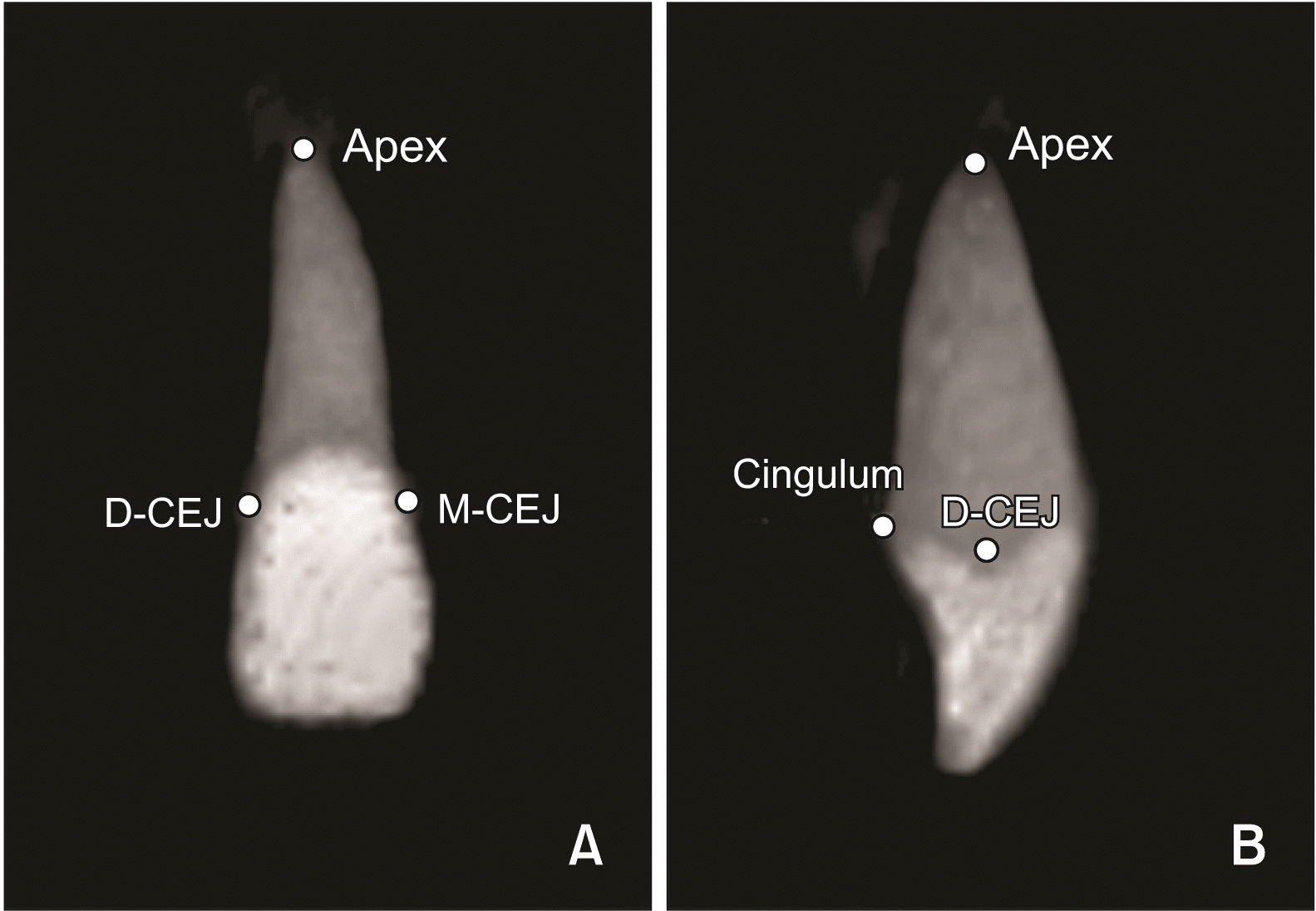
Figure 1, root apex, midpoints of the mesial cementoenamel junction (M-CEJ) and distal cementoenamel junction (D-CEJ), and the most prominent point of the cingulum were used as landmarks for orientation and measurement.
 | Figure 1Four reference points: apex, mesial cementoenamel junction (M-CEJ), distal cementoenamle junction (D-CEJ), and cingulum. A, Coronal view. B, Proximal view. 
|
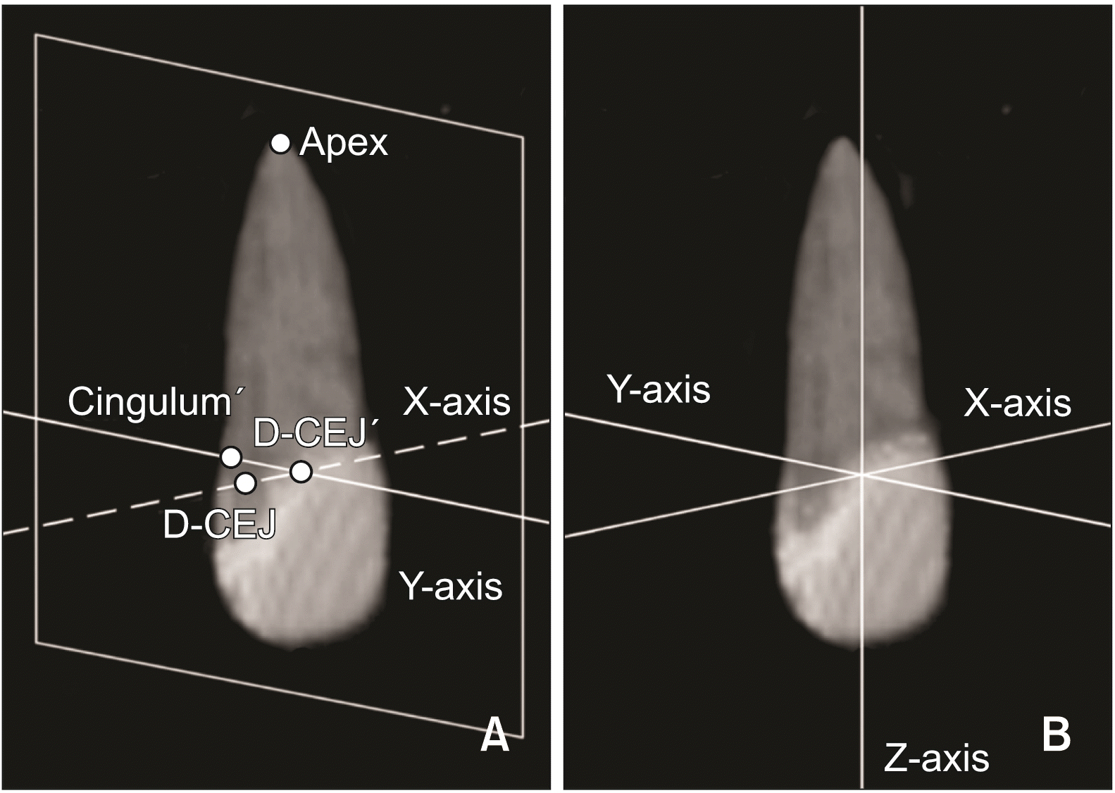
X-axis was the line passing through M-CEJ and D-CEJ (
Figure 2). The sagittal reference plane was defined as a plane perpendicular to the X-axis and passing through the apex. Y-axis was the line passing the projected distal CEJ point (D-CEJ’) and cingulum point (Cingulum’) on the sagittal reference plane. Z-axis was the cross product of the X-axis and Y-axis.
 | Figure 2Three-dimensional orientation of tooth. A, X-axis is the line passing through mesial cementoenamel junction (M-CEJ) and distal cementoenamle junction (D-CEJ). Y-axis is the line passing through the projected distal CEJ (D-CEJ’) and cingulum point (Cingulum’) on the sagittal reference plane. B, Z-axis is the cross product of the X-axis and Y-axis. 
|
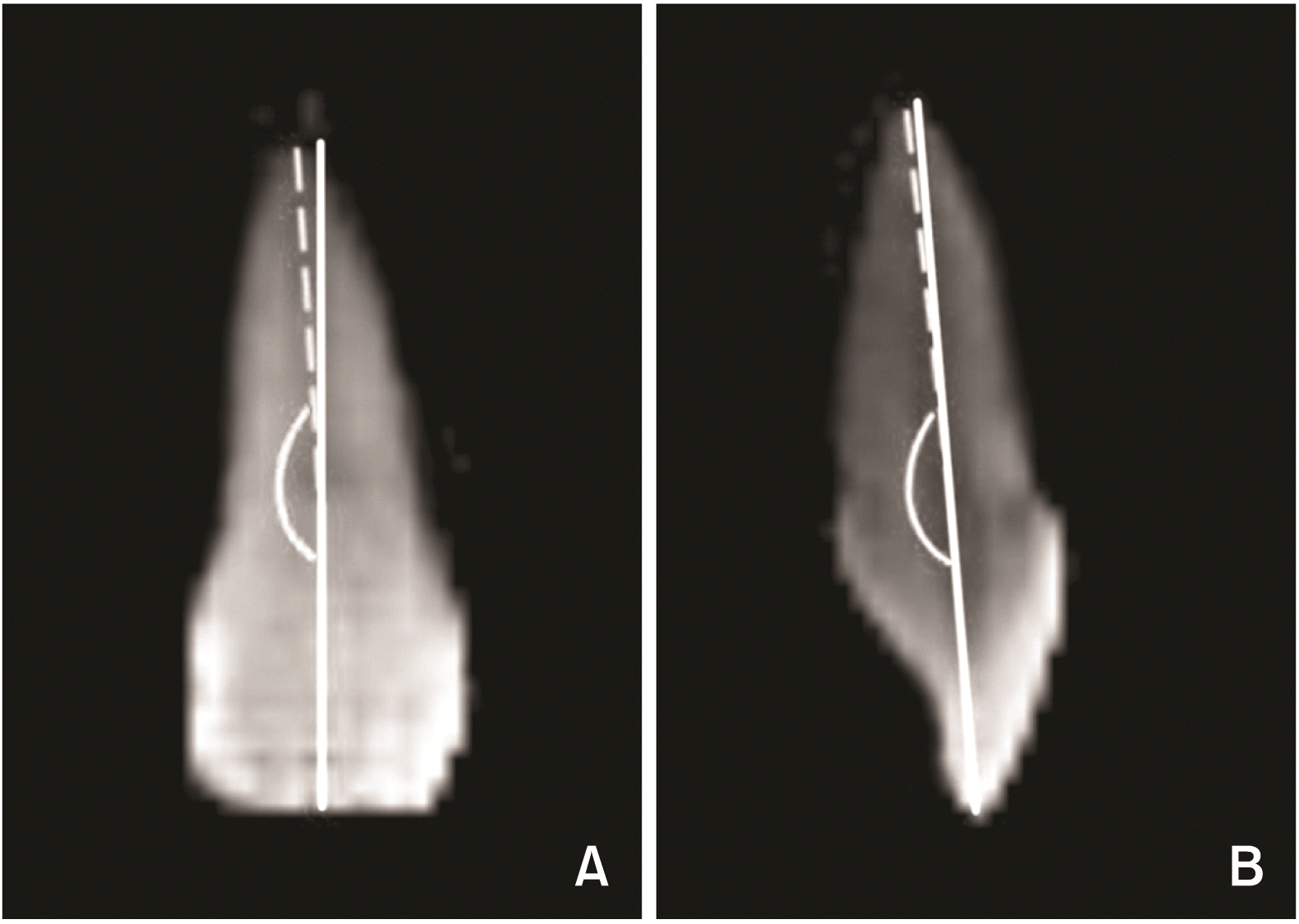
After the orientation of each tooth, 2D coronal and sagittal images such as periapical radiographs were generated using the function in the OnDemand software. The crown and root axis were created on each coronal and sagittal 2D images. MDCRA was measured on the coronal image and LLCRA was measured on the sagittal one (
Figure 3). The root axis was the line passing the root apex and the midpoint of M-CEJ and D-CEJ. The crown axis was the line passing the midpoint of M-CEJ and D-CEJ and the midpoint of incisor edges for incisors and the cusp tip for the canine. The obtuse angle between the root axis and the crown axis was measured to evaluate the CRA (
Figure 3). An angle of less than 180° represented distal angulation of the root on sagittal images and lingual angulation on coronal images. All technical procedure, orientations, and measurements of all teeth were carried out by one examiner (Lee K. H.) with a blind procedure to minimize the examiner bias.
 | Figure 3Measurement of the crown-root angulation (CRA). A, Mesiodistal CRA (MDCRA) in the coronal plane. B, Labiolingual CRA (LLCRA) in the sagittal plane. 
|
Method errors
Ten of 60 CBCT images were randomly selected. Landmark identifications, orientations, and measurements were repeated by the same examiner (Lee K. H.) to test method errors. The method error was calculated using Dahlberg's formula (ME = √(∑d2/2n)), where d was the difference between measurements and n was the sample size. Method errors of repeated measurement were 0.58°, 0.37°, and 0.39° for the MDCRA and 0.50°, 0.64°, and 0.58° for the LLCRA of the central incisor, lateral incisor, and canine, respectively.
Statistical analysis
The null hypothesis was that the CRA of each maxillary anterior tooth was not significantly different among skeletal Class I, Class II, and Class III groups. A nonparametric statistical test was used because variables did not show a normal distribution in the Shapiro–Wilk test. Kruskal–Wallis test was used to test differences among three groups. Mann–Whitney U test was then used to evaluate any significant difference between the two groups, and the alpha value was set at 0.017 according to Bonferroni correction (0.05/3). All statistical analyses were performed using SPSS software (PASW Statistics 18.0; IBM Co., Armonk, NY, USA).
Go to :

RESULTS
Table 3 shows the mean and standard deviation of CRAs of the maxillary anterior teeth in each skeletal malocclusion group. Mean MDCRAs of the maxillary central incisors were 177.9°, 177.2°, and 178.7° in Class I, Class II, and Class III groups, respectively. MDCRAs for the lateral incisors were 177.8°, 177.6°, and 178.3° in Class I, Class II, and Class III groups, respectively. MDCRAs for the canines were 174.0°, 173.0°, and 173.9° in Class I, Class II, and Class III group, respectively. The mean MDCRAs of the maxillary anterior teeth showed no significant difference among groups.
Table 3
Mean and standard deviation (SD) of CRA of three groups
|
CRA |
Class I (n = 20) |
|
Class II (n = 20) |
|
Class III (n = 20) |
|
Kruskal–Wallis test |
|
Mann–Whitney U test |
|
Mean |
SD |
Mean |
SD |
Mean |
SD |
Sig. |
Sig. |
|
MDCRA |
|
|
|
|
|
|
|
|
|
|
|
|
|
#11 |
177.9 |
2.3 |
|
177.2 |
2.7 |
|
178.7 |
3.0 |
|
0.231 |
|
NS |
|
#12 |
177.8 |
3.8 |
|
177.6 |
3.1 |
|
178.3 |
3.7 |
|
0.836 |
|
NS |
|
#13 |
174.0 |
3.2 |
|
173.0 |
3.3 |
|
173.9 |
4.3 |
|
0.528 |
|
NS |
|
LLCRA |
|
|
|
|
|
|
|
|
|
|
|
|
|
#11 |
178.2 |
3.0 |
|
178.4 |
3.7 |
|
174.0 |
3.5 |
|
0.001***
|
|
I, II > III |
|
#12 |
175.9 |
1.8 |
|
175.0 |
2.3 |
|
174.0 |
2.4 |
|
0.030*
|
|
I > III |
|
#13 |
184.3 |
3.6 |
|
182.6 |
2.2 |
|
183.5 |
3.0 |
|
0.181 |
|
NS |

The mean LLCRA of the maxillary central incisors was significantly lower in skeletal Class III group (174.0°) than that in Class I group (178.2°) or Class II group (178.4°). The mean LLCRA of the lateral incisors was also significantly lower in skeletal Class III group (174.0°) than that in Class I group (175.9°). Mean LLCRAs of the canines were 184.3°, 182.6°, and 183.5° in Class I, Class II, and Class III groups, respectively, showing no significant difference among groups.
Results of statistical comparisons among CRAs of the maxillary anterior teeth for each malocclusion group are shown in
Table 4. The mean MDCRA of the canines was significantly lower than that of the central or lateral incisors in all groups. The mean LLCRA of the maxillary canines was the greatest, followed by that of the central and lateral incisors in Class I and Class II group. However, Class III group showed no significant difference in LLCRA between the central and lateral incisors.
Table 4
Statistical comparison of the maxillary anterior teeth in each group
|
CRA |
Kruskal–Wallis test |
Mann–Whitney U test |
|
#11 vs. #12 |
#11 vs. #13 |
#12 vs. #13 |
|
MDCRA |
|
|
|
|
|
Class I |
< 0.001***
|
0.547 |
0.002**
|
< 0.001***
|
|
Class II |
< 0.001***
|
0.904 |
< 0.001***
|
< 0.001***
|
|
Class III |
< 0.001***
|
0.738 |
< 0.001***
|
< 0.001***
|
|
LLCRA |
|
|
|
|
|
Class I |
< 0.001***
|
0.002**
|
< 0.001***
|
< 0.001***
|
|
Class II |
< 0.001***
|
< 0.001***
|
< 0.001***
|
< 0.001***
|
|
Class III |
< 0.001***
|
0.968 |
< 0.001***
|
< 0.001***
|

Go to :

DISCUSSION
Tooth morphology is one of the most interesting issues in orthodontic treatment. And CRA is considered as an important feature of the tooth morphology, particularly in single rooted anterior teeth. In anatomical studies about tooth morphology, some authors have stated that the LLCRA of the maxillary central incisors is lower in deep overbite (“deckbiss”) cases.
14,15,23 Many studies have investigated LLCRAs of the maxillary central incisors using the extracted teeth or 2D cephalometric radiograph.
1,11-15,18 These previous studies have shown various ranges of LLCRAs in maxillary central incisors. However, no study has tried to measure MDCRAs of the maxillary central incisors which is might due to methodological limitations for measuring MDCRAs. This was the first study trying to assess not only LLCRAs, but also MDCRAs of the maxillary anterior teeth using CBCT.
The present study showed that MDCRAs of the maxillary anterior teeth were not significantly different among skeletal malocclusion groups (
Table 3). In this study, samples were divided only by skeletal malocclusion based on sagittal relationships. However, other possible factors that might influence MDCRAs of the maxillary anterior teeth should be considered in the future. For example, during root development and tooth eruption, crowding due to small apical base of the maxilla and relationship between the tooth germs might influence MDCRAs.
In the present study, the MDCRA of the maxillary central incisors was like that of the maxillary lateral incisors in each skeletal malocclusion. The MDCRA of the maxillary canines was significantly lower than that of the maxillary central or lateral incisors (
Table 4). This indicate that orthodontists do not need to give different esthetic bend according to skeletal malocclusions in edgewise appliance technique. In addition, during bracket positioning on the canine, more crown distal tipping than might be better for establishing root parallelism.
The present study showed that the mean LLCRA of the maxillary central incisors in Class III malocclusion was lower than that in Class I or Class II division 1 malocclusion. This result is like the result of 2D cephalometric study conducted by Harris et al.
12 This configuration might be explained in two ways. First, Harris et al.
12 suggested that the bending phenomenon leading low LLCRAs of the maxillary central incisors in Class III malocclusion with anterior crossbite might have resulted from maxillary central incisors being trapped within the lower arch. Second, there might be relatively increased effect of the upper lip due to low tongue position in skeletal Class III malocclusion. Because teeth are generally on equilibrium state among surrounding environments, the bending phenomenon of the maxillary central incisors might be induced by the relative increased pressure of the upper lip in skeletal Class III malocclusion.
In some previous studies using 2D radiograph images, the relationship between the crown and root of the maxillary central incisors has been determined according to malocclusions.
1,12-14,18 Most of these studies reported that LLCRAs of maxillary central incisors in Class II division 2 were lower than those in Class I, Class II division 1, or Class III. Backlund
23 suggested that the lower lip can influence the maxillary incisors during eruption, causing bending phenomenon of the crown relative to the root because the lip line in Angle Class II division 2 is often higher than that in other malocclusions.
Different LLCRAs of the maxillary incisors suggest some clinical implications during orthodontic treatment. In camouflage treatment of skeletal Class III malocclusion with non-extraction, maxillary incisors generally become proclined. In such situation, orthodontists must be aware of the possibility of contact between the root and palatal cortical bone. Delivanis and Kuftinec
14 and Bryant et al.
1 also suggested similar clinical complication of low LLCRAs of the maxillary central incisors in Angle Class II division 2. McIntyre and Millett
13 suggested that when maxillary incisors with pronounced low LLCRAs are observed, a prediction template tracing can be used to ascertain whether the expected tooth movements are feasible or whether the incisors apex would touch the palatal cortical plate.
In a FEM study conducted by Heravi et al.,
24 the equivalent force was heavier while retracting maxillary incisors with low LLCRAs. Therefore, the amount of retraction force must be paid attention in skeletal Class III surgery with maxillary premolar extraction.
For the maxillary lateral incisors, although LLCRAs in skeletal Class III malocclusion tended to be lower than those in skeletal Class I and Class II malocclusions, the extent was not remarkable, unlike that of the maxillary central incisors (
Table 3). The mean LLCRA of the maxillary lateral incisors was like that of the maxillary central incisors (
Table 4). These results might mean that LLCRAs of the maxillary central incisors are more influenced by skeletal Class III malocclusion than those of the maxillary lateral incisors.
Until recent times, there has been very little agreement on what factor, either environmental or genetic, has the most effect on the bending phenomenon of the maxillary incisors. Some studies have suggested that the bending phenomenon of the maxillary incisors among malocclusions is due to multiplicity of environmental forces such as lip, tongue, and overbite relationship.
12,23 However, Logan
25 indicated that genetic factors also can be a primary cause of the relationship between the crown and root of the maxillary incisors.
There have been some previous studies about LLCRAs of the maxillary anterior teeth using 2D cephalometric radiographs.
1,12-14,18 However, studies using 2D cephalometric radiographs generally have inherent tracing errors due to overlapping of the anterior teeth, various 3D positions of the teeth or blunt images.
26,27 Some classical studies have also used extracted incisors to measure LLCRAs.
11,15 Clinically speaking, it is difficult to collect sound extracted anterior teeth. Therefore, this present study was the first study trying to investigate CRAs of the anterior teeth after orientation of individual teeth via reference lines using CBCT.
This study has several limitations. To assess CRAs of maxillary anterior teeth according to skeletal malocclusions, environmental factors that might influence CRAs were not considered. Also, because of sample shortage, Angle Class II division 2 malocclusion could not be evaluated. A further direction of this study will be to elucidate various environmental factors and assess CRAs of not only anterior teeth, but also posterior teeth with sufficient sample size.
Go to :


 PDF
PDF Citation
 Print
Print


 XML Download