Journal List > Korean J Physiol Pharmacol > v.26(6) > 1516080096

Hu, Zhang, Fan, Zhang, Wang, and Wang: Lactate promotes vascular smooth muscle cell switch to a synthetic phenotype by inhibiting miR-23b expression

Abstract

Recent research indicates that lactate promotes the switching of vascular smooth muscle cells (VSMCs) to a synthetic phenotype, which has been implicated in various vascular diseases. This study aimed to investigate the effects of lactate on the VSMC phenotype switch and the underlying mechanism. The CCK-8 method was used to assess cell viability. The microRNAs and mRNAs levels were evaluated using quantitative PCR. Targets of microRNA were predicted using online tools and confirmed using a luciferase reporter assay. We found that lactate promoted the switch of VSMCs to a synthetic phenotype, as evidenced by an increase in VSMC proliferation, mitochondrial activity, migration, and synthesis but a decrease in VSMC apoptosis. Lactate inhibited miR-23b expression in VSMCs, and miR-23b inhibited VSMC's switch to the synthetic phenotype. Lactate modulated the VSMC phenotype through downregulation of miR-23b expression, suggesting that overexpression of miR-23b using a miR-23b mimic attenuated the effects of lactate on VSMC phenotype modulation. Moreover, we discovered that SMAD family member 3 (SMAD3) was the target of miR-23b in regulating VSMC phenotype. Further findings suggested that lactate promotes VSMC switch to synthetic phenotype by targeting SMAD3 and downregulating miR-23b. These findings suggest that correcting the dysregulation of miR-23b/SMAD3 or lactate metabolism is a potential treatment for vascular diseases.

INTRODUCTION

Unlike terminally differentiated muscle cells, including skeletal muscle cells and cardiomyocytes, the contractile state of vascular smooth muscle cells (VSMCs) can switch to a synthetic phenotype in response to various stimuli [1]. The switch of VSMCs to a synthetic phenotype has been implicated in various vascular diseases, including atherosclerosis [2-4]. VSMCs with a synthetic phenotype are less able to contract, migrate to the intima, proliferate, and produce extracellular matrix proteins, leading to vascular dysfunction [5]. Thus, understanding the underlying mechanisms of VSMC phenotypic regulation will lead to developing a novel strategy for preventing and treating vascular diseases.
Various factors, including growth factors, mechanical injury, reactive oxygen species, and metabolites [6,7], regulate the VSMC phenotypic switch. Under normal, well-oxygenated conditions, VSMCs exhibit unusually high glycolysis rates, relying heavily on glycolytically generated ATP to sustain various cell functions [8]. Lactate, the end product of glycolysis, has been considered a metabolic waste product for a very long time. However, accumulating evidence suggests that lactate is taken up by numerous cells and functions as a signal in numerous biological processes [9-12]. Additionally, lactate levels rise in response to ischemia, which is linked to various vascular diseases [13]. Recent research has demonstrated that lactate promotes the synthetic phenotype of VSMCs, establishing a link between glycolysis and VSMC phenotypic switch [14]. However, the mechanism underlying lactate's regulation of VSMC phenotype is largely unknown.
MicroRNAs (miRNAs) are a cluster of non-coding small RNAs with ~22 nucleotides long mature products [15-17] at play a significant role in cellular processes [18]. Recent research demonstrates that microRNAs play a crucial role in regulating VSMC differentiation and phenotype switch [19]. Tang et al. [20] reported that miR-124 was significantly correlated with the contractile VSMC phenotype and that activation of SP1 could significantly reverse the antiproliferative effect of miR-124. miR-23b is significantly downregulated after vascular injury, and overexpression of miR-23b inhibited the migration markedly by elevating smooth muscle α-actin and smooth muscle myosin-injured arteries; additional analyses revealed that miR-23b modifies the phenotype of VSMCs by targeting SMAD family member 3 and transcription factor forkhead box O4 [21]. In addition, miR-143 and miR-145 stimulate the migration of pulmonary arterial smooth muscle cells by targeting ABCA1 [22]. Despite these, several miRNAs have been identified as regulators of the VSMC phenotype. These include miR-22 [19], miR-100 [23], miR-133 [24], miR-146a [25], miR-221/222 [26], and miR-424 [27]. However, the precise mechanism of VSMC phenotype switching is not yet completely understood.
Here, we investigated whether these miRNAs are involved in lactate's regulation of VSMC phenotype and discovered that lactate promotes VSMC's switch to the synthetic phenotype by inhibiting miR-23b expression.

METHODS

Cell culture

As described previously, primary VSMCs were isolated from the thoracic aortas of standard deviation (SD) rats (170–200 g, male) [28]. Briefly, after anesthetization with the intraperitoneal administration of pentobarbital sodium (60 mg/kg), thoracic aortas were removed and washed three times with phosphate buffer saline. The media layer of the aorta was dissected, cut into pieces, and transplanted into a six-well culture plate. Cells were growing at 37°C in a humidified atmosphere containing 5% CO2 in Dulbecco's modified eagle medium (DMEM) supplemented with 10% fetal bovine serum (FBS), penicillin, and streptomycin for 2 weeks. The experiments used VSMCs between passages 3 and 5. All animal procedures were conducted in accordance with the National Institutes of Health's Guidelines for the Care and Use of Laboratory Animals and with the approval of the Second Affiliated Hospital of Xi'an Jiaotong University's Ethics Committee.

Detection of cell viability

Next, using the CCK-8 Kit, cell viability was determined (Dojindo, Kumamoto, Japan). Briefly, cells were seeded in 96-well plates and 10 μl of CCK-8 (5 mg/ml) was added to the culture medium in each well. The absorbance was measured at 450 nm using an Exl 800 microplate reader (Bio-tek, Winooski, VT, USA). Cell viability (%) = (experimental group OD value − zero group OD value) / (control group OD value − zero group OD value) × 100%.

Tunel assay

VSMC apoptosis was determined using the Tunel assay with a Tunel-specific detection kit (Roche, Mannheim, Germany) according to the manufacturer's instructions. Briefly, cells were fixed with 4% paraformaldehyde at room temperature for 10 min, permeabilized with 0.1% Triton X-100, and then fragmented DNA in VSMCs was end-labeled with FITC. In addition, the Tunel-positive cells were examined using a confocal microscope.

JC-1 determination

Mitochondrial membrane potential was determined using JC-1 staining (KeyGEN biotechnology, Jiangsu, China) according to the manufacturer's instructions, followed by flow cytometry evaluation (BD Bioscience, San Jose, CA, USA). Specifically, Q2 represents the mitochondrial cells that are healthy, whereas Q3 represents the mitochondrial membrane decline cells.

Transwell assay

The ability of VSMCs to migrate was evaluated using the Transwell assay. VSMCs were seeded in the upper chamber of the transwell at a concentration of 1.0 × 10⁵ cells/well in 300 μl. The lower chamber was filled with 600 μl of 10% FBS-containing DMEM. The cells in the upper chamber migrated to the lower chamber after 24 h of incubation. The cells on the surface of the lower chambers were then fixed with 20% methanol for 10 min at room temperature and stained with 1% crystal violet (diluted in methanol) for 15 min at room temperature. Under a light microscope, the migrated cells were then quantified.

Real-time reverse transcription PCR

Total RNA was isolated using RNAiso Plus (Takara, Shiga, Japan) reagent as directed by the manufacturer. cDNA was synthesized from 500 ng of RNA per sample using the Prime Script Master Mix (Takara). Then, quantitative PCR was conducted using a SYBR Green PCR kit (Takara) in a CFX200 (Bio-Rad, Hongkong, China). Each gene's mRNA level was normalized to those of the housekeeping gene GAPDH. The sequences of the primers are listed in Supplementary Table 1.

Cell transfection

In addition, miRNA mimics and inhibitors, in addition to their respective negative controls (NC), Empty vector (NC), and SMAD3 overexpression (SMAD3 OE) plasmids, were acquired from RIBOBIO Co. Ltd. (Guangzhou, China). NC mimics (100 nM; #miR1N0000001-1-10); miR-23b mimics (100 nM; 5-UGGGUUCCUGGCAUGCUGAUUU-3); NC inhibitors (200 nM; #miR2N0000001-1-10); miR-222 inhibitors (200 nM; 5´-AGGAUCUACACUGGCUACUGAG-3´), miR-23b inhibitors (200 nM; 5'-AAAUCAGCAUGCCAGGAACCCA-3´), miR-133a inhibitors (200 nM; 5´-CAGCUGGUUGAAGGGGACCAAA-3´), NC (2 μg), or SMAD3 OE (2 μg) were transfected using Lipofectamine 3000 (Invitrogen, Carlsbad, CA, USA) according to manufacturer’s instructions. After 48 h of transfection, cells were harvested and used in subsequent experiments.

Dual-luciferase reporter assay

Wt and Mt SMAD3 3' UTR sequences were cloned into the SpeI and HindIII sites of the pMir-Report Luciferase vector following PCR amplification using template and primers (Applied Biosystems, Foster City, CA, USA). Following the manufacturer's instructions, 5 ng of the resulting construct was transfected into 293T cells with 20 nM control mimics or 20 nM miR-23b mimics using Lipofectamine-2000 (Invitrogen). After 24 h of transfection, luciferase activity in the cells was determined using a Luciferase Assay System (Promega, Madison, WI, USA).

Western blot

Subsequently, using the RIPA buffer, proteins were extracted from cells for immunoblotting. 15–50 μg of total protein extracts were subjected to sodium dodecyl sulfate-polyacrylamide gel electrophoresis and transferred to a polyvinylidene fluoride membrane. After blocked with 5% skimmed milk, membranes were then probed with anti-Bax (1:2,000 dilution; #ab3203; Abcam, Cambridge, UK), anti-Bcl-2 (1:1,000; #ab32124; Abcam), anti-cleaved-caspase3 (1:500; #ab32042; Abcam), anti-cleaved-caspase9 (1:1,000; #ab2324; Abcam), anti-α-SMA (1:1,000; #ab5694; Abcam), anti-SM22 (1:1,000; #ab14106; Abcam), anti-SM-MHC (1:2,000; #ab133567; Abcam), anti-vimentin (1:2,000; #ab92547; Abcam), anti-collagen I (1:1,000; #ab270993; Abcam), anti-SMAD3 (1:2,000; #ab40854; Abcam), and anti-β-actin (1:5,000; #ab8226; Abcam) at room temperature for 1.5 h. Then, membranes were incubated with the appropriate secondary antibody conjugated to HRP. Then, the BM chemiluminescence blotting system (Thermo Fisher Scientific, Waltham, MA, USA) was used to visualize protein bands, and ImageJ Software (NIH, Bethesda, MD, USA) was used to quantify protein bands.

Statistical analysis

All values are presented using the mean ± SD format. The data were compared using the unpaired t-test, or one-way ANOVA followed by Tukey's test, as appropriate. The normal distribution of data was analyzed using the Kolmogorov–Smirnov normality test. Using Bonferroni's correction for multiple comparisons. When p < 0.05, differences were considered significant.

RESULTS

Lactate switched VSMCs to a synthetic phenotype

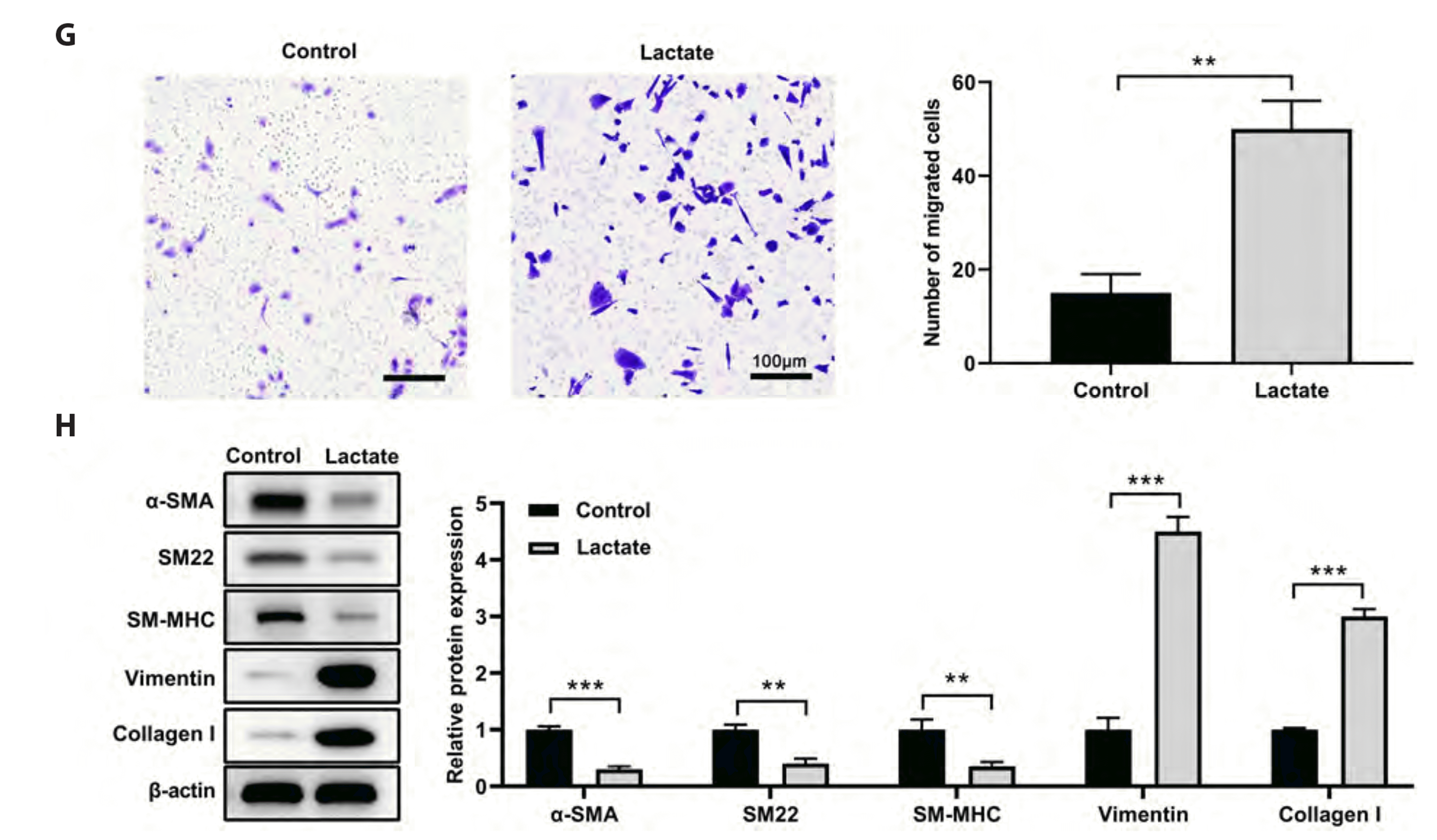
VSMCs were treated with lactate (0, 2, 4, or 8 mM) for 3 days. Lactate treatment caused the VSMCs to become less spindle-shaped and to develop the irregular morphology characteristic of synthetic VSMCs (Fig. 1A). The analysis of cell viability revealed that lactate treatment decreased the viability of VSMCs after 24 h of treatment and then significantly increased cell viability in a dose-dependent manner, despite the presence of 6 mM lactate (Fig. 1B). We hypothesized that the cytotoxicity induced by a high lactate concentration (6 mM) may have contributed to this persistent reduction in cell viability. Lactate (4 mM) treatment significantly decreased the apoptotic rate in VSMCs (Fig. 1C), whereas it significantly increased JC-1 signaling in VSMCs (Fig. 1D, E). Furthermore, Western blotting revealed that lactate treatment (4 mM) significantly decreased the levels of Bax, cleaved-caspase-3, and cleaved-caspase-9 in VSMCs, while increasing the level of Bcl-2 (Fig. 1F). These results suggest that lactate treatment may promote the proliferation of VSMCs. In addition, lactate treatment (4 mM) increased VSMC migration as measured by the transwell assay (Fig. 1G). Moreover, lactate treatment (4 mM) for 3 days decreased mRNA levels for markers of the contractile phenotype, including α-SMA, SM22, and SM-MHC, while increasing mRNA levels for markers of the synthetic phenotype, such as vimentin and collagen I (Fig. 1H). These findings suggest that lactate promotes the synthetic phenotype switch of VSMC.

Lactate decreased miR-23b expression in VSMCs

In order to determine whether miRNA is involved in lactate-induced regulation of the VSMC phenotype switch, the reported miRNAs involved in the VSMC phenotype switch were screened. As shown in Fig. 2A, 15 miRNAs were detected, and miR-222, miR-23b, and miR-133a were the top three miRNAs that decreased in lactate-treated VSMCs relative to untreated VSMCs. VSMCs were then transfected with their respective miRNA inhibitors to inhibit miR-222, mi-23b, and miR-133a expression (Fig. 2B). Then, phenotypic markers, including α-SMA, SM22, SM-MHC, vimentin, and collagen I, were then detected in VSMCs transfected with miRNA inhibitors. The qRT-PCR revealed that miR-222, miR-23b, and miR-133a inhibitors inhibited the levels of contractile markers (α-SMA, SM22, and SM-MHC) but increased the levels of synthetic markers (vimentin and collagen I), with the miR-23b inhibitor having the most significant effect (Fig. 2C–E). Consequently, miR-23b was chosen for the subsequent experiments. Overexpression of miR-23b inhibitors significantly increased the viability and migration of VSMCs compared to the NC group, as determined by additional analyses (Fig. 2F, G). These findings suggested that lactate may promote the VSMCs’ switch to a synthetic phenotype via miR-23b downregulation.

miR-23b mimic attenuated the effects of lactate on VSMC phenotype switch

A miR-23b mimic was used to determine whether miR-23b contributes to lactate's effects on VSMC phenotype switch. Figure 3A demonstrates that the miR-23b mimic increased miR-23b levels in VSMCs. Thus, miR-23b mimic diminished the effects of lactate (4 mM) on cell viability (Fig. 3B). In addition, miR-23b mimics inhibited lactate-induced apoptosis and JC-1 signaling (Fig. 3C, D). In addition, overexpression of the miR-23b mimic attenuated the lactate-induced migration enhancement (Fig. 3E). In addition, Western blot analysis demonstrated that the overexpression of the miR-23b mimics significantly reversed the effects of lactate on reducing the expression of α-SMA, SM22, and SM-MHC while increasing the expression of vimentin and collagen I (Fig. 3F). These findings suggest that inhibition of miR-23b contributes to the effects of lactate on VSMC synthetic phenotype switch promotion.

SMAD3 was the target of miR-23b

miR-23b target genes were predicted by miRDB, TargetScan, ENCORI, and GO 0007050 (cell cycle arrest). Four candidates were screened using a Venn analysis (Fig. 4A). Compared to the NC group, the miR-23b mimic reduced the mRNA levels of WEE1, HMGA2, and SMAD3, with VSMCs showing the greatest reduction (Fig. 4B). Fig. 4C indicates that SMAD3 is a potential target of miR-23b based on the correlation between the protein levels detected by the Western blot and the mRNA levels. Dual-luciferase reporter assay was used to confirm this result. Fig. 4D depicts the complementary sequences between the 3'UTR of SMAD3 and miR-23b. Either wild-type or mutant 3'UTRs containing putative miR-23b binding sites were cloned into a reporter plasmid, and their responsiveness to miR-23b in cells was evaluated. The results demonstrated that miR-23b inhibited luciferase activity in SMAD3 wild-type 3'UTR constructs but had no effect when the miR-23b binding sites were mutated (Fig. 4E, F). Western blot analysis revealed that lactate treatment significantly increased SMAD3 protein expression, whereas miR-23b mimic clearly reversed this upregulation (Fig. 4G). These results indicated that SMAD3 is a miR-23b target.

Lactate regulates the miR-23b/SMAD3 axis to modulate the VSMC phenotype switch

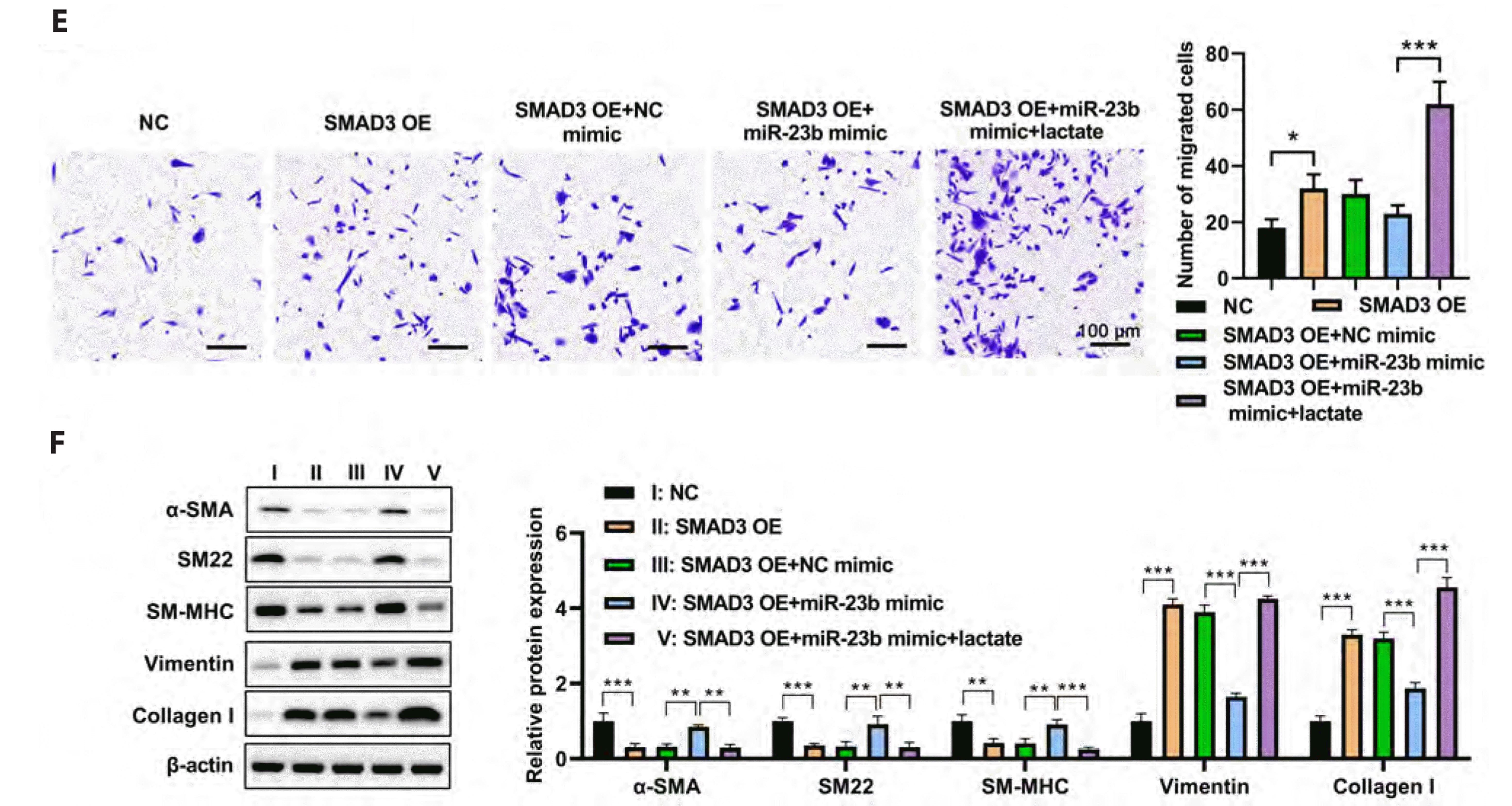
In order to determine whether it contributes to the effects of lactate on the VSMC phenotype switch, VSMCs were transfected with SMAD3, and miR-23b mimics individually. miR-23b was significantly upregulated in miR-23b mimic overexpressed VSMCs, whereas lactate treatment significantly reversed this upregulation (Fig. 5A). Moreover, SMAD3 expression was significantly increased after transfection with SMAD3 OE plasmid, miR-29b mimic significantly reversed this accumulation, and lactate significantly attenuated miR-29b's effects, thereby increasing SMAD3 expression (Fig. 5A). Lactate treatment reversed the effect of miR-23b to increase the cell viability of VSMCs. In contrast, SMAD overexpression significantly increased the cell viability of VSMCs (Fig. 5B). Meanwhile, SMAD3 decreases the apoptotic rate and promotes JC-1 signaling in VSMCs; however, miR-29b mimic significantly reverses these changes; and lactate treatment clearly reverses the effects of miR-23b to enhance SMAD3 effects on apoptosis and JC-1 signaling in VSMCs (Fig. 5C, D). In addition, miR-23b mimic inhibited the migration enhancement of VSMCs induced by SMAD3 expression, whereas lactate treatment abolished miR-23b's effects on SMAD3-mediated migration enhancement (Fig. 5E). Additionally, the expression of α-SMA, SM22, SM-MHC, vimentin, and collagen I detect the effects of lactate on phenotype switch. The findings revealed that miR-23b significantly inhibited the SMAD3-mediated decrease of α-SMA, SM22, and SM-MHC, as well as the increase of vimentin and collagen I in VSMCs. Conversely, lactate could reverse the effect of miR-23b and enhance the effect of SMAD3 in VSMCs (Fig. 5F). These findings supported the hypothesis that lactate promotes the switch of VSMC to a synthetic phenotype via regulation of the miR-23b/SMAD3 axis.

DISCUSSION

Lactate is a metabolic byproduct which has recently been shown to function as a signal in various processes, such as wound healing, inflammation, angiogenesis, and cancer development [8-10,29]. Recent research indicates that lactate [14] may play a role in the pathogenesis of vascular diseases, as it promotes the switch of VSMC to the synthetic phenotype. In this study, we discovered that lactate promotes VSMC switch to synthetic phenotype via downregulation of miR-23b, indicating that correcting the dysregulation of the miR-23b/SMAD3 axis may be a potential treatment for vascular diseases.
Unlike striated muscle cells, VSMCs exhibit unusually high glycolysis rates even under normal, well-oxygenated conditions, relying heavily on ATP from glycolysis rather than glucose oxidation to maintain their biological activity [30]. It is estimated that only 30% of ATP comes from mitochondrial oxidation, whereas at least 90% of glycolysis flux results in lactate production [31]. Thus, VSMCs produce a considerable amount of lactate. In addition, lactate concentrations rise in response to a variety of stimuli, including ischemia, exercise, cardiac arrest, shock, trauma, and burns [11-13,32]. Myocytes, endothelial cells, and human cytotoxic T lymphocytes take up lactate, inhibiting phosphofructokinase [33], altering gene expression in L6 muscle cells [8], contributing to T-cell migration [29], and promoting tumor growth [9]. As evidenced by the fact that lactate promotes VSMC viability, migration, and expression of synthetic phenotype markers, we discovered that lactate promotes VSMC's switch to the synthetic phenotype. Our findings and those of others link glucose metabolism to VSMC phenotype modulation, suggesting that metabolic disturbance plays a role in VSMC phenotype switching regulation. These findings may explain the role of metabolic dysfunction in inducing vascular dysfunction in vascular diseases.
Recent research indicates that microRNAs play an essential role in regulating VSMC differentiation and phenotype switch, and miR-23b is one of the microRNAs that inhibits VSMC switch to synthetic phenotype [21]. Previous research demonstrated that lactate transport was significantly altered in hypoxic muscle and that miR-124 regulated lactate transport by targeting MCT1 [34]. It had also been reported that lactate was significantly upregulated in gastric cancer tumor-infiltrating T cells and was associated with the decreased miR-34a and the increased lactate dehydrogenase A, thus impacting the hypoxic tumor environment, which was tightly correlated with the phenotype control in the development of cancer [35]. Therefore, we hypothesized that lactate may regulate the phenotype switching of cells by targeting multiple miRNAs or by being regulated by miRNAs. We discovered that lactate promotes the switch of VSMC to a synthetic phenotype by downregulating miR-23b. Lactate dose-dependently suppressed the expression of miR-23b in VSMCs. Overexpression of miR-23b promoted the switch of VSMC to a synthetic phenotype and inhibited the effects of lactate on this switch. miR-23b, miR-27b, and miR-24-1 are all expressed from the same primary transcript [36]. It has been demonstrated that miR-23b is associated with cancer development. For instance, miR-23b expressions are reduced in human prostate tumor samples and have an inverse correlation with cell proliferation and migration [37]. In addition, miR-23b inhibits the pathogenesis of multiple autoimmune diseases by targeting cytokine-mediated pro-inflammatory signaling [38]. miR-23b also plays a role in the regulation of VSMC phenotype switching, with miR-23b downregulation promoting VSMC phenotype switching [21].
Several miR-23b targets have been implicated in the regulation of VSMC function, with urokinase-type plasminogen activator (uPA) and SMAD3 being of particular importance. Furthermore, uPA is an indispensable regulator of neointimal growth and vascular remodeling. Studies indicate that increased uPA expression contributes to VSMC proliferation, migration, and neointima formation following injury [39,40]. SMAD3 participates in TGF-β signaling. It was observed to be overexpressed in several vascular diseases [41]. Furthermore, SMAD3 overexpression has been reported to stimulate VSMC proliferation and phenotypic switching [21,42]. Targeting SMAD3, we found that miR-23b regulates VSMC phenotypic switching. The inhibition of SMAD3 diminished the modulatory effects of miR-23b and lactate on the VSMC phenotype. These findings suggested that the miR-23b/SMAD3 axis plays an important role in VSMC phenotypic modulation, and correcting the dysregulation of miR-23b/SMAD3 or lactate metabolism may be an effective treatment for vascular diseases.
We discovered that lactate promotes the switch of VSMC to a synthetic phenotype by downregulating miR-23b expression. Additionally, miR-23b promotes the synthetic phenotype of VSMC by targeting SMAD3. The regulation of VSMC phenotype by lactate metabolism may contribute to the maintenance of vascular health and the prevention of vascular diseases, as suggested by these findings.

SUPPLEMENTARY MATERIALS

Supplementary data including one Table can be found with this article online at https://doi.org/10.4196/kjpp.2022.26.6.519.

ACKNOWLEDGEMENTS

We are very grateful for the equipment and technical support provided by The Second Affiliated Hospital of Xi'an Jiaotong University.

Notes

FUNDING

This work was supported by the 2020 Key Research and Development Plan of Shaanxi Province the: role of urotensin II and transforming growth factorβ1 in the left ventricular remodeling in chronic heart failure (2021SF-321).

CONFLICTS OF INTEREST

The authors declare no conflicts of interest.

REFERENCES

1. Basatemur GL, Jørgensen HF, Clarke MCH, Bennett MR, Mallat Z. 2019; Vascular smooth muscle cells in atherosclerosis. Nat Rev Cardiol. 16:727–744. DOI: 10.1038/s41569-019-0227-9. PMID: 31243391. PMID: https://www.scopus.com/inward/record.uri?partnerID=HzOxMe3b&scp=85068318311&origin=inward.
crossref
2. Bennett MR, Sinha S, Owens GK. 2016; Vascular smooth muscle cells in atherosclerosis. Circ Res. 118:692–702. DOI: 10.1161/CIRCRESAHA.115.306361. PMID: 26892967. PMCID: PMC4762053. PMID: https://www.scopus.com/inward/record.uri?partnerID=HzOxMe3b&scp=84959048825&origin=inward.
crossref
3. Morrow D, Guha S, Sweeney C, Birney Y, Walshe T, O'Brien C, Walls D, Redmond EM, Cahill PA. 2008; Notch and vascular smooth muscle cell phenotype. Circ Res. 103:1370–1382. DOI: 10.1161/CIRCRESAHA.108.187534. PMID: 19059839. PMID: https://www.scopus.com/inward/record.uri?partnerID=HzOxMe3b&scp=58149379347&origin=inward.
crossref
4. Lacolley P, Regnault V, Avolio AP. 2018; Smooth muscle cell and arterial aging: basic and clinical aspects. Cardiovasc Res. 114:513–528. DOI: 10.1093/cvr/cvy009. PMID: 29514201. PMID: https://www.scopus.com/inward/record.uri?partnerID=HzOxMe3b&scp=85042943530&origin=inward.
crossref
5. Grootaert MOJ, Moulis M, Roth L, Martinet W, Vindis C, Bennett MR, De Meyer GRY. 2018; Vascular smooth muscle cell death, autophagy and senescence in atherosclerosis. Cardiovasc Res. 114:622–634. DOI: 10.1093/cvr/cvy007. PMID: 29360955. PMID: https://www.scopus.com/inward/record.uri?partnerID=HzOxMe3b&scp=85042944408&origin=inward.
crossref
6. Chistiakov DA, Orekhov AN, Bobryshev YV. 2015; Vascular smooth muscle cell in atherosclerosis. Acta Physiol (Oxf). 214:33–50. DOI: 10.1111/apha.12466. PMID: 25677529. PMID: https://www.scopus.com/inward/record.uri?partnerID=HzOxMe3b&scp=84927640251&origin=inward.
crossref
7. Salabei JK, Hill BG. 2015; Autophagic regulation of smooth muscle cell biology. Redox Biol. 4:97–103. DOI: 10.1016/j.redox.2014.12.007. PMID: 25544597. PMCID: PMC4309847. PMID: https://www.scopus.com/inward/record.uri?partnerID=HzOxMe3b&scp=84919782591&origin=inward.
crossref
8. Hashimoto T, Hussien R, Oommen S, Gohil K, Brooks GA. 2007; Lactate sensitive transcription factor network in L6 cells: activation of MCT1 and mitochondrial biogenesis. FASEB J. 21:2602–2612. DOI: 10.1096/fj.07-8174com. PMID: 17395833. PMID: https://www.scopus.com/inward/record.uri?partnerID=HzOxMe3b&scp=34547790342&origin=inward.
crossref
9. Hirschhaeuser F, Sattler UG, Mueller-Klieser W. 2011; Lactate: a metabolic key player in cancer. Cancer Res. 71:6921–6925. DOI: 10.1158/0008-5472.CAN-11-1457. PMID: 22084445. PMID: https://www.scopus.com/inward/record.uri?partnerID=HzOxMe3b&scp=81155126061&origin=inward.
crossref
10. Dhup S, Dadhich RK, Porporato PE, Sonveaux P. 2012; Multiple biological activities of lactic acid in cancer: influences on tumor growth, angiogenesis and metastasis. Curr Pharm Des. 18:1319–1330. DOI: 10.2174/138161212799504902. PMID: 22360558. PMID: https://www.scopus.com/inward/record.uri?partnerID=HzOxMe3b&scp=84857716739&origin=inward.
crossref
11. Müller P, Duderstadt Y, Lessmann V, Müller NG. 2020; Lactate and BDNF: key mediators of exercise induced neuroplasticity? J Clin Med. 9:1136. DOI: 10.3390/jcm9041136. PMID: 32326586. PMCID: PMC7230639. PMID: 76c733ff1dc9428887e259897e30f192. PMID: https://www.scopus.com/inward/record.uri?partnerID=HzOxMe3b&scp=85087787055&origin=inward.
crossref
12. Baltazar F, Afonso J, Costa M, Granja S. 2020; Lactate beyond a waste metabolite: metabolic affairs and signaling in malignancy. Front Oncol. 10:231. DOI: 10.3389/fonc.2020.00231. PMID: 32257942. PMCID: PMC7093491. PMID: 2b0659710b5a4cafb82419c662eae7cc. PMID: https://www.scopus.com/inward/record.uri?partnerID=HzOxMe3b&scp=85082649332&origin=inward.
crossref
13. Pereira-Nunes A, Afonso J, Granja S, Baltazar F. 2020; Lactate and lactate transporters as key players in the maintenance of the Warburg effect. Adv Exp Med Biol. 1219:51–74. DOI: 10.1007/978-3-030-34025-4_3. PMID: 32130693. PMID: https://www.scopus.com/inward/record.uri?partnerID=HzOxMe3b&scp=85081043083&origin=inward.
crossref
14. Yang L, Gao L, Nickel T, Yang J, Zhou J, Gilbertsen A, Geng Z, Johnson C, Young B, Henke C, Gourley GR, Zhang J. 2017; Lactate promotes synthetic phenotype in vascular smooth muscle cells. Circ Res. 121:1251–1262. DOI: 10.1161/CIRCRESAHA.117.311819. PMID: 29021296. PMCID: PMC5681426. PMID: https://www.scopus.com/inward/record.uri?partnerID=HzOxMe3b&scp=85037676960&origin=inward.
crossref
15. Gomez-Cabrera MC, Domenech E, Viña J. 2008; Moderate exercise is an antioxidant: upregulation of antioxidant genes by training. Free Radic Biol Med. 44:126–131. DOI: 10.1016/j.freeradbiomed.2007.02.001. PMID: 18191748. PMID: https://www.scopus.com/inward/record.uri?partnerID=HzOxMe3b&scp=37849021505&origin=inward.
crossref
16. Griffiths-Jones S. 2004; The microRNA Registry. Nucleic Acids Res. 32:D109–D111. DOI: 10.1093/nar/gkh023. PMID: 14681370. PMCID: PMC308757.
crossref
17. Griffiths-Jones S, Grocock RJ, van Dongen S, Bateman A, Enright AJ. 2006; miRBase: microRNA sequences, targets and gene nomenclature. Nucleic Acids Res. 34:D140–D144. DOI: 10.1093/nar/gkj112. PMID: 16381832. PMCID: PMC1347474. PMID: https://www.scopus.com/inward/record.uri?partnerID=HzOxMe3b&scp=33644750115&origin=inward.
crossref
18. Pasquinelli AE, Reinhart BJ, Slack F, Martindale MQ, Kuroda MI, Maller B, Hayward DC, Ball EE, Degnan B, Müller P, Spring J, inivasan A Sr, Fishman M, Finnerty J, Corbo J, Levine M, Leahy P, Davidson E, Ruvkun G. 2000; Conservation of the sequence and temporal expression of let-7 heterochronic regulatory RNA. Nature. 408:86–89. DOI: 10.1038/35040556. PMID: 11081512. PMID: https://www.scopus.com/inward/record.uri?partnerID=HzOxMe3b&scp=0034597777&origin=inward.
crossref
19. Yang F, Chen Q, He S, Yang M, Maguire EM, An W, Afzal TA, Luong LA, Zhang L, Xiao Q. 2018; miR-22 is a novel mediator of vascular smooth muscle cell phenotypic modulation and neointima formation. Circulation. 137:1824–1841. DOI: 10.1161/CIRCULATIONAHA.117.027799. PMID: 29246895. PMCID: PMC5916488. PMID: https://www.scopus.com/inward/record.uri?partnerID=HzOxMe3b&scp=85051770266&origin=inward.
crossref
20. Tang Y, Yu S, Liu Y, Zhang J, Han L, Xu Z. 2017; MicroRNA-124 controls human vascular smooth muscle cell phenotypic switch via Sp1. Am J Physiol Heart Circ Physiol. 313:H641–H649. DOI: 10.1152/ajpheart.00660.2016. PMID: 28667053. PMID: https://www.scopus.com/inward/record.uri?partnerID=HzOxMe3b&scp=85029370012&origin=inward.
crossref
21. Iaconetti C, De Rosa S, Polimeni A, Sorrentino S, Gareri C, Carino A, Sabatino J, Colangelo M, Curcio A, Indolfi C. 2015; Down-regulation of miR-23b induces phenotypic switching of vascular smooth muscle cells in vitro and in vivo. Cardiovasc Res. 107:522–533. DOI: 10.1093/cvr/cvv141. PMID: 25994172. PMID: https://www.scopus.com/inward/record.uri?partnerID=HzOxMe3b&scp=84948129366&origin=inward.
crossref
22. Yue Y, Zhang Z, Zhang L, Chen S, Guo Y, Hong Y. 2018; miR-143 and miR-145 promote hypoxia-induced proliferation and migration of pulmonary arterial smooth muscle cells through regulating ABCA1 expression. Cardiovasc Pathol. 37:15–25. DOI: 10.1016/j.carpath.2018.08.003. PMID: 30195228. PMID: https://www.scopus.com/inward/record.uri?partnerID=HzOxMe3b&scp=85052849462&origin=inward.
crossref
23. Grundmann S, Hans FP, Kinniry S, Heinke J, Helbing T, Bluhm F, Sluijter JP, Hoefer I, Pasterkamp G, Bode C, Moser M. 2011; MicroRNA-100 regulates neovascularization by suppression of mammalian target of rapamycin in endothelial and vascular smooth muscle cells. Circulation. 123:999–1009. DOI: 10.1161/CIRCULATIONAHA.110.000323. PMID: 21339483. PMID: https://www.scopus.com/inward/record.uri?partnerID=HzOxMe3b&scp=79952739991&origin=inward.
crossref
24. Torella D, Iaconetti C, Catalucci D, Ellison GM, Leone A, Waring CD, Bochicchio A, Vicinanza C, Aquila I, Curcio A, Condorelli G, Indolfi C. 2011; MicroRNA-133 controls vascular smooth muscle cell phenotypic switch in vitro and vascular remodeling in vivo. Circ Res. 109:880–893. DOI: 10.1161/CIRCRESAHA.111.240150. PMID: 21852550.
crossref
25. Luo Y, Xiong W, Dong S, Liu F, Liu H, Li J. 2017; MicroRNA-146a promotes the proliferation of rat vascular smooth muscle cells by downregulating p53 signaling. Mol Med Rep. 16:6940–6945. DOI: 10.3892/mmr.2017.7477. PMID: 28901447. PMID: https://www.scopus.com/inward/record.uri?partnerID=HzOxMe3b&scp=85030107419&origin=inward.
crossref
26. Liu X, Cheng Y, Zhang S, Lin Y, Yang J, Zhang C. 2009; A necessary role of miR-221 and miR-222 in vascular smooth muscle cell proliferation and neointimal hyperplasia. Circ Res. 104:476–487. DOI: 10.1161/CIRCRESAHA.108.185363. PMID: 19150885. PMCID: PMC2728290. PMID: https://www.scopus.com/inward/record.uri?partnerID=HzOxMe3b&scp=61949252089&origin=inward.
crossref
27. Merlet E, Atassi F, Motiani RK, Mougenot N, Jacquet A, Nadaud S, Capiod T, Trebak M, Lompré AM, Marchand A. 2013; miR-424/322 regulates vascular smooth muscle cell phenotype and neointimal formation in the rat. Cardiovasc Res. 98:458–468. DOI: 10.1093/cvr/cvt045. PMID: 23447642. PMCID: PMC3656613. PMID: https://www.scopus.com/inward/record.uri?partnerID=HzOxMe3b&scp=84877949558&origin=inward.
crossref
28. Tang R, Mei X, Wang YC, Cui XB, Zhang G, Li W, Chen SY. 2019; LncRNA GAS5 regulates vascular smooth muscle cell cycle arrest and apoptosis via p53 pathway. Biochim Biophys Acta Mol Basis Dis. 1865:2516–2525. DOI: 10.1016/j.bbadis.2019.05.022. PMID: 31167125. PMCID: PMC6663079. PMID: https://www.scopus.com/inward/record.uri?partnerID=HzOxMe3b&scp=85067497328&origin=inward.
crossref
29. Haas R, Smith J, Rocher-Ros V, Nadkarni S, Montero-Melendez T, D'Acquisto F, Bland EJ, Bombardieri M, Pitzalis C, Perretti M, Marelli-Berg FM, Mauro C. 2015; Lactate regulates metabolic and pro-inflammatory circuits in control of T cell migration and effector functions. PLoS Biol. 13:e1002202. DOI: 10.1371/journal.pbio.1002202. PMID: 26181372. PMCID: PMC4504715. PMID: 23101ff17da749d2a6752ee7354ed8ab. PMID: https://www.scopus.com/inward/record.uri?partnerID=HzOxMe3b&scp=84938702403&origin=inward.
crossref
30. Butler TM, Siegman MJ. 1985; High-energy phosphate metabolism in vascular smooth muscle. Annu Rev Physiol. 47:629–643. DOI: 10.1146/annurev.ph.47.030185.003213. PMID: 3158271.
crossref
31. Paul RJ. 1983; Functional compartmentalization of oxidative and glycolytic metabolism in vascular smooth muscle. Am J Physiol. 244:C399–409. DOI: 10.1152/ajpcell.1983.244.5.C399. PMID: 6846528. PMID: https://www.scopus.com/inward/record.uri?partnerID=HzOxMe3b&scp=0020582997&origin=inward.
crossref
32. Andersen LW, Mackenhauer J, Roberts JC, Berg KM, Cocchi MN, Donnino MW. 2013; Etiology and therapeutic approach to elevated lactate levels. Mayo Clin Proc. 88:1127–1140. DOI: 10.1016/j.mayocp.2013.06.012. PMID: 24079682. PMCID: PMC3975915. PMID: https://www.scopus.com/inward/record.uri?partnerID=HzOxMe3b&scp=84888627079&origin=inward.
crossref
33. Leite TC, Coelho RG, Da Silva D, Coelho WS, Marinho-Carvalho MM, Sola-Penna M. 2011; Lactate downregulates the glycolytic enzymes hexokinase and phosphofructokinase in diverse tissues from mice. FEBS Lett. 585:92–98. DOI: 10.1016/j.febslet.2010.11.009. PMID: 21074528. PMID: https://www.scopus.com/inward/record.uri?partnerID=HzOxMe3b&scp=78650911575&origin=inward.
crossref
34. Zhao LL, Wu H, Sun JL, Liao L, Cui C, Liu Q, Luo J, Tang XH, Luo W, Ma JD, Ye X, Li SJ, Yang S. 2020; MicroRNA-124 regulates lactate transportation in the muscle of largemouth bass (micropterus salmoides) under hypoxia by targeting MCT1. Aquat Toxicol. 218:105359. DOI: 10.1016/j.aquatox.2019.105359. PMID: 31765944. PMID: https://www.scopus.com/inward/record.uri?partnerID=HzOxMe3b&scp=85075308374&origin=inward.
crossref
35. Ping W, Senyan H, Li G, Yan C, Long L. 2018; Increased lactate in gastric cancer tumor-infiltrating lymphocytes is related to impaired T cell function due to miR-34a deregulated lactate dehydrogenase A. Cell Physiol Biochem. 49:828–836. DOI: 10.1159/000493110. PMID: 30165351. PMID: https://www.scopus.com/inward/record.uri?partnerID=HzOxMe3b&scp=85052941175&origin=inward.
crossref
36. Bang C, Fiedler J, Thum T. 2012; Cardiovascular importance of the microRNA-23/27/24 family. Microcirculation. 19:208–214. DOI: 10.1111/j.1549-8719.2011.00153.x. PMID: 22136461. PMID: https://www.scopus.com/inward/record.uri?partnerID=HzOxMe3b&scp=84859125017&origin=inward.
crossref
37. Majid S, Dar AA, Saini S, Arora S, Shahryari V, Zaman MS, Chang I, Yamamura S, Tanaka Y, Deng G, Dahiya R. 2012; miR-23b represses proto-oncogene Src kinase and functions as methylation-silenced tumor suppressor with diagnostic and prognostic significance in prostate cancer. Cancer Res. 72:6435–6446. DOI: 10.1158/0008-5472.CAN-12-2181. PMID: 23074286. PMCID: PMC3940348. PMID: https://www.scopus.com/inward/record.uri?partnerID=HzOxMe3b&scp=84871222985&origin=inward.
crossref
38. Zhu S, Pan W, Song X, Liu Y, Shao X, Tang Y, Liang D, He D, Wang H, Liu W, Shi Y, Harley JB, Shen N, Qian Y. 2012; The microRNA miR-23b suppresses IL-17-associated autoimmune inflammation by targeting TAB2, TAB3 and IKK-α. Nat Med. 18:1077–1086. DOI: 10.1038/nm.2815. PMID: 22660635. PMID: https://www.scopus.com/inward/record.uri?partnerID=HzOxMe3b&scp=84863686039&origin=inward.
crossref
39. Plekhanova OS, Parfyonova YV, Bibilashvily RSh, Stepanova VV, Erne P, Bobik A, Tkachuk VA. 2000; Urokinase plasminogen activator enhances neointima growth and reduces lumen size in injured carotid arteries. J Hypertens. 18:1065–1069. DOI: 10.1097/00004872-200018080-00011. PMID: 10953998. PMID: https://www.scopus.com/inward/record.uri?partnerID=HzOxMe3b&scp=0033851467&origin=inward.
crossref
40. Clowes AW, Clowes MM, Au YP, Reidy MA, Belin D. 1990; Smooth muscle cells express urokinase during mitogenesis and tissue-type plasminogen activator during migration in injured rat carotid artery. Circ Res. 67:61–67. DOI: 10.1161/01.RES.67.1.61. PMID: 2114227. PMID: https://www.scopus.com/inward/record.uri?partnerID=HzOxMe3b&scp=0025295728&origin=inward.
crossref
41. Kundi R, Hollenbeck ST, Yamanouchi D, Herman BC, Edlin R, Ryer EJ, Wang C, Tsai S, Liu B, Kent KC. 2009; Arterial gene transfer of the TGF-beta signalling protein Smad3 induces adaptive remodelling following angioplasty: a role for CTGF. Cardiovasc Res. 84:326–335. DOI: 10.1093/cvr/cvp220. PMID: 19570811. PMCID: PMC2761202. PMID: https://www.scopus.com/inward/record.uri?partnerID=HzOxMe3b&scp=71549123218&origin=inward.
crossref
42. Tsai S, Hollenbeck ST, Ryer EJ, Edlin R, Yamanouchi D, Kundi R, Wang C, Liu B, Kent KC. 2009; TGF-beta through Smad3 signaling stimulates vascular smooth muscle cell proliferation and neointimal formation. Am J Physiol Heart Circ Physiol. 297:H540–H549. DOI: 10.1152/ajpheart.91478.2007. PMID: 19525370. PMCID: PMC2724222. PMID: https://www.scopus.com/inward/record.uri?partnerID=HzOxMe3b&scp=68049103323&origin=inward.

Fig. 1

Lactate-induced synthetic phenotype in vascular smooth muscle cells (VSMCs).

(A) Lactate treatment caused VSMCs to lose their spindle shape and acquire an irregular morphology. Scale bar = 200 μm. (B) CCK-8 assay reveals the cell viability of VSMCs treated with the indicated lactate. (C) VSMC apoptosis was determined using the Tunel assay after treatment with 4 mM lactate (magnification 200×). After treatment with 4 mM lactate, the mitochondrial membrane potential of VSMCs was measured by flow cytometry (E) and quantified (D). (F) The protein levels of apoptosis-associated markers in VSMCs treated with 4 mM lactate were measured. (G) A representative image of the transwell assay was used to determine the migration of VSMCs following treatment with 4 mM lactate (magnification 400×). (H) Using Western blotting, the protein levels of contractile phenotype and synthetic phenotype markers were determined in lactate-treated VSMCs. Values in this figure are presented with mean ± SD. n = 3. *p < 0.05, **p < 0.01, and ***p < 0.001.
kjpp-26-6-519-f1a.tifkjpp-26-6-519-f1b.tif
Fig. 2

Lactate decreased miR-23b expression in vascular smooth muscle cells (VSMCs).

(A) Lactate treatment (4 mM) altered the expression of microRNAs in VSMCs. (B) qRT-PCR was used to determine the expression of the top three downregulated miRNAs: miR-222, miR-23b, and miR-133a in VSMCs transfected with miRNA inhibitors. (C–E) qRT-PCR analysis of the expression of VSMC phenotypic markers in VSMCs transfected with miR-222 (C), miR-23b (D), or miR-133a (E) inhibitors. (F) VSMC proliferation was measured with CCK-8 after transfection with miR-23b inhibitors. (G) After transfection with miR-23b inhibitors, the migration of VSMC was measured using the transwell assay (magnification 400×). Values in this figure are presented with mean ± SD. n = 3. NC, negative control. *p < 0.05, **p < 0.01, and ***p < 0.001.
kjpp-26-6-519-f2.tif
Fig. 3

miR-23b mimic attenuated the effects of lactate on vascular smooth muscle cell (VSMC) phenotype switch.

(A) qRT-PCR analysis of the expression of miR-23b in VSMCs after transfection with a miR-23b mimic. (B–F) VSMCs cells were treated with 4 mM lactate with or without overexpression of a miR-23b mimic. (B) VSMC cell viability was determined using CCK-8. (C) The Tunel assay was used to determine the apoptosis of VSMSCs (magnification 200×). (D) Using JC-1 staining, the mitochondrial membrane potential of VSMCs was determined. (E) The migration of VSMCs was determined using a transwell assay, and the quantitative results were presented (magnification 400×). (F) The effects of lactate on the protein levels of α-SMA, SM22, SM-MHC, vimentin, and collagen 1 in VSMCs were attenuated using a Western blot assay. Values in this figure are presented with mean ± SD. n = 3. NC, negative control. *p < 0.05, **p < 0.01, and ***p < 0.001.
kjpp-26-6-519-f3a.tifkjpp-26-6-519-f3b.tif
Fig. 4

miR-23b targeting of SMAD3.

(A) Target genes of miR-23b have been predicted using bioinformatics. (B) The mRNA expression of WEE1, CNOT2, HMGA2, and SMAD3 in vascular smooth muscle cells (VSMCs) after miR-23b mimic transfection. (C, D) The protein expression of WEE1, CNOT2, HMGA2, and SMAD3 in VSMCs after miR-23b mimic transfection. The representative image was displayed (C), followed by statistical analysis in triplicate (D). (E) Complementary sequences of Wt/Mut-SMAD3 3UTR and miR-23b. (F) Dual-luciferase reporter assay demonstrating that SMAD3 is an miR-23b target. (G) In VSMCs, the level of SMAD3 protein was decreased by the miR-23b mimic and increased by lactate. Values in this figure are presented with mean ± SD. n = 3. NC, negative control. *p < 0.05, **p < 0.01, and ***p < 0.001.
kjpp-26-6-519-f4.tif
Fig. 5

Lactate modulates vascular smooth muscle cell (VSMC) phenotype switch via regulating miR-23b/SMAD3 axis.

Following transfection with SMAD3 OE or/and miR-23b mimic, VMSCs were treated with lactate. (A) Expression of miR-23b and SMAD3 in VSMCs as determined by qRT-PCR and Western blotting, respectively. (B) CCK-8 assay determines the B cell viability of VSMCs. (C) The VSMC apoptosis as determined by the Tunel assay (magnification 200×). (D) By JC-1 staining, the mitochondrial membrane potential of VSMCs is determined. (E) A transwell assay was used to determine VSMC migration (magnification 400×). (F) The Western blot-detected expression levels of phenotype switch-associated makers. Values in this figure are presented with mean ± SD. n = 3. *p < 0.05, **p < 0.01, and ***p<0.001.
kjpp-26-6-519-f5a.tifkjpp-26-6-519-f5b.tif
TOOLS
Similar articles