Abstract
Population aging and the usage of small devices in implant prosthetic procedures have led to many incidents of dental aspiration and ingestion. Various preventive measures have been introduced to prevent these accidents. However, accidents can occur at any time. Dental aspiration and ingestion lead to fatal consequences if the issue is not promptly and appropriately dealt with. Preparing a collaborative system for dealing with accidents before they occur can prevent further sequelae. This study involves ingestion and aspiration accidents that occurred during dental treatment: two ingestion cases and one aspiration case. All dental foreign bodies were removed according to the guidelines presented in our review. With the cooperation of other medical departments, the issues were quickly resolved. Simple and accurate protocols should be provided to all dentists and dental staff to respond to such dental emergencies. In addition, collaboration among other medical departments should be established before any accidental ingestion and aspiration events occur.
Dental-related foreign body ingestion and aspiration accidents have been variously reported across all age groups for decades1. The incidence of ingestion and aspiration accidents in dental schools in Japan was reported to be 0.00412. As the usage of dental implants has increased with the number of elderly patients, the possibility of related accidents increases as well. Even though several preventive measures against such ingestion and aspiration accidents, they continue to occur. Appropriate diagnosis and treatment can be implemented through simple removal and observation. If a patient without symptoms is treated conservatively, bigger issues may result. Complications resulting from aspirated or ingested prostheses and instruments include intestinal perforation, abscess or fistula formations, obstruction, respiratory damage, and death3. Cooperation with other departments is critical to properly address accidents that have already occurred. This study shows three cases of the removal of foreign bodies with collaboration with otorhinolaryngology, respiratory medicine, gastroenterology, and radiology departments.
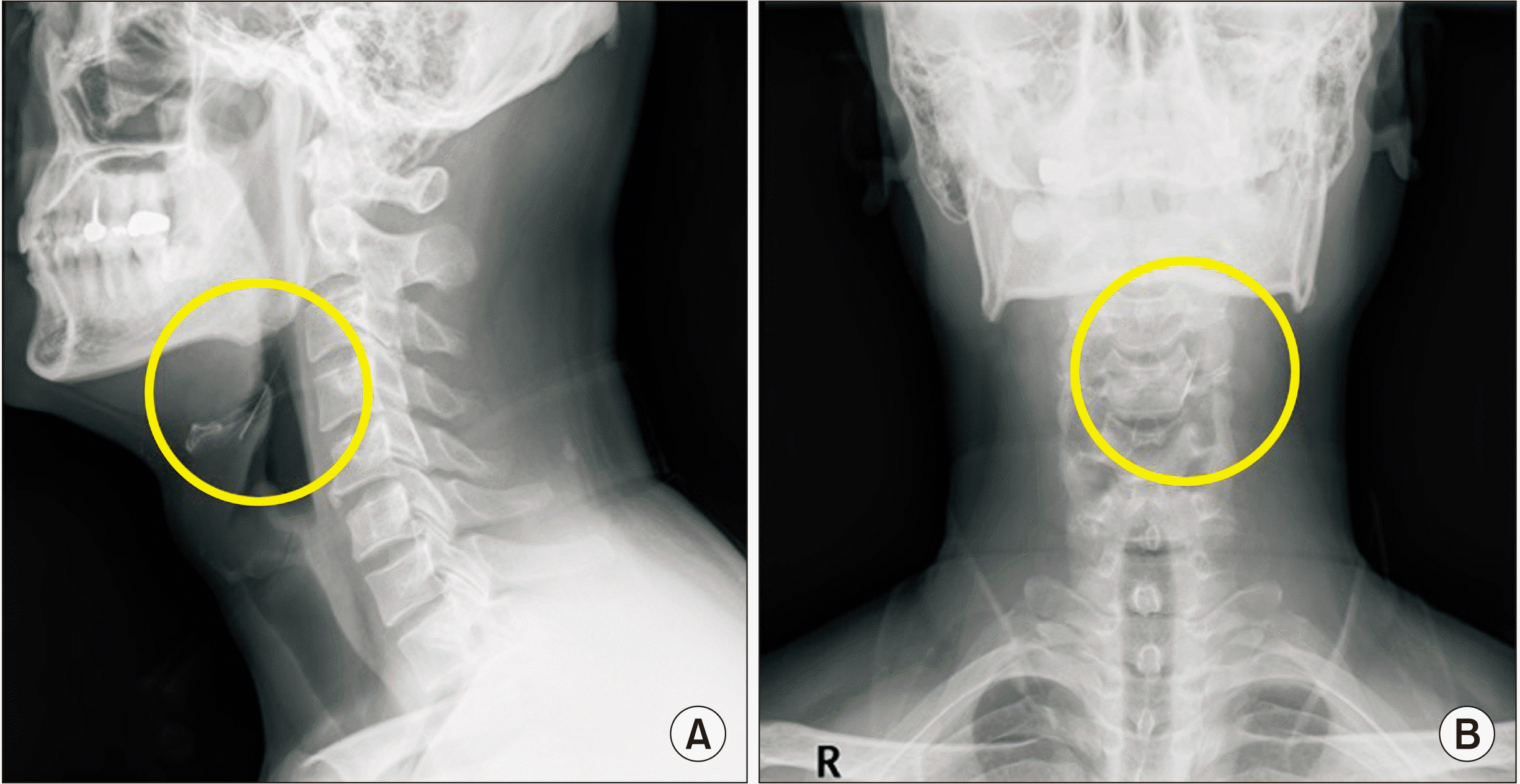
A 44-year-old male with no specific medical history presented to our hospital with lower left molar spontaneous pain and underwent root canal treatment on the mandibular left second molar. During root canal treatment, after accessing the pulp cavity, a 25-mm K-file was inserted into the tooth to measure the length of the root canal system. The patient ingested the file while moving to the radiology room. A rubber dam was placed, but the frame was temporarily removed in order to obtain the radiograph. The patient did not exhibit any symptoms of coughing, dyspnea, drooling, or other respiratory abnormalities. After confirming the patient’s respiratory stability, radiographs were taken. A file was observed in the glottal region of the C3-C4 level, and the patient was immediately referred to an otolaryngologist.(Fig. 1) The file was subsequently removed with a nasopharyngeal endoscope. The issue was solved immediately through the emergency phone line that had been prepared in advance. Afterwards, root canal therapy was completed and the patient was discharged from the hospital that same day.
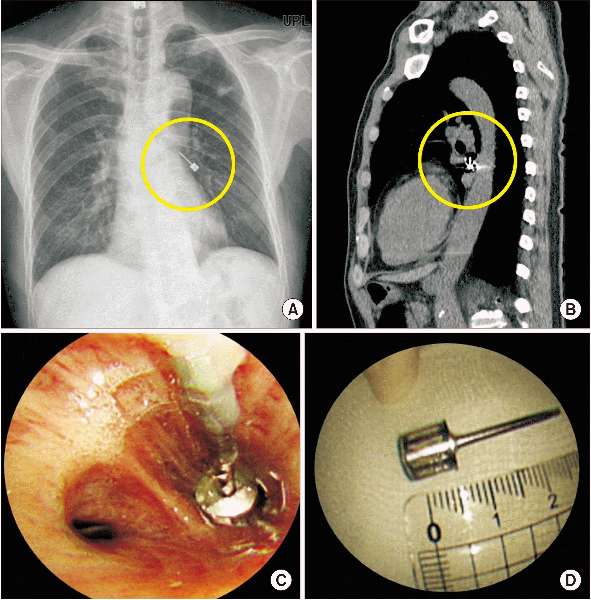
A 79-year-old male with no specific medical history presented to our hospital due to the ingestion of an implant driver after implant surgery. The patient did not exhibit any symptoms of coughing, dyspnea, drooling, or other respiratory abnormalities. After confirming the patient’s respiratory stability, radiographs were taken.(Fig. 2. A, 2. B) It was determined that a foreign body had entered the bronchus on the chest PA (posteroanterior) and the was admitted to the respiratory emergency department for an emergency bronchoscopy. The implant driver was embedded in the entrance of the left lower lobe and was removed using a snare.(Fig. 2. C) The length of the removed implant screwdriver was 28 mm.(Fig. 2. D) After the removal of the foreign substance, the patient was placed on ceftriaxone prophylactically in case bronchitis occurred. No specific complications and no respiratory symptoms occurred, so the patient was discharged the very next day.
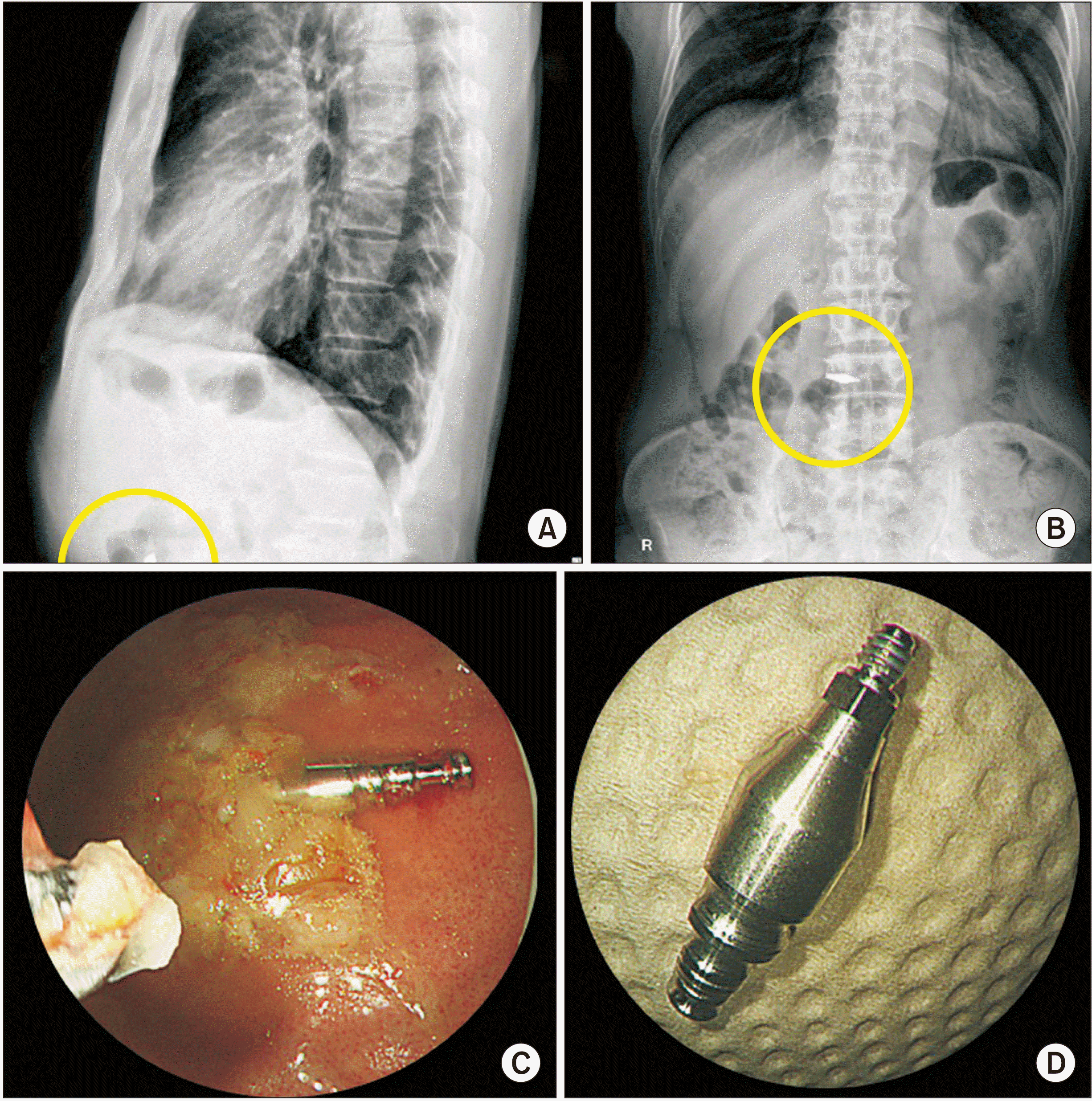
A 61-year-old male with no specific medical history presented to our hospital for a left maxillary second molar implant. The implant coping fell into the mouth while taking the impression for the prosthesis. The patient immediately tilted his head to the side and attempted to remove the object, but ingestion occurred. The patient did not exhibit any symptoms of coughing, dyspnea, drooling, or other respiratory abnormalities. After confirming the patient’s respiratory stability, radiographs were taken. The presence of the foreign body was confirmed in the midline abdomen on the abdomen X-ray.(Fig. 3. A, 3. B) Immediately after referral to a gastroenterologist, the impression coping was removed using a non-sleeping gastroscopy.(Fig. 3. C, 3. D) No specific complications were observed, so the patient was discharged that same day.
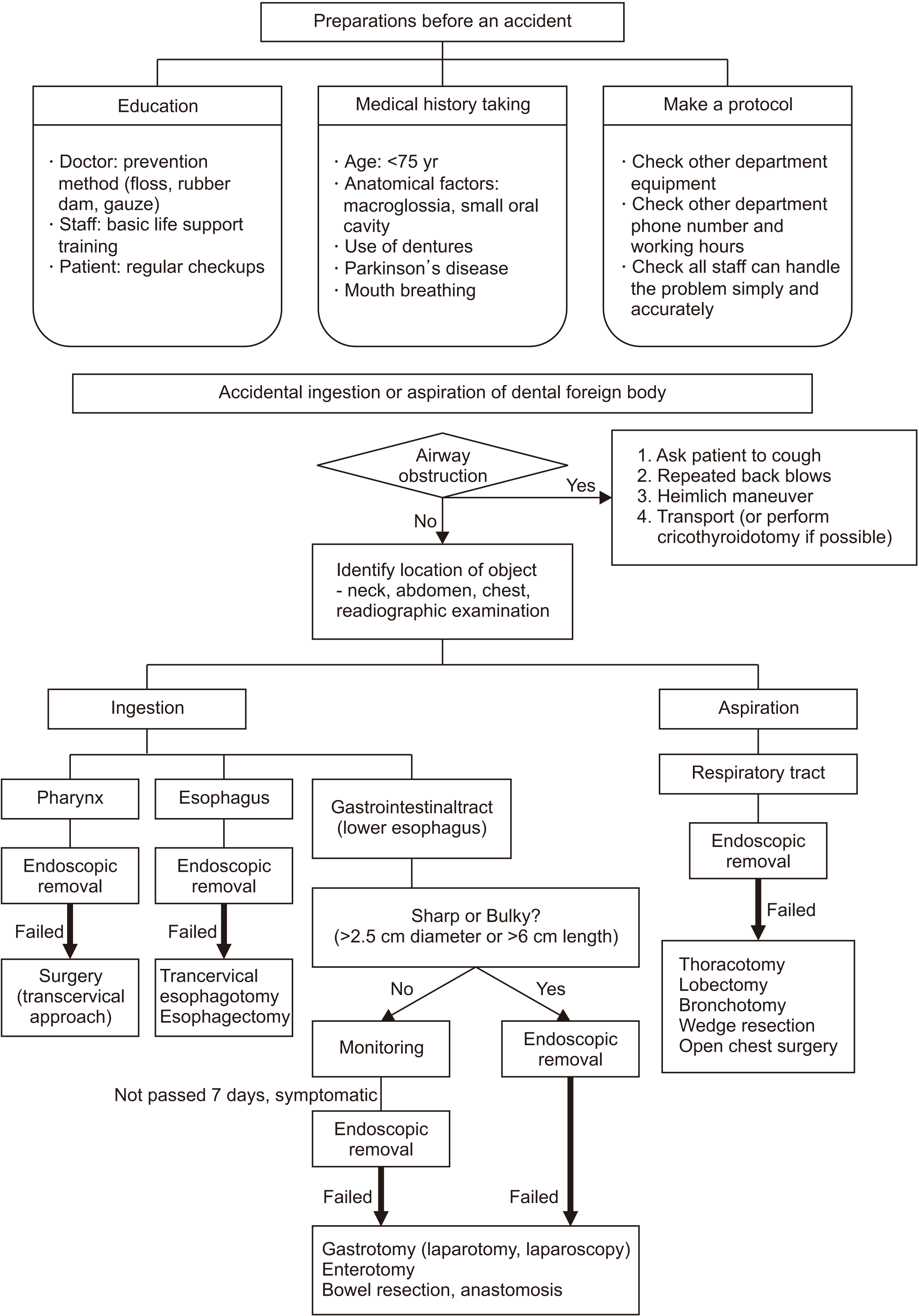
Dental-related aspiration and ingestion accidents can occur at any time and place, inside or outside the dental office. Although the incidence of such accidents is low, they should not be overlooked because of the potential serious complications as well as legal liability4. In addition to preventing such accidents from occurring, preparations should be made to take appropriate measures in case of such accidents.(Fig. 4)
Prevention is the gold standard for avoiding accidents. Preventive equipment such as the use of rubber dams and dental floss tied to instruments must always be used5. Before dental procedures, patients with a high probability of an accident should be prepared for thoroughly with a medical history review. Issues have been reported to occur more frequently in elderly or pediatric patients, denture users, alcoholics, mentally ill individuals, and patients with a lot of anxiety6. It is also important to check the fasting time of the patient because an endoscopy may require emergency general anesthesia.
Cooperation with other departments is essential to properly solve issues without delay. Pre-preparation with the departments of otolaryngology, radiology, gastroenterology, and respiratory internal medicine is key to ensuring that medical requests and treatments are made promptly. Each department should have a pre-determined emergency protocol that suits their needs. All staff should be prepared to perform the protocol and be able to secure an airway in case of an emergency.
After an accident, the first thing to do is to assess the patient’s breathing, check for any airway obstructions, confirm apnea, and measure oxygen saturation. If the foreign body obstructs the airway, the Heimlich maneuver should be performed4. If the airway obstruction is life-threatening and the foreign body cannot be removed, the placement of a laryngeal mask airway or endotracheal intubation should be performed, and a tracheotomy should be performed immediately if an oral and maxillofacial surgeon is available.
After confirming that a respiratory emergency is not occurring, the location of the dental foreign body should be confirmed. For an accurate diagnosis, anterior-posterior and lateral views are recommended during radiographic imaging. Depending on the location of the accident, cervical, chest, and abdominal images are also required. For a more accurate diagnosis, a computed tomography image can also be taken to specifically check for perforations and abscess formations7.
If foreign body aspiration is certain, a physician must be consulted immediately for an urgent bronchoscopy. In the case of ingestion, if the foreign body is small (less than 2 cm) in size and not sharp, natural discharge can be an option. If the foreign body is sharp or large (more than 2 cm) in size, it should be removed with an endoscope as early as possible when it is in the upper gastrointestinal tract3. Since most dental instruments are sharp or large, removal is recommended for dental ingestion and aspiration materials.
In cases of aspiration or ingestion accidents, most of the accidents are resolved only by removal through an endoscope and observation. However, if the removal of the foreign body through the endoscope fails or if the foreign body is impacted into the digestive tract, the issue has to be resolved surgically. When a rigid bronchoscopy failed after an aspiration accident, a bronchotomy, lobectomy, and thoracotomy were performed5. When an endoscopy failed after an ingestion accident, an esophagotomy, gastrotomy, laparotomy, and resection were performed8.
To investigate and evaluate cases that required surgery due to the occurrence of an accident, a comprehensive literature review was conducted in 1988 and compared in tables to provide data on the cause of the accident, the initial visit time, location, result, and treatment6-19.(Tables 1, 2) A PubMed search of the terms “dental ingestion and aspiration” and “surgery” was performed from 1988 to 2022. The focus of the review was to investigate any serious sequelae or complications caused by aspiration and ingestion accidents. Therefore, cases involving a simple removal with an endoscope were excluded. Overall, a total of 14 papers with 7 patients with aspiration and 7 patients with ingestion who required surgery were selected for review according to the study criteria.
Most patients with aspiration complain of coughing, shortness of breath, foreign body sensation, and chest pain, and those who ingested exhibited symptoms such as dysphagia, vomiting, sore throat, cyanosis, and coughing5. Asymptomatic cases after an initial event occurred in 64.29% of the cases in the literature9-13. Ulkü et al.12 reported a case where a simple observation was performed because there were no symptoms after a dental material aspiration accident. Three days later, a male patient visited the emergency room complaining of coughing and dyspnea symptoms. He was diagnosed with aspiration, and a thoracotomy was performed after a rigid bronchoscopy failed. Therefore, simple observation even if there are no symptoms can lead to a bigger accident.
Only 40% of patients were referred to emergency department immediately after an accident in the literature6,9,13,14,16. As time passes, the removal of foreign bodies can become more difficult. Foreign bodies entering the esophagus may be discharged naturally, but they can become impacted, causing compression necrosis and possible perforation. If a foreign body aspirated into the airway is diagnosed late, it can create inflammation and severe swelling, making it difficult to remove by bronchoscopy. In the event of an accident, diagnosis and treatment should be carried out immediately.
Shreshtha et al.18 and Gachabayov et al.8 reported two patients who were unaware that they had ingested a denture. Both patients were only aware that their dentures had disappeared. The missing dentures were present in the esophagus in one patient and in the sigmoid colon in the other patient, resulting in a neck abscess and perforation on the pelvic side wall, respectively. To solve these issues, an esophagostomy and sigmoid colectomy had to be performed. Therefore, patients who wear dentures must be made aware of the possibility of ingesting the dentures when their dentures disappear. Because this issue has the potential to cause patient death, regular check-ups should be performed to ensure that the dentures do not become loose.
There were also two deaths due to ingestion and aspiration accidents. According to the report by Kim et al.13, after 4 attempts of rigid bronchoscopies, the tooth aspirated during the tooth extraction procedure was successfully removed, but the patient died 7 days after surgery due to worsening delirium and pneumonia. Funayama et al.20 reported on a patient who wore partial dentures who presented to the emergency room due to pharynx discomfort and dysphagia. The ingested partial denture was not found on the radiograph and no special treatment was performed. The next morning, the patient lost consciousness while eating and was taken to the hospital. Food residue and the partial denture were removed from the esophagus, but the patient died 12 days later. Based on these experiences, if proper prevention and treatment of aspiration and ingestion accidents of dental foreign substances are not performed, the patient not only has to undergo a major operation but can potentially die as a result.
Simple and accurate protocols should be provided for all dental members to respond to dental emergencies. Collaboration among other medical departments should be established before the event of an accidental ingestion and aspiration. Regular dental checkups for denture patients and radiographs after the accidents can be lifesaving.
Notes
Authors’ Contributions
Y.S. has conceived and drafted the manuscript. Y.J., S.o.H., and R.K. reviewed the paper. All authors read and approved the final manuscript.
Ethics Approval and Consent to Participate
No ethical approval or consent to participate was necessary since the data collected did not include information of personal identification.
This case report was approved for exemption from Institutional Review Board (IRB) review by the IRB of Kyung Hee University Hospital at Gangdong (IRB No. KHNMC 2022-03-065).
References
1. Bilder L, Hazan-Molina H, Aizenbud D. 2011; Medical emergencies in a dental office: inhalation and ingestion of orthodontic objects. J Am Dent Assoc. 142:45–52. https://doi.org/10.14219/jada.archive.2011.0027. DOI: 10.14219/jada.archive.2011.0027. PMID: 21193766.

2. Hisanaga R, Hagita K, Nojima K, Katakura A, Morinaga K, Ichinohe T, et al. 2010; Survey of accidental ingestion and aspiration at Tokyo Dental College Chiba Hospital. Bull Tokyo Dent Coll. 51:95–101. https://doi.org/10.2209/tdcpublication.51.95. DOI: 10.2209/tdcpublication.51.95. PMID: 20689240.

3. Li J, Choi BH, Kim YH, Kim HS, Ko CY. 2007; Ingestion of orthodontic anchorage screws: an experimental study in dogs. J Korean Assoc Oral Maxillofac Surg. 33:121–3. DOI: 10.1016/j.ajodo.2007.01.014. PMID: 17561055.
4. Yadav RK, Yadav HK, Chandra A, Yadav S, Verma P, Shakya VK. 2015; Accidental aspiration/ingestion of foreign bodies in dentistry: a clinical and legal perspective. Natl J Maxillofac Surg. 6:144–51. https://doi.org/10.4103/0975-5950.183855. DOI: 10.4103/0975-5950.183855. PMID: 27390487. PMCID: PMC4922223.

5. Araujo SCS, Bustamante JED, de Souza AAB, Peixoto LC, Amaral MBF. 2022; Aspiration of dental items: case report with literature review and proposed management algorithm. J Stomatol Oral Maxillofac Surg. 123:452–8. https://doi.org/10.1016/j.jormas.2021.10.009. DOI: 10.1016/j.jormas.2021.10.009. PMID: 34687948.

6. Cossellu G, Farronato G, Carrassi A, Angiero F. 2015; Accidental aspiration of foreign bodies in dental practice: clinical management and prevention. Gerodontology. 32:229–33. https://doi.org/10.1111/ger.12068. DOI: 10.1111/ger.12068. PMID: 24102914.

7. Patel PH, Slesser AA, Idaikkadar P, Kostourou I, Awad RW. 2012; Delayed presentation of a small bowel perforation secondary to an ingested denture. JRSM Short Rep. 3:60. https://doi.org/10.1258/shorts.2012.012014. DOI: 10.1258/shorts.2012.012014. PMID: 23323200. PMCID: PMC3545345.

8. Gachabayov M, Isaev M, Orujova L, Isaev E, Yaskin E, Neronov D. 2015; Swallowed dentures: two cases and a review. Ann Med Surg (Lond). 4:407–13. https://doi.org/10.1016/j.amsu.2015.10.008. DOI: 10.1016/j.amsu.2015.10.008. PMID: 26635957. PMCID: PMC4637341.

9. Delap TG, Dowling PA, McGilligan T, Vijaya-Sekaran S. 1999; Bilateral pulmonary aspiration of intact teeth following maxillofacial trauma. Endod Dent Traumatol. 15:190–2. https://doi.org/10.1111/j.1600-9657.1999.tb00800.x. DOI: 10.1111/j.1600-9657.1999.tb00800.x. PMID: 10815570.

10. Welcker K, Nakashima M, Branscheid D. 2005; [Aspiration of dental implant -- reasons, management and prevention]. Pneumologie. 59:174–7. German. https://doi.org/10.1055/s-2004-830210. DOI: 10.1055/s-2004-830210. PMID: 15756630.

11. Başoglu OK, Buduneli N, Cagirici U, Turhan K, Aysan T. 2005; Pulmonary aspiration of a two-unit bridge during a deep sleep. J Oral Rehabil. 32:461–3. https://doi.org/10.1111/j.1365-2842.2005.01472.x. DOI: 10.1111/j.1365-2842.2005.01472.x. PMID: 15899026.

12. Ulkü R, Başkan Z, Yavuz I. 2005; Open surgical approach for a tooth aspirated during dental extraction: a case report. Aust Dent J. 50:49–50. https://doi.org/10.1111/j.1834-7819.2005.tb00085.x. DOI: 10.1111/j.1834-7819.2005.tb00085.x. PMID: 15881306.

13. Kim E, Noh W, Panchal N. 2017; Mortality from an aspiration of dental crown during extraction. Gerodontology. 34:498–500. DOI: 10.1111/ger.12288. PMID: 29094438.

14. Sadda R, Sadda AR. 2019; Prevention and management of life-threatening complications during dental implant surgery: a clinical case series. Gen Dent. 67:52–6. PMID: 31199745.
15. Furihata M, Tagaya N, Furihata T, Kubota K. 2004; Laparoscopic removal of an intragastric foreign body with endoscopic assistance. Surg Laparosc Endosc Percutan Tech. 14:234–7. https://doi.org/10.1097/01.sle.0000136682.69871.db. DOI: 10.1097/01.sle.0000136682.69871.db. PMID: 15472556.

16. Chua YK, See JY, Ti TK. 2006; Oesophageal-impacted denture requiring open surgery. Singapore Med J. 47:820–1. PMID: 16924368.
17. Singh P, Singh A, Kant P, Zonunsanga B, Kuka AS. 2013; An impacted denture in the oesophagus-an endoscopic or a surgical emergency-a case report. J Clin Diagn Res. 7:919–20. https://doi.org/10.7860/JCDR/2013/5337.2976. DOI: 10.7860/JCDR/2013/5337.2976. PMID: 23814744. PMCID: PMC3681071.

18. Shreshtha D, Sikka K, Singh CA, Thakar A. 2013; Foreign body esophagus: when endoscopic removal fails…. Indian J Otolaryngol Head Neck Surg. 65:380–2. https://doi.org/10.1007/s12070-013-0662-6. DOI: 10.1007/s12070-013-0662-6. PMID: 24427604. PMCID: PMC3851502.

19. Flanagan M, Clancy C, Riordain MGO. 2018; Impaction of swallowed dentures in the sigmoid colon requiring sigmoid colectomy. Int J Surg Case Rep. 47:89–91. https://doi.org/10.1016/j.ijscr.2018.04.033. DOI: 10.1016/j.ijscr.2018.04.033. PMID: 29753276. PMCID: PMC5994734.

20. Funayama K, Fujihara J, Takeshita H, Takatsuka H. 2016; An autopsy case of prolonged asphyxial death caused by the impacted denture in the esophagus. Leg Med (Tokyo). 23:95–8. https://doi.org/10.1016/j.legalmed.2016.10.006. DOI: 10.1016/j.legalmed.2016.10.006. PMID: 27890112.

Fig. 1
Radiograph showing an endodontic file (yellow circles) in the glottal region of the C3-C4 level. A. Lateral view of the neck. B. Posterior-anterior view of the neck.

Fig. 2
Radiograph showing an implant screwdriver (yellow circles) in the lower lobe of the left lung. A. Lateral view of the chest radiograph. B. Posterior-anterior view of the chest computed tomography. C. Implant screwdriver visualized and retrieved from the lower lobe of the left lung during the bronchoscopy. D. Removed implant screwdriver.

Fig. 3
Radiograph showing an impression coping (yellow circles) in the stomach. A. Lateral view of the abdominal radiograph. B. Posterior-anterior view of the abdominal radiograph. C. Impression coping visualized and retrieved from the stomach during the gastroscopy. D. Removed impression coping.

Fig. 4
Flowchart diagram: coping protocols before and after the dental ingestion and aspiration accidents.

Table 1
Summary of dental aspirations with serious adverse events: 7 studies sorted by year of publication
| Study | Age (yr)/sex | Material | Cause | Symptoms | Time1 | Location | Result | Treatment |
|---|---|---|---|---|---|---|---|---|
| Delap et al.9 (1999) | 20/M | Teeth | Assault | Absence | 0 hr | Right lower lobe | Deeply embedded in tissue |
Rigid bronchoscopy failed Right thoracotomy |
| Welcker et al.10 (2005) | 77/M | Cast Post | Dental procedure |
Absence Later chronic cough |
3 yr | Lower right bronchus |
Retention pneumonia Embedded in granulation tissue |
Lobectomy |
| Başoglu et al.11 (2005) | 37/M | Bridge | Deep sleep |
Absence Later cough, fever, dyspnea |
1 yr | Lower right bronchus | Embedded in granulation tissue |
Rigid bronchoscopy failed Vertical bronchotomy |
| Ülkü et al.12 (2005) | 8/M | Tooth | Dental extraction |
Absence Later cough, dyspnea |
3 days | Right main bronchus | Deeply embedded in tissue |
Rigid bronchoscopy failed Thoracotomy |
| Cossellu et al.6 (2015) | 80/M | Dental bur | Dental procedure | N/A | 0 hr | Right lower lung | Deeply embedded in tissue |
Rigid bronchoscopy failed Wedge resection by thoracoscopic approach |
| Kim et al.13 (2017) | ≥90/M | Crown | Dental extraction | Cough, dyspnea | 0 hr | Right main bronchus |
Worsening delirium Pneumonia Death |
Rigid bronchoscopy succeeded after 4 attempts |
| Sadda and Sadda14 (2019) | 62/M | Screwdriver | Implant procedure | N/A | 0 hr | N/A | Deeply embedded in tissue |
Rigid bronchoscopy failed Open chest surgery |
Table 2
Summary of dental ingestion with serious adverse events: 7 studies sorted by year of publication
| Study | Age (yr)/sex | Material | Cause | Symptoms | Time1 | Location | Result | Treatment |
|---|---|---|---|---|---|---|---|---|
| Furihata et al.15 (2004) | 57/F | Valplast | Accidental swallow |
Absence Later epigastric discomfort |
2 days | Antrum of the stomach |
Mediastinal emphysema Subcutaneous emphysema |
Laparoscopic approach combined with endoscope |
| Chua et al.16 (2006) | 36/M | Flipper | Sleeping |
Odynophagia Blood sputum |
0 hr | Esophagus | Impacted at the C7 level | Esophagostomy |
| Patel et al.7 (2012) | 64/F | Bridge | N/A |
Absence Later abdominal pain |
90 days | Sigmoid colon | Ileal perforation | Laparotomy |
| Singh et al.17 (2013) | 40/M | Flipper | Accidental swallow |
Absence Later fever |
5 days | Esophagus | Esophagus wall necrotized | Laparotomy |
| Shreshtha et al.18 (2013) | 42/F | Denture | Accidental swallow | Dysphagia, odynophagia for 1 year | N/A | Esophagus | Neck abscess | Esophagostomy |
| Gachabayov et al.8 (2015) | 54/F | Flipper | Accidental fall |
Absence Later nausea, vomit |
22 hr | Ileum | Bowel obstruction | Enterotomy |
| Flanagan et al.19 (2018) | 67/F | Denture | N/A |
Absence Later abdominal cramping |
3 yr | Sigmoid colon | Perforation on the pelvic side wall | Sigmoid colectomy |


 PDF
PDF Citation
 Print
Print


 XML Download