

 PDF
PDF Citation
 Print
Print


I. Introduction
Osseous metaplasia is the formation of lamellar bone inside soft tissue structures where bone normally does not exist1. It forms as a result of the transformation of non-osseous connective tissue into mature bone. Osseous metaplasia is usually caused by osteoblasts differentiating from fibroblasts secondary to inflammation, tissue damage, exposure to substances such as bone morphogenetic proteins released from neoplastic cells, or dystrophic calcification in necrotic tissue. Several cases of osseous metaplasia has been reported to occur in thyroid gland, endometrium, breast, etc. It occurs as a common complication after orthopedic surgery, but it is rare in the oral and maxillofacial region and only a few cases have been reported in the literature. Here we present a case of osseous metaplasia of the palate that did not result from an ill-fitting dental prosthesis, trauma, or intra-oral carcinoma.
II. Case Report
A 60-year-old male patient visited our clinic complaining of a palpable mass on the right palate area. He did not complain of any pain from the mass, and reported no history of trauma. The patient had no relevant diseases other than hypertension.
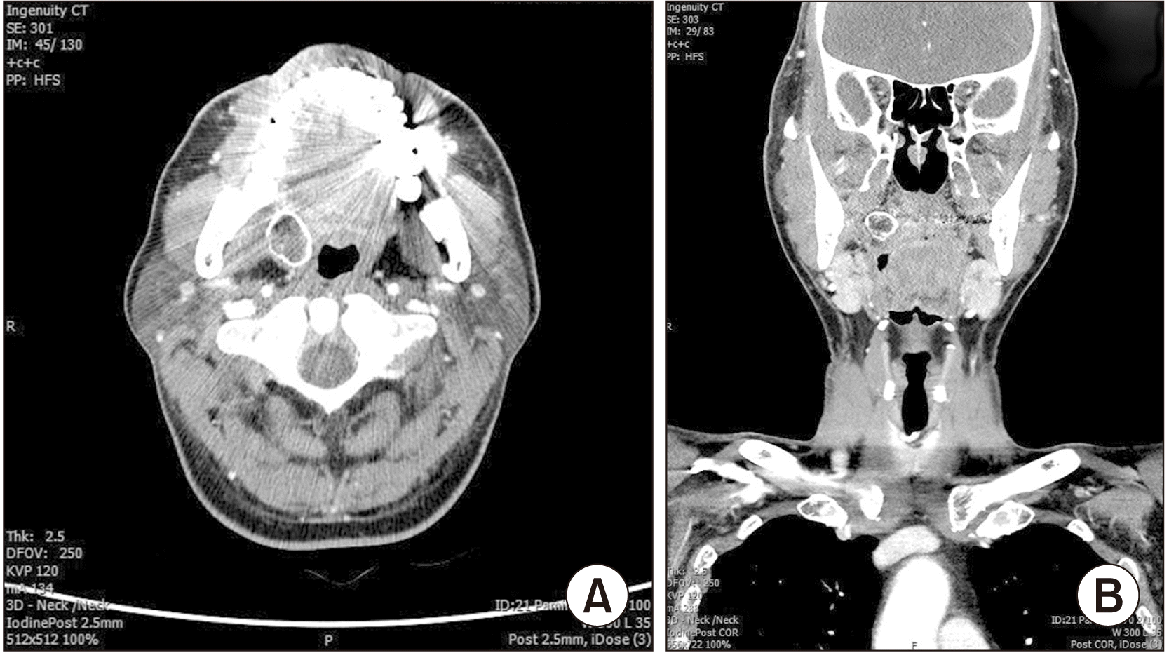
Clinical examination revealed a large, round, firm mass on the right palate area that was also observed on panoramic view. On computed tomography, there was a round hard-tissue mass approximately 2 cm in diameter on the right palate area.(Fig. 1)
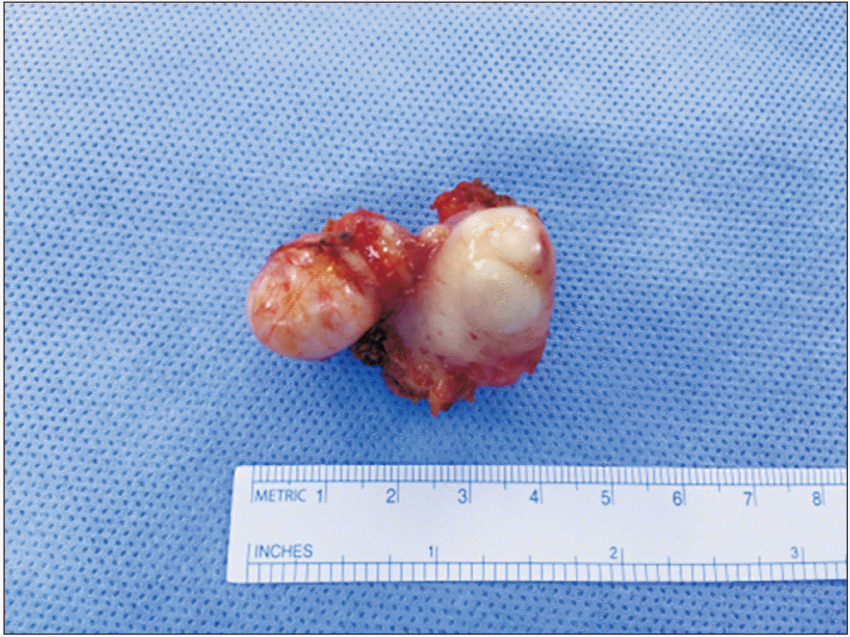
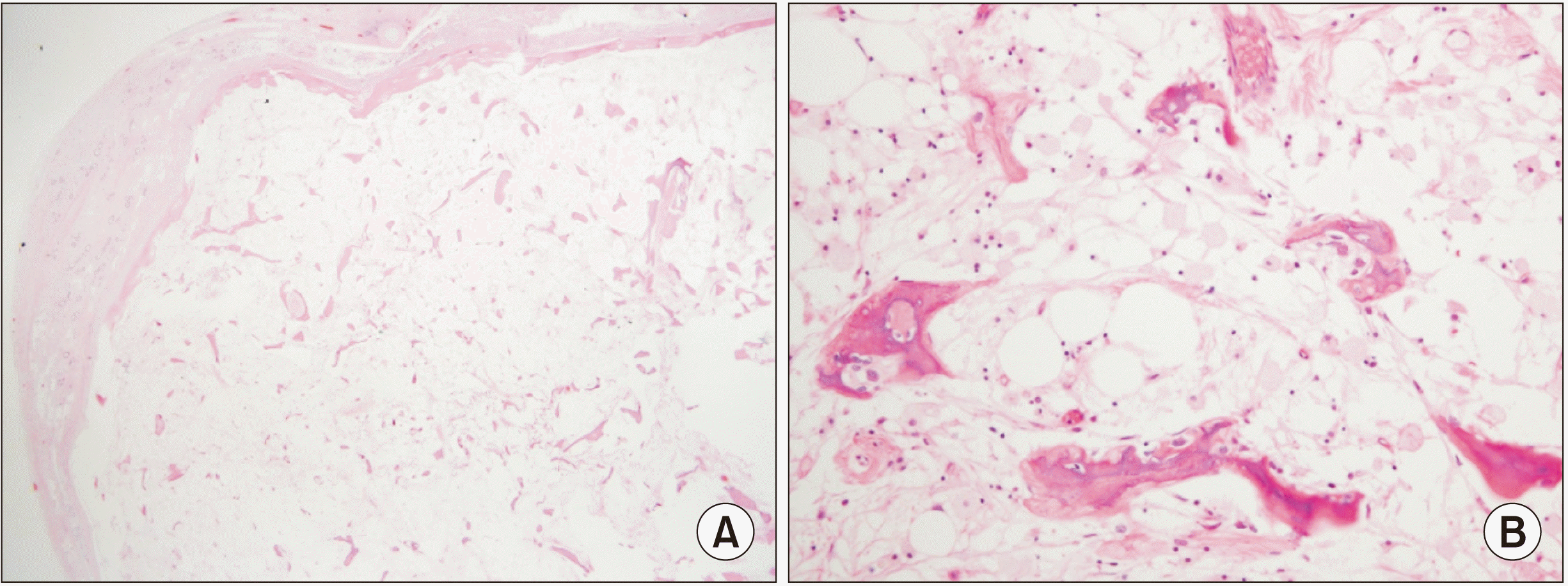
Based on clinical and radiographic examination, the clinical diagnosis was osseous metaplasia of the right palate, and we planned to excise the mass under general anesthesia. The surgical procedure for excision was performed intraorally and biopsy was performed.(Fig. 2, 3)
The patient was treated with IV antibiotics and was discharged two days after surgery. Histological examination revealed osseous metaplasia.
III. Discussion
Osseous metaplasia has been reported to occur systemically other than intra-oral portion. Chun et al.2 reported three cases of osseous metaplasia of the thyroid gland in female patients with thyroid nodules who were diagnosed with nodular hyperplasia with osseous metaplasia and mature bone formation. Lee et al.3 reported a case of endometrial osseous dysplasia in a 25-year-old female.
There are many papers reporting osseous metaplasia, but few studies have described osseous metaplasia in the oral and maxillofacial region. These cases were associated with ill-fitting dental prosthesis, surgical intervention of the paranasal sinus, and reactive conditions due to intra-osseous carcinoma. Cutright4 reported 42 cases of osseous and chondromatous metaplasia related to ill-fitting dentures that occurred in the loose fibrous connective tissue beneath denture. First, osseous metaplasia occurred two times more often in women. Second, it occurred more often in the maxilla than in mandible. Third, it occurred most often in the fifth, sixth, and seventh decades of life. Fourth, it was more commonly found in anterior areas rather than posterior areas4. Maitra et al.5 reported osseous metaplasia of the maxillary sinus with formation of a well-developed Haversian system and bone marrow. The usual cause of the formation was surgical trauma of the sinus such as endoscopic sinus surgery5. Bennett et al.6 reported that intra-osseous odontogenic squamous cell carcinoma was characterized by marked osseous/dentinoid metaplasia in the surrounding connective tissue stroma.
As osseous metaplasia can occur due to reactive conditions of intraosseous carcinoma, differential diagnosis and clinical follow-up are needed. Vencio et al.7 reported that the fibro-osseous component in heterotrophic ossification may resemble osteochondroma or chondrosarcoma.
In our case, the overlying gingiva was clean and a solid, firm mass was isolated from the surrounding soft and hard tissue. There was no dental prosthesis, other tumorous condition, or trauma in this patient. This case suggests the need to further explore possible causes of osseous metaplasia.
Also, as Cutright4 reported, osseous metaplasia has been shown in the anterior region of the alveolar ridge more often than the posterior regions. In our case, osseous metaplasia was found in the posterior maxilla region, which is unusual.
In conclusion, osseous metaplasia frequently occurs systemically but rarely in the oral and maxillofacial region. The most common causes of osseous metaplasia are trauma or surgery. In the oral cavity, it most commonly occurs due to ill-fitting dental prostheses. Sinus trauma due to endoscopic sinus surgery can also be a cause. Additionally, osseous metaplasia in the surrounding tissue stroma is characteristic of odontogenic squamous cell carcinoma. This case of osseous metaplasia did not occur due to any trauma or surgery, and it occurred in the posterior maxilla, which is an uncommon site in the oral cavity. For these reasons, it should be considered as a unique case. Because osseous metaplasia rarely occurs in the oral cavity and is a rare condition in the oral and maxillofacial region that can resemble other osseous benign or malignant lesions, histopathologic study and differential diagnosis are needed.
 XML Download