

 PDF
PDF Citation
 Print
Print


I. Introduction
Surgical extraction of impacted teeth is one of the most common operations in oral and maxillofacial surgery. Anatomically, extraction surgery has a high possibility of additional damage to the adjacent tissues, which can cause a range of complications both during and after surgery. Hemorrhage, swelling, and pain are predictable complications, but injuries to the oral tissue, such as adjacent teeth injuries, alveolar bone fractures, nerve injuries, and temporomandibular joint disorders, are reported often1. Analysis of the cause of these complications is important not only for prevention, but also for avoiding legal disputes by correctly explaining the diagnosis, treatment, and prognosis to the patient.
Ophthalmic complications after tooth extraction are rare but can cause critical dysfunctions. Since the first cases with local anesthesia were recorded in 1936, several accounts of ocular complications after local anesthesia, including diplopia, visual loss, and ischemic optic neuropathy, have been published2-4. Horowitz et al.3 reviewed 39 ophthalmic complications after dental local anesthesia, most of which were diplopia caused by lateral rectus palsy. These authors suggested the possibility of an anesthetic solution spreading through the blood vessels. In some review articles, a mechanism was suggested in that the anesthetic solution could have been injected into the arteries or veins, causing diplopia due to ophthalmoplegia, visual loss due to ischemic optic neuropathies, and tonic pupil4,5. Ischemic optic neuropathies can lead to permanent visual loss due to optic atrophy and can be diagnosed by the clinical findings of optic disc edema, afferent pupillary defect, and crowded small optic nerve head or by fat-suppressed contrast-enhanced magnetic resonance imaging (MRI) findings of compressive optic neuropathy or inflammatory optic neuritis6. In 2016, Alamanos et al.4 published a systematic review of ophthalmic complications correlated with dental local anesthesia reported between 1954 and 2013 and suggested the following causes: i) diffusion of anesthetic solution, ii) intra-arterial injection, iii) intra-venous injection, iv) direct trauma of the peri-arterial sympathetic plexus, v) interpretation of signs from the oculo-autonomic pathway, vi) parasympathetic reactions, and vii) sympathetic reactions. On the other hand, few of the ophthalmic complication reports available have examined or evaluated the patient during the blurred vision event. In 2002, Helfman7 reported a positive result of a lidocaine allergic reaction in a patient with blurred vision, but there was a limitation to confrim their diagnosis due to the lack of medical records at the time of the ophthalmic symptom.
Pseudomyopia, also termed a spasm of accommodation, is defined as transient myopic shift in refraction power caused by a spasm of the ciliary muscle. Clinically, accommodative spasm can cause blurring of vision, headache, ocular pain, micropsia, macropsia, and intermittent diplopia8. Pseudomyopia is a component of spasm occurring the near reflex and that can be accompanied by convergent strabismus or miosis9. The etiology of spasm of the near reflex is divided into functional and organic causes8. Other causes, such as excessive near work and interference in binocular vision, are also potential causes10. Transient myopia has been reported to occur with the use of certain drugs, such as topiramate and sulfonamide11.
This report presents a case of transient blurring of vision after tooth extraction surgery in a 20-year-old male patient. With additional examination of the MRI and ophthalmic test results, an oral and maxillofacial surgeon, anesthesiologist, and ophthalmologist discussed the reasons for and diagnosis of this case.
II. Case Report
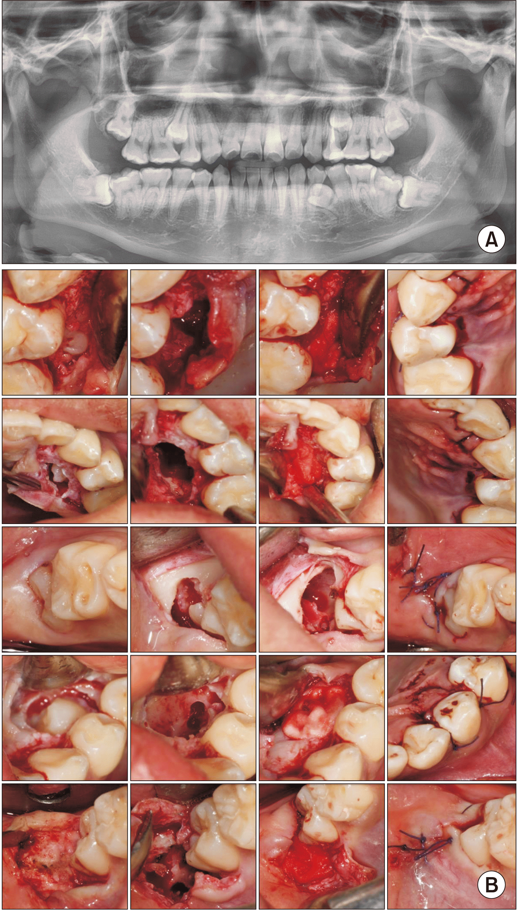
This study was approved by the Institutional Review Board of the Armed Forces Capital Hospital (No. AFCH-19-IRB-004). A 20-year-old male (161 cm, 69.5 kg) without a specific medical history underwent extraction of seven impacted teeth under general anesthesia.(Fig. 1. A) The patient was transported to the operation room without premedication and monitored by non-invasive blood pressure measurement, pulse oximetry, and three-channel electrocardiography. Anesthesia was induced by intravenous injection of remifentanil using a target-controlled infusion (TCI) system (Orchestra Base Primea; Fresenius Kabi, Bad Homburg, Germany), 60 mg of lidocaine, 160 mg of propofol, and 50 mg of rocuronium. The patient was intubated with a nasal endotracheal tube (portex polar preformed tracheal tube, north nasal, profile, soft seal, cuff, ivory; Smiths Medical International, Hythe, England) with an inner diameter of 7.0 mm. Oxygen and air were supplied at a fraction of inspired oxygen (FiO2) of 0.5 with sevoflurane. During anesthetic maintenance, the concentration of sevoflurane was controlled so that the blood pressure and heart rate were held within 20% of the preoperative measurements; remifentanil was injected continuously for analgesia as necessary.
Local anesthesia with 7.2 mL of lidocaine (2% lidocaine HCl; Huons, Seongnam, Korea) was infiltrated on the buccal vestibule of the right maxillary third molar (#18), left maxillary third molar (#28), left mandibular third molar (#38), and right mandibular third molar (#48), as well as on the palatal mucosa of right and the left maxillary second premolars (#15 and #25) and lingual mucosa of the left mandibular supernumerary impacted premolar. Aspiration was performed prior to delivery. A crevicular incision was made on the palatal sides of #14 and #24 and the lingual side of #35, and a crevicular incision with a distal incision was made on #18, #28, #38, and #48. A mucoperiosteal flap was elevated, and the surrounding bone was ground smooth. The impacted #15, #18, #25, #28, #34, #38, and #48 teeth were extracted during odontectomy. Following curettage of granulomatic tissue, profuse saline was irrigated. No severe complications, such as bleeding or nerve damage, were encountered, but the sinus Schneiderian membrane was perforated while extracting the left maxillary premolar. The surgical area was covered with Surgicel (Johnson & Johnson, New Brunswick, NJ, USA) to close the perforation, and the extracted socket was packed with Ateloplug (SK Bioland, Cheonan, Korea) on #15, #18, #25, and #28. The mandibular extraction socket was packed with Bio BMP2 (Cellumed, Seoul, Korea) covering the Ateloplug before primary closure.(Fig. 1. B)
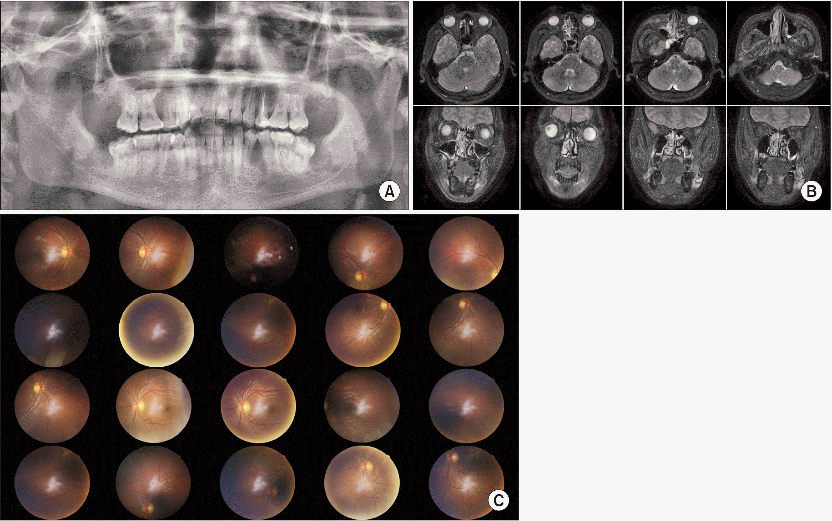
After surgery, sevoflurane and air were discontinued. In the immediate postoperative period, 30 mg of ketorolac was injected as a rescue analgesic. Pyridostigmine (15 mg) and glycopyrrolate (0.4 mg) were injected to reverse muscle relation. When the patient could breathe spontaneously and respond to verbal commands, he was extubated and transferred to the post-anesthesia care unit. After recovery from general anesthesia, the patient complained of blurred vision in both eyes and diplopia. He denied any prior use of spectacles and said that he had never experienced such discomfort in vision. Following radiography and MRI (Fig. 2. A, 2. B), he underwent comprehensive ophthalmic examinations, including visual acuity, refraction, slit-lamp examination, and indirect ophthalmoscopy. His unaided distant visual acuity was 20/1,000 in both eyes. The refraction showed a myopic shift of –3.25 diopters sphere (DS)/–0.75 diopter cylinder (DC)×30° in the right eye and –2.75 DS/–0.5 DC×150° in the left eye. After correction with a –3.00 DS lens, the visual acuity in both eyes improved to 20/20. The pupil size was normal, and the light reflex was intact. No deviation of the eyeball was observed at near and far distances. The slit-lamp examination revealed a clear cornea, cloudless lens, and deep and quiet anterior chamber. In the fundus examination, the retina was clear and flat, and the shape of the optic disc was normal.(Fig. 2. C) Through cycloplegic refraction with 1% cyclopentolate, the refraction showed a slight hypermetropic shift of +1.5 DS/–0.75 DC×30° in the right eye and +1.25 DS/–0.5 DC×150° in the left. The pseudomyopia was believed to have been induced by a spasm of the ciliary muscle. Because it was thought to be a temporary phenomenon related to the tooth-extraction surgery performed under general anesthesia, a decision was made to observe the patient’s progress without treatment.
On the next day, he reported slight blurring of vision, but his unaided visual acuity was 20/20, and refraction was observed in both eyes. The visual discomfort was attributed to mydriasis caused by the cycloplegic agent administered on the previous day. Two days later, the pupils had returned to their original size on examination, and the patient did not report any visual discomfort. Lidocaine allergy tests were performed and yielded negative results on skin prick, intradermal testing, and lidocaine provocation testing using a standardized quality unit tablet given by an allergy and clinical immunologist.
III. Discussion
The authors searched PubMed from 1936 to 2019 for relevant articles regardless of country of origin using the following search string: (“dental” AND “ophthalmic” AND “complication”) OR (“blurring of vision”) OR (“accommodation disturbance”). The search was conducted on September 1, 2019, and 1,352 articles were identified from the database. Of these, 270 articles were selected for further assessment via title and abstract screening. Among these, articles that were not related to the field of dentistry, not published in English, and/or that did not reveal ophthalmic symptoms in detail were excluded. Finally, five studies (six patients) reporting similar symptoms to those of the present patient, such as blurring of vision or accommodation disturbance after dental intervention, were analyzed. With the addition of the present case, seven patients were selected for data extraction and a detailed review3,7,12-14.(Table 1)
All six previously reported patients were female, while our patient is male. The age of all seven patients, including the present case, ranged from 20 to 35 years (mean±standard deviation, 26.57±6.35 years). Among the included studies, an additional examination was conducted in only one published in 2002, which reported the case of a 35-year-old female. Two previous incidences of blurred vision lasting for several hours triggered an antigen/antibody reaction (allergic response) to 2% lidocaine7.
In 2010, Boynes et al.14 reported a 27-year-old female who complained of ipsilateral complete loss of vision after inferior alveolar nerve block anesthesia with 1.8 mL of 2% lidocaine but who recovered after approximately 15 minutes. Despite her being uncooperative during the examination, the authors presumed an accommodation disturbance. In 2012, Steenen et al.15 reported a 22-year-old female who complained of ipsilateral blurred vision and diplopia without blanching of the skin after local anesthesia for a right impacted maxillary and mandibular third molar with 5.1 mL of articaine. She suffered ophthalmoplegia for approximately six hours with no abduction of the right eye and was assessed by an oral and maxillofacial surgeon. In 2014, von Arx et al.2 reviewed ophthalmologic complications after local anesthesia and reported four cases of blurred vision or loss of accommodation. Among the four case reports, these authors observed that the 1990 report by Goldenberg16 showed a loss of accommodation, but Goldenberg16 reported diplopia due to oculomotor disturbances after a xylocaine injection. In 2016, Alamanos et al.4 reviewed the ophthalmic complications after dental local anesthesia and reported seven cases of blurred vision or loss of accommodation. Among these seven case reports, they reported that the 1983 report by Goldenberg17 included blurred vision and the 2010 report by Al-Sandook and Al-Saraj18 included a loss of accommodation. However, Goldenberg17 concluded diplopia due to lateral rectus muscle palsy, and Al-Sandook and Al-Saraj18 reported dizziness, confusion, numbness of the infraorbital region, and diplopia after 2% lidocaine injection.
Most ophthalmic complication studies have suggested that local anesthetic solutions reach the orbital area via the blood vessels, nervous system, muscle fibers, or lymphatic network19. Although most dental anesthesia is performed without severe complications20, care should be taken not to inject into the blood vessels. Even with aspiration, however, the needle can be moved during administration or the injection pressure can cause anesthesia to flow back through the arteries5.
In the present case, our patient was diagnosed with pseudomyopia after tooth extraction under general anesthesia by an ophthalmologist, although he complained of diplopia. Local anesthesia was limited to the alveolus levels because surgery was performed under general anesthesia, and MRI, completed during the ophthalmic event, showed that the diffused range of local anesthetics or postoperative edema did not invade the ocular area. The absence of symptoms such as tonic pupil, ptosis, and diplopia suggested a lower likelihood of anesthetic delivery to the ocular area via diffusion or the vascular system. In addition, the absence of allergic reactions to the medications was confirmed by an allergy and clinical immunologist. However, 204 mg of lidocaine was administrated with 60 mg of general anesthesia and 144 mg of local anesthesia. Because this is not a small amount relative to the weight of our patient, it is possible that the temporarily high venous concentration of lidocaine in the ocular area could have had an adverse effect. Further, the prospect of other central nervous system pathways was low because of the absence of physical symptoms, such as dizziness or skin color changes, except for blurred vision. MRI was performed to confirm the organic causes, and no specific findings were noted. During general anesthesia, various drugs, such as anesthetics, anti-muscarinic drugs, sedative and analgesic perioperative drugs, muscle relaxants, and anticholinesterases, were administered intravenously. Since the ciliary muscle is dominated by parasympathetic fibers during accommodation, the authors classified pyridostigmine and glycopyrrolate as anticholinesterases.
Two possibilities were hypothesized. Lidocaine toxicity with an unknown etiology or the temporary effect of medications such as glycopyrrolate (used to reduce excessive saliva) or pyridostigmine (used to reverse the effect of muscle relaxants) induced a spasm of the ciliary muscle, resulting in pseudomyopia. Considering other studies have shown similar symptoms with local anesthesia, the possibility of lidocaine toxicity was strengthened. Within the limitations of this study, the mechanism of pseudomyopia was not revealed. Through MRI results analysis and literature review, however, this symptom appeared more weakly correlated with ischemic complications due to delivery of local anesthesia. Consequently, all reported patients who complained of blurred vision or loss of accommodation after dental intervention, as with the present patient, experienced only temporary symptoms. Therefore, patients with blurred vision (loss of accommodation; pseudomyopia) could be observed without immediate intervention, and their myopia should improve without further treatment.
In conclusion, temporary deterioration of vision can occur after surgical dental extraction. If blurred vision or a myopic shift of refraction is present, pseudomyopia should be suspected, and cycloplegic refraction is essential for diagnosis. The recovery from pseudomyopia within two days is an important finding in this case report.
 XML Download