

 PDF
PDF Citation
 Print
Print


I. Introduction
Mandibular condylar fractures are common, and their most frequent causes are traffic accidents, violence, slip-down, fall-down, and sports. Mandibular condylar fractures are accompanied by dental trauma or concomitant fractures on the contralateral side. Clinically, complications such as malocclusion, anterior open bite, residual pain, mouth opening limitation (MOL), pathological changes in the temporomandibular joint (TMJ), osteonecrosis, facial asymmetry, and ankylosis may occur due to condylar fractures, and appropriate treatment is required to avoid these complications1,2.
Mandibular condylar fractures can be treated using open reduction (OR) or closed reduction (CR). During OR, three-dimensional stability of the mandible can be obtained through proper reduction of the fracture fragment, action of the lateral pterygoid muscle, a relatively fast recovery for a normal diet, and a short treatment period; however, there is the potential for damage to the facial nerve, blood vessels, and joint capsules or infection with this approach. Conversely, CR carries the advantages of being non-invasive and less likely to damage anatomical structures but has its own disadvantages, such as malocclusion, MOL, facial asymmetry, chronic pain, ankylosis, and a prolonged treatment period3-6. Depending on the presence of malocclusion, displacement of the fracture fragment, dislocation of the condyle, and unilateral or bilateral fracture, treatment for condylar fractures can vary.
Although there are reports that OR is preferred when the fracture fragment is displaced and CR otherwise, there is significant controversy about which of the two methods is more effective7,8. In this study, the treatment method for condylar fractures was investigated to determine the indications for OR or CR.
Go to : 
II. Patients and Methods
Patients who were treated for mandibular condylar fractures at the Department of Oral and Maxillofacial Surgery, Chonnam National University Hospital, from 2011 to 2020 with a follow-up period ≥3 months were included in this study. Analysis was performed on patients >12 years of age, who were expected to have permanent dentition. This study followed the medical protocols and ethics guidelines outlined in the Declaration of Helsinki (2013). This study was approved by the Institutional Review Board (IRB) of Chonnam National University Hospital (IRB No. CNUH-2022-164), and the informed consent was waived by the IRB. Mandibular condylar fractures were classified as condylar head, condylar neck, or subcondylar fractures. Medical records of the patients were reviewed for sex, age, fracture site, treatment method (OR or CR), and postoperative intermaxillary fixation (IMF). To compare the complications, amount of mouth opening, temporomandibular disorder (TMD), malocclusion, and facial nerve weakness were evaluated through a review of the medical records. MOL was defined as an opening <40 mm5.
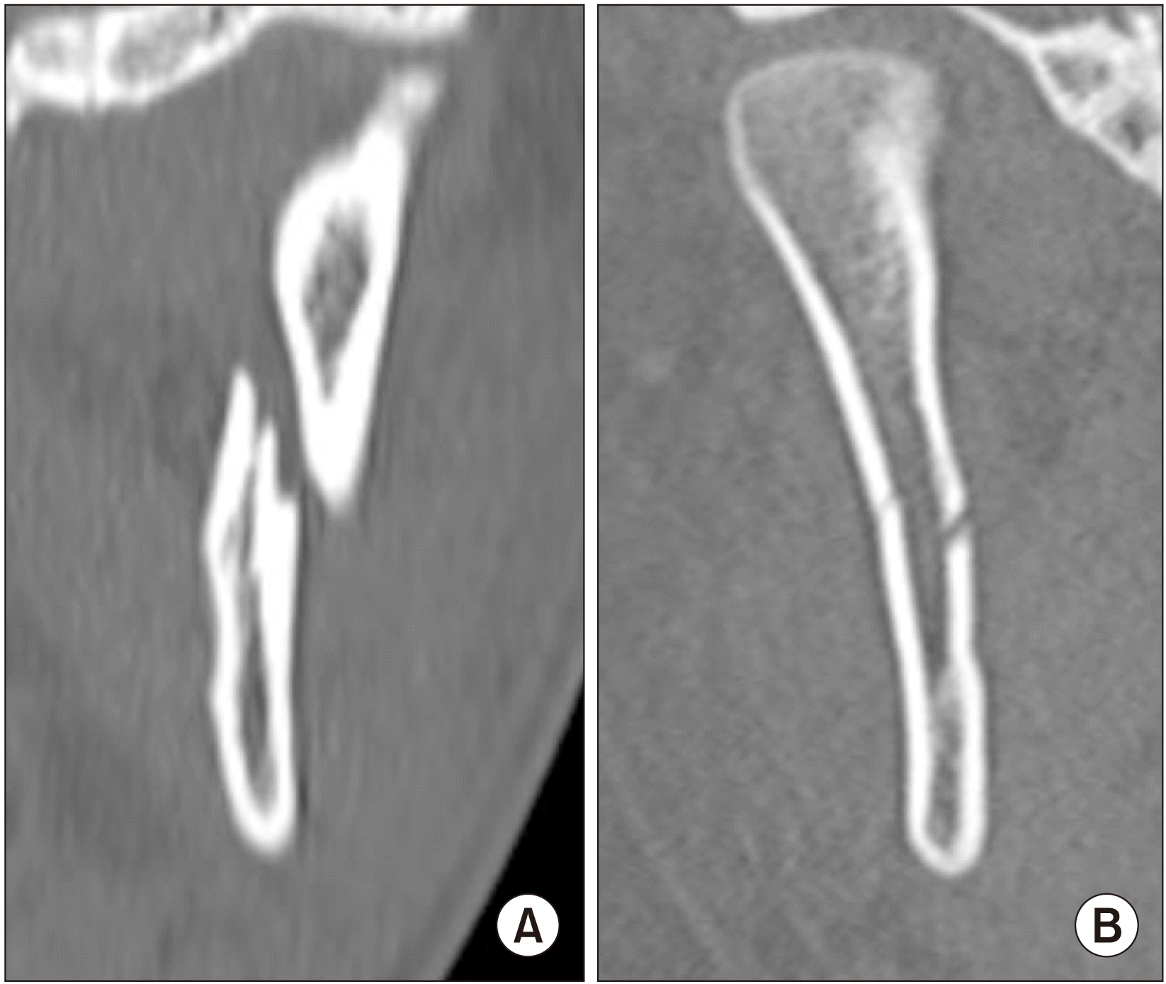
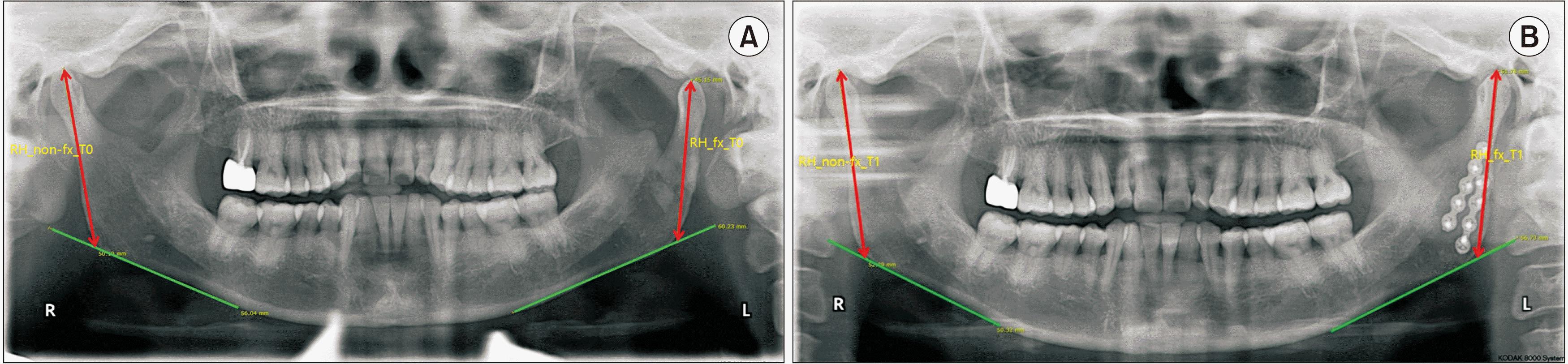
Radiological analysis of fracture fragment displacement was performed using computed tomography (CT) and panoramic radiography. Displacement of the fracture fragment was defined as abnormal position of the fractured condylar fragment in relation to the distal segment bone in the CT images.(Fig. 1) Among patients treated for unilateral condylar fractures, we investigated the relationships between fracture characteristics and treatment method using binary logistic regression analysis. In addition, among patients treated for unilateral condylar fractures with displacement, the difference in ramal height of the fractured and the non-fractured sides before treatment and ≥3 months after treatment was measured using panoramic radiography. This difference was investigated between the CR and OR groups. Ramal height on panoramic image was defined as the distance from the highest point of the mandibular condyle to the intersection of the tangent to the mandibular inferior border and the posterior edge of the ramus. This was measured on both the fractured and non-fractured sides9.(Fig. 2) Statistical analysis was performed using IBM SPSS Statistics software (ver. 26; IBM, Armonk, NY, USA), and P-values less than 0.05 were considered statistically significant.
Go to :
III. Results
A total of 273 patients with mandibular condylar fractures was treated at the study hospital for 10 years. Among them, 62 patients with a follow-up period less than three months were excluded. Of the 211 patients enrolled, 13 were ≤12 years of age; 1 of these pediatric patients underwent surgery, while the rest were placed in the CR group. A total of 198 patients >12 years of age was investigated, of whom 48.0% (n=95) were treated with CR and 52.0% (n=103) underwent OR. There was no significant correlation between method and patient sex, age, or follow-up period.(Table 1)
Among the postoperative complications, malocclusion was found in four patients in the CR group and in two patients in the OR group. Whereas all four patients in the CR group had bilateral condylar fractures, there was no bilateral case among the malocclusion cases in the OR group. TMD was found in five patients (by TMJ sound in three and pain in two patients) in the CR group and in one patient (TMJ pain) in the OR group. Three of the five patients with TMD in the CR group had bilateral fractures; the one patient with TMD in the OR group did not. Ten of 14 patients in the CR group and two of 11 patients in the OR group with MOL showed bilateral fractures. There was no significant difference between incidence of complications and treatment method. The average period of postoperative IMF was 1.9±1.2 weeks in the OR group and 3.2±1.2 weeks in the CR group. The postoperative IMF period was significantly shorter in the OR group than in the CR group.(Table 2)
Table 2
Complications and IMF periods of the patients
| Variable | CR (n=95) | OR (n=103) | P-value |
|---|---|---|---|
| Occlusion | 0.3041 | ||
| Favorable | 91 | 101 | |
| Malocclusion | 4 | 2 | |
| Temporomandibular joint | 0.1071 | ||
| No symptoms | 90 | 102 | |
| TMD | 5 | 1 | |
| Mouth opening limitation (<40 mm) | 0.4022 | ||
| No | 81 | 92 | |
| Yes | 14 | 11 | |
| Amount of mouth opening in patients with MOL (mm) | 30.7±4.2 | 30.5±4.7 | 0.9053 |
| IMF period (wk) | 3.2±1.2 | 1.9±1.2 | <0.0014 |
![]()
Among the 139 patients with unilateral condylar fracture or with other concomitant mandibular fractures that were surgically reduced without affecting the occlusion, 50 were treated with CR and 89 were treated with OR. In the OR group, 83, 6, and 0 patients had fractures in the subcondylar region, condyle neck region, and condylar head region, respectively. In contrast, among the patients treated with CR, fracture in the subcondylar or condylar neck region was were observed in 17 patients. The remaining 33 patients had fractures in the condylar head region.(Table 3)
Table 3
Fracture site and fragment displacement in patients with unilateral condylar fractures
| Variable |
CR (n=50) |
OR (n=89) |
P-value1 |
|---|---|---|---|
| Fracture site | <0.001 | ||
| Condyle head | 33 | 0 | |
| Condyle neck | 8 | 6 | |
| Subcondyle | 9 | 83 | |
| Condylar fracture with | 0.284 | ||
| Displacement of the fractured fragment | 20 | 44 | |
| No displacement of the fractured fragment | 30 | 45 |
![]()
Binary logistic regression analysis showed a significantly higher odds ratio in patients with subcondylar fracture than in those with condylar head area fracture. There was no significant correlation between group and fracture fragment displacement.(Table 4)
Table 4
Binary logistic regression analysis of differences in surgical treatment according to fracture site and fractured fragment displacement
![]()
Of the 65 patients with changes in ramal height due to displaced fractured fragment, CR and OR were conducted in 21 and 44, respectively. In the 21 patients treated with CR, the difference in ramal height between the fractured and non-fractured sides was 5.8±3.5 mm before treatment and 6.6±3.4 mm after treatment. In 44 patients who were treated with OR, the difference in ramal height was 5.0±2.9 mm before treatment and 1.5±2.4 mm after treatment. There was a significant difference in the change in ramal height difference between the fractured and non-fractured sides during treatment between the OR and CR groups.(Table 5)
Table 5
Comparison of the difference in ramal height of the fractured and non-fractured sides before and after treatment measured on panoramic radiographs in unilateral condyle fracture patients
| Variable | CR | OR | P-value |
|---|---|---|---|
| Difference in ramal height before treatment (mm) | 5.8±3.5 | 5.0±2.9 | 0.2531 |
| Difference in ramal height after treatment (mm) | 6.6±3.4 | 1.5±2.4 | <0.0011 |
| P-value | 0.2642 | <0.0012 | |
| Changes of ramal height difference before and after treatment (mm) | –0.7±3.1 | –3.5±2.5 | <0.0013 |
![]()
Go to :
IV. Discussion
Condylar fracture treatment is a subject of debate. Patients with condylar fractures without displacement, dislocation, or derangement of occlusion seem to be best treated with medication alone. Slight derangement of occlusion and mild-to-moderate displacement appear to be best treated by CR and IMF. Grossly displaced dislocated fractures are best treated with OR and internal fixation7. It is believed that fractures with a deviation >10º or shortening of the ascending ramus >2 mm should be treated with OR and fixation irrespective of the level8. In patients with fractures in the condylar head region, CR is conducted when there is no fracture fragment displacement or when the fracture line is close to the medial pole, while OR is conducted when there is more than one fracture line or when the fracture line is close to the lateral pole10. In the present study, CR was significantly performed when the fracture site was the condyle head, while OR was performed frequently for subcondylar fractures. However, a difference in preference for treatment method was not observed depending on displacement of the fracture fragment. In this study, there were many patients who underwent surgery even though there was no displacement of the fractured fragment. If there is no displacement of the fractured fragment, surgery likely is not necessary. However, the absence of displacement of the fractured fragment does not automatically exclude the presence of malocclusion. A fractured fragment may be displaced while waiting for surgery, and long-term IMF may be selected instead of surgery to prevent this. For these reasons, there were many cases in this study in which surgery was performed even though there was no displacement. In patients with fractures in the condyle neck site, significance was not observed in choice of treatment method. It seems that treatment method is determined considering various factors, such as condition of displacement or dislocation of the fractured fragment, patient general condition and age, and surgeon preference.
Several indices have been used for the pre- and post-treatment evaluation of mandibular condylar fractures, such as ramal height, fracture fragment angulation, and resorption pattern of condylar fracture fragments. There are studies reporting better results when surgical treatment was conducted with respect to TMJ morphology—such as less resorption of the condylar fracture fragment and proper reduction of the condyle in the fossa—than those achieved with conservative treatment11-14. Studies have documented significant recovery of ramal height over time, even in patients who underwent adequate CR15. Malocclusion, TMD, MOL, mandibular dysfunctional movement, infection, and nerve damage are clinical indicators of mandibular condylar fractures and are expected to improve after treatment2,16,17. In the present study, the amount of fracture fragment reduction was significantly greater in displaced condylar fractures treated surgically than that in the conservative treatment group.
In our study, malocclusion, TMD, and MOL were investigated as clinical complications. A previous study suggested that some of the most important factors for development of malocclusion are condylar dislocation and bilateral fractures18. In this study, malocclusion occurred in four bilateral fracture cases in the CR group. However, complications related to condylar dislocation could not be considered separately in this study. The remodeling and regenerative capacity of the condyle is impaired following dislocation, and the prognosis is difficult to predict19. Therefore, it is important to consider management of TMJ capsular injury to prevent complications.
Although complications according to specific fracture site was not statistically investigated, no significant difference was found between the OR and CR groups. Proper clinical results can be obtained by selecting an appropriate treatment method and postoperative management.
In both OR and CR, postoperative IMF was performed for a certain period after treatment until appropriate reduction. During the postoperative IMF period, patients experienced inconveniences such as pain and difficulty with oral hygiene. The average IMF period is shorter in OR cases than in CR cases, and postoperative IMF is not performed in some OR cases17,20. The possibility of complications increases with intraoperative time21; therefore, efforts, including use of customized three-dimensional printed plates and semi-rigid fixation, are being made to reduce intraoperative time9,22. Although OR requires a longer operation time than CR, the IMF period of OR procedures was significantly shorter than that of CR procedures in the present study. Discomfort due to IMF and the possibility of postoperative complications were also considered when selecting the appropriate treatment method for mandibular condylar fractures.
In previous studies, the sex ratio of patients with mandibular condylar fractures has been reported to skew significantly male (3:1). This is because fractures from traffic accidents, violence, industrial accidents, and sports are more commonly of the mandibular condylar type23. In this study, among the 211 patients enrolled with mandibular condylar fractures, there were 156 male patients and 55 female patients, showing a sex trend similar to those of other studies. In treating pediatric mandibular condylar fracture cases, CR is preferred because surgical treatment can cause postoperative complications, such as facial nerve damage, scarring, and asymmetry of the mandible due to growth disorders. When OR is necessary in pediatric patients with severe bone fragment displacement, use of absorbable plates should be considering24. When mandibular condylar fractures occur with little displacement of fracture fragments, few functional and growth disorders are present; however, when the displacement is severe between the bone fragments and the TMJ capsule, complications such as facial asymmetry and functional and growth disorders show high incidence in pediatric patients25. Among the 13 pediatric patients aged ≤12 years with mandibular condylar fractures in our study, CR was performed in 12 cases and OR was performed in one case and was conducted with an absorbable plate. In this study, treatment of mandibular condylar fractures in pediatric patients was mainly conservative.
This study has several limitations. First, it uses a retrospective design. The need for prospective studies of patients with mandibular condylar fractures is emphasized. In addition, to find more accurate indications for OR and CR, more studies are needed to further analyze the implications for fracture site; TMJ capsular injury type, such as dislocation of the condyle head; unilateral or bilateral fracture type; and degree of displacement. Next, radiologic analysis using panoramic views may be inaccurate compared to three-dimensional analysis using CT imaging. Three-dimensional analysis is thought to provide more accurate information about condylar changes.
Go to :
V. Conclusion
In this study, no significant clinical differences were found between OR and CR in patients with mandibular condylar fractures. OR was performed more frequently in patients with subcondyle fractures than in those with condyle head area fractures. There was no significant relationship between fracture fragment displacement and treatment method. In terms of morphological changes at the fracture site, change in ramal height on the fractured side after treatment was significant in the OR group.
Go to :
 XML Download