

 PDF
PDF Citation
 Print
Print


I. Introduction
Le Fort I osteotomy, performed to improve skeletal malocclusion in the course of orthodontic treatment, is a commonly performed surgical method because it can achieve dramatic results by enabling free movement of the maxilla1,2. This is a surgery that is separated by crossing from the sinus wall level of the maxillary bone. The front and sides can be visibly sawed, while the separation of the pterygomaxillary junction (PMJ) located at the posterior part is invisible, which is a difficult procedure for most surgeons3,4. For this reason, the pterygoid vein plexus, descending palatal artery, posterior wall of the maxillary sinus, pterygoid plate, etc. are carefully separated using an osteotome and mallet to minimize postoperative complications3,4.
When the cross-sectional separation at the sinus wall level of the maxilla is finished, the shape and volume of the maxillary sinus change according to the three-dimensional movement of the maxilla5,6. After surgery, the maxillary sinus is filled with blood, and the change in mucosal thickness, swelling, and other inflammatory reactions appear during the healing process7. Mucosal thickness usually increases after surgery and then gradually decreases7.
Maxillary sinus mucous thickening does not necessarily cause any inflammatory symptoms or discomfort to the patient7, and is sometimes asymptomatic. However, a persistently thickened mucous membrane can result in chronic sinusitis, which can cause symptoms such as headache and nasal congestion, and this can make it difficult to perform subsequent sinus-related surgery, including a sinus graft.
Many studies have been conducted to investigate the relationship between various factors that can affect the maxillary sinus after Le Fort I osteotomy. However, there are insufficient studies on the effect of the pterygomaxillary disjunction type, which constitutes the posterior part of the maxilla, on the healing pattern of the maxillary sinus after surgery. In this paper, to investigate the difference in changes in the maxillary sinus healing according to different separation patterns of PMJ, cutting types were classified into two groups and the thickness changes of the maxillary sinus mucosa were investigated. Based on this, we can think about the precautions to be taken in the PMJ separation process and whether the effort and time to cleanly separate the PMJ is reasonable.
II. Materials and Methods
The study was approved by the Institutional Review Board (IRB) of Kyung Hee University Dental Hospital (IRB No. KH-DT22019) and the informed consent was waived by the IRB.
This study is a retrospective cohort study in which 100 patients who underwent Le Fort I osteotomy from 2013 to 2020 at Kyung Hee University Dental Hospital were followed-up by cone-beam computed tomography (CBCT) for one year. The operation was performed by only one oral and maxillofacial surgeon.
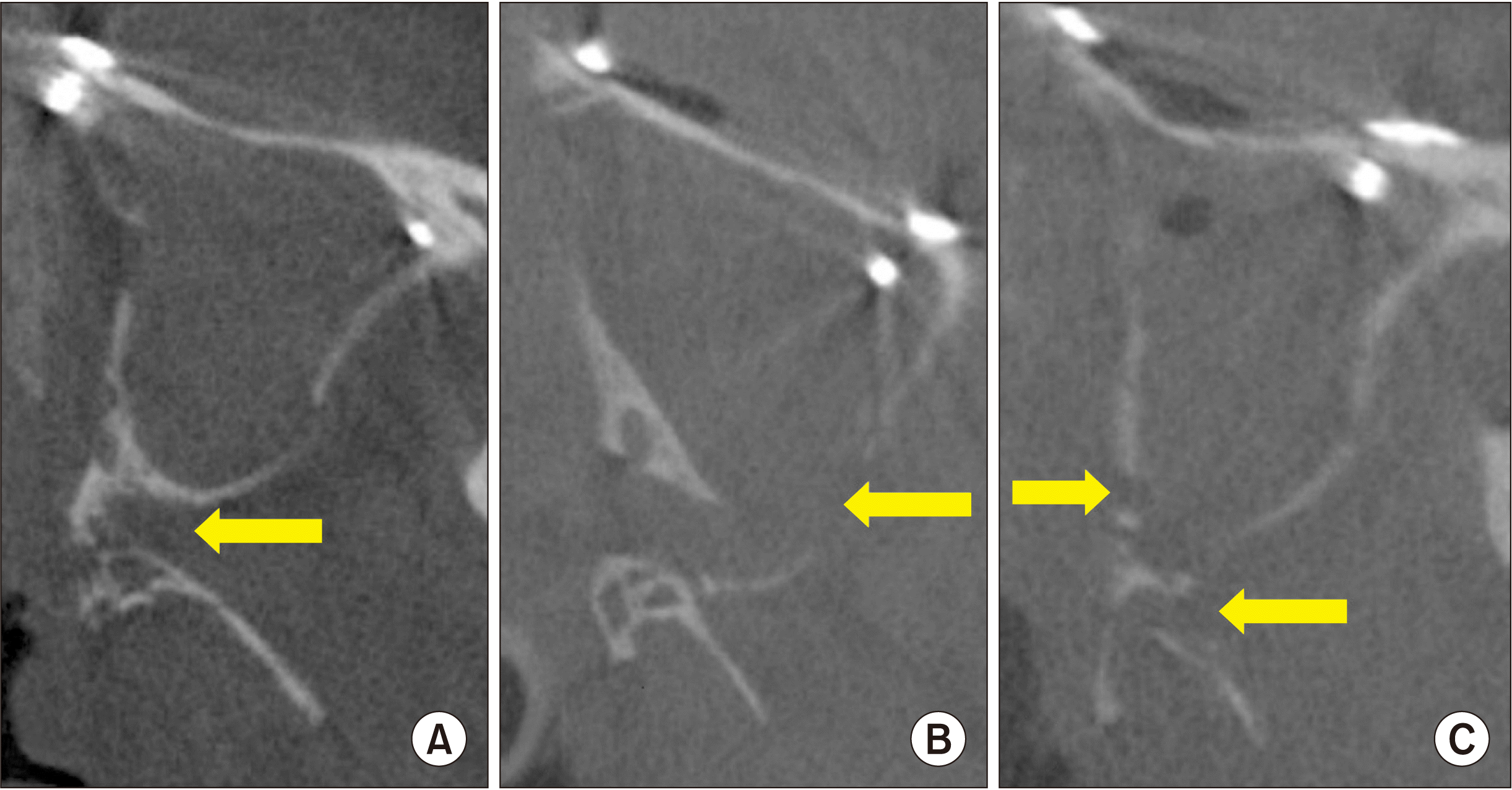
Through 100 CBCTs, we classified the PMJ cutting type into two groups, clean cut type (Type I) (Fig. 1. A) and fractured type (Type II).(Fig. 1. B, 1. C) To compare the changes in mucosal thickness of the maxillary sinus over time, we measured it in preoperative, postoperative three months, and postoperative one year.
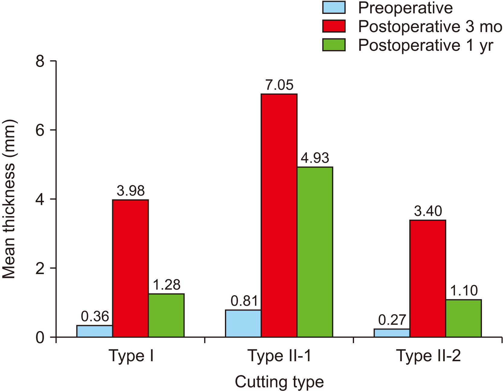
In addition, the fractured type was classified into the posterior wall type (Type II-1) in which the posterior wall of the maxillary sinus was significantly separated, and the pterygoid fracture type (Type II-2) in which a part of the posterior wall of the maxillary sinus and the pterygoid plate were damaged, respectively, and measured as seen in Fig. 1. B and 1. C.
For PMJ cutting, the right maxilla was mainly selected from CBCT taken one day postoperative. However, in the evaluation of factors related to mucosal thickening, the presence of disease in the maxillary sinus was investigated before surgery, and if there was a history of surgery in the right maxillary sinus or a change in the right sinus mucosal thickness was detected, the contralateral left side was classified and measured.
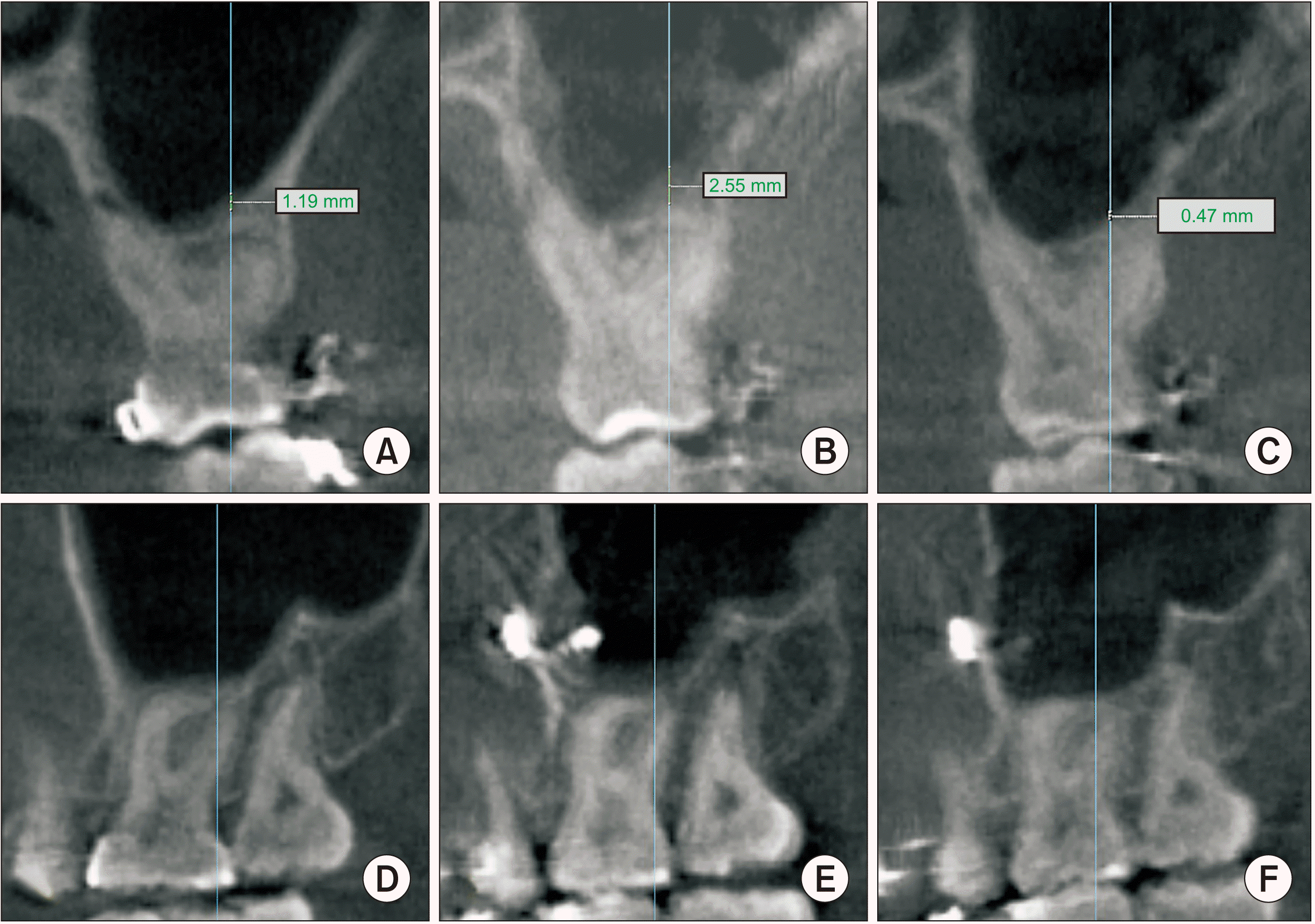
Therefore, PMJ cutting investigation and classification of areas with no history of maxillary sinus disease and related surgery were performed on 100 males and females with a mean age of 22.8 years (range, 18-43 years) at the time of surgery and sinus mucosal thickness was measured.(Table 1) Mucosal thickness over time was measured at the same point (approximately the first molar distobuccal root site) of the maxillary sinus on the side where the PMJ cutting type was observed.(Fig. 2)
Method error was defined as the reproducibility of double judgment and calculated as the standard error of measurement. The method error of repeated linear measurements was within 0.3 mm. A statistically significant difference was not detected in linear measurements.
Since the mucosal thickening of each group did not follow a normal distribution, statistical analysis was performed using non-parametric tests, Mann–Whitney U test, and Kruskal–Wallis test. Statistical analysis was performed using IBM SPSS Statistics (ver. 26.0; IBM, Armonk, NY, USA). P-values less than 0.05 were statistically significant.
III. Results
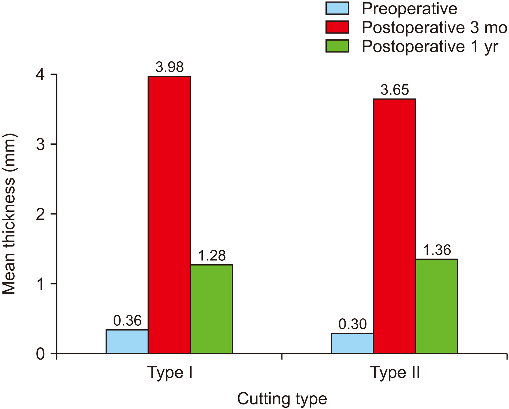
The Type I cases numbered 28 out of 100 cases and the Type II represented 72 cases. Among fractured types, the Type II-1 was detached in three cases.(Table 2) After surgery, the sinus mucosal thickness increased by more than 3 mm on mean (3.74 mm) at three months and decreased to less than 2 mm after one year.(Table 3) The Type I and Type II cases were measured and compared as seen in Fig. 3, and Type II was classified into Type II-1 and Type II-2, measured, and compared as seen in Fig. 4.
There was no difference in the standard deviation of sinus mucosal thickness at postoperative three months and one year between Type I and Type II, and no statistical significance was noted. So, there was no difference in sinus mucosal thickening at postoperative three months and one year between the two groups.(Table 4) However, there was a significant difference in sinus mucosal thickness one year after surgery in Type II-1 compared to the other types, as seen in Tables 5 and 6.
IV. Discussion
Separation of the PMJ during the Le Fort I osteotomy is a challenging procedure for most surgeons because it is not directly visible3,4. So, it is difficult to separate the PMJ as intended, and posterior structures such as the pterygoid vein plexus, descending palatal artery, posterior wall of the maxillary sinus, and pterygoid plate may be damaged in this process4. Due to this damage, some bleeding may be observed, thereby prolonging the operation time. So, the surgeon spends effort and time to separate the PMJ cleanly in the hope that it will preserve the adjacent structures and reduce the possibility of postoperative complications.
However, if bleeding is controlled, posterior and lateral maxillary movement becomes easier by fracturing posterior hard tissue structure, including the pterygoid plate. As a result, it is possible to shorten the overall operation time by skipping the process of trimming the hard tissue structures. In addition, it can be expected that the incidence of postoperative complications can be further reduced.
Further, if the posterior wall of the maxillary sinus is not detached, the PMJ separation pattern does not significantly affect the occurrence of internal problems or recovery patterns after Le Fort I osteotomy. Therefore, it may not be reasonable to spend time, even at the risk of postoperative complications, due to the extension of the operation time.
Of course, whether only damage to the posterior wall of the maxillary sinus, or the gap caused by the three-dimensional movement of the maxilla also affected the maxillary sinus mucosal thickening could be considered. During Le Fort I osteotomy, the amount of three-dimensional movement including impaction, advance, and setback of the maxilla was all within 5 mm, and soft tissue proliferated and penetrated the gap in the transversely separated sinus of the maxilla. However, most cases were not accompanied by an overall thickening of the maxillary sinus mucosa. The infiltrated soft tissue was usually observed in a limited area up to one year after surgery, but no specificity was found in the maxillary sinus. In other words, it can be considered that the gap in the maxillary sinus wall caused by the movement of the maxilla has no effect on complications within the maxillary sinus after surgery. However, additional research is needed on whether the gap generated by the maxillary movement of more than 5 mm affects the maxillary sinus mucosal thickening.
In addition, we can see from these data that there is little effect on the inside of the maxillary sinus due to penetrating screws through the maxillary cortical bone and sinus membrane for plate fixation after the maxilla movement. Based on this, even if the implant fixture penetrates the sinus membrane during placement, it can be considered that this does not cause problems inside the sinus, such as sinusitis.
V. Conclusion
The PMJ separation process during Le Fort I osteotomy is invisible, so it may take some time because it is difficult to cleanly separate. Although there is some damage to posterior structures of the PMJ, we found that there is no significant difference inside the sinus in the postoperative complication compared to the clean cut case. Therefore, spending significant time on the PMJ cutting process may not be a good choice considering the overall postoperative complications.
 XML Download