

 PDF
PDF Citation
 Print
Print


I. Introduction
The inferior alveolar nerve (IAN) is a branch of the trigeminal nerve responsible for sensation of the mandibular teeth, lower lip, and chin. IAN injuries can be caused by iatrogenicity due to anatomical variations, treatment based on improper diagnosis, or unskilled performance during procedures such as surgical extraction of the mandibular third molar1, dental implant surgery2, root canal treatment3, IAN block anesthesia4, and orthognathic surgery5. Neuropathies like hypoesthesia, paresthesia, and dysesthesia may be present in patients with IAN injury. These symptoms can impair essential functions such as speech, swallowing, and mastication, reducing quality of life6. In addition, injuries to the IAN are rare but can potentially cause chronic neuropathic pain syndrome7.
A series of studies previously reported that the most frequent cause of injury to the IAN is surgical extraction of a mandibular third molar. However, as dental implant surgery has become more common, the incidence of related injury to the IAN has increased8. According to several recent studies, sensory changes after surgical extraction of a mandibular third molar occurred in 0.3%-8.4% of cases9, temporary sensory changes in 0.8%-5.7%10, and permanent sensory changes in 0.7%11. In other studies on dental implant surgery, the frequency of sensory change due to injury to the IAN was reported to be 0.0%-13.0%12. The possibility of permanent sensory change after dental implant surgery was approximately 3.4 times higher than that occurring after surgical extraction of a mandibular third molar13. These results demonstrate the large impact of sensory changes after dental implant surgery.
Sensory changes after dental implant surgery can be caused by pressure on the IAN due to bone compression14 when the implant fixture is placed within 2 mm of the IAN canal or to hematoma formation due to dental implant surgery. Such nerve damage belongs to the neurapraxia type of Seddon and Sunderland’s classification of nerve damage, and complete recovery of sensation can be expected. If over-drilling occurs during dental implant surgery or the implant fixture invades the IAN canal, nerve damage, such as axonotmesis or neurotmesis, can occur. Although some degree of sensory recovery can be expected, such an injury often results in permanent damage13. However, IAN block anesthesia or mental nerve injury due to flap elevation during dental implant surgery can also cause sensory change with the possibility of permanence4,15.
In general, if sensory changes occur after dental implant surgery, a non-surgical approach such as medication should be used as treatment16. While several reports have suggested that the implant fixture be removed within 36 hours17, cases with sensory recovery without removal of the implant fixture have been reported16.
This study aimed to 1) analyze the data on nerve damage in patients who complained of sensory changes after dental implant surgery, 2) evaluate the prognosis of sensory changes according to proximity of the implant fixture to the IAN canal, and 3) identify the factors affecting recovery of sensation.
II. Materials and Methods
From 2011 to 2021, raw data of 293 patients who visited the Department of Oral and Maxillofacial Surgery at Gangnam Severance Hospital with sensory changes (hypoesthesia, paresthesia, dysesthesia, etc.) were collected from Yonsei Medical Center’s Severance Open BIG data. In total, 64 patients satisfied the inclusion criteria. This retrospective study complied with the tenets of the Declaration of Helsinki and was approved by the Institutional Review Board (IRB) of Gangnam Severance Hospital (IRB approval No. 2021-0958-001).
The inclusion criteria were: (1) adult patients aged >19 years, 2) complaint of sensory changes after dental implant surgery of the mandible, and 3) computed tomography (CT) showing a relationship between dental implant surgery and the IAN canal. The exclusion criteria were: 1) patients with a medical history or syndrome associated with sensation, 2) patients with abnormal neural symptoms in the facial area before dental treatment, and 3) patients who complained of osteomyelitis of the jaw.
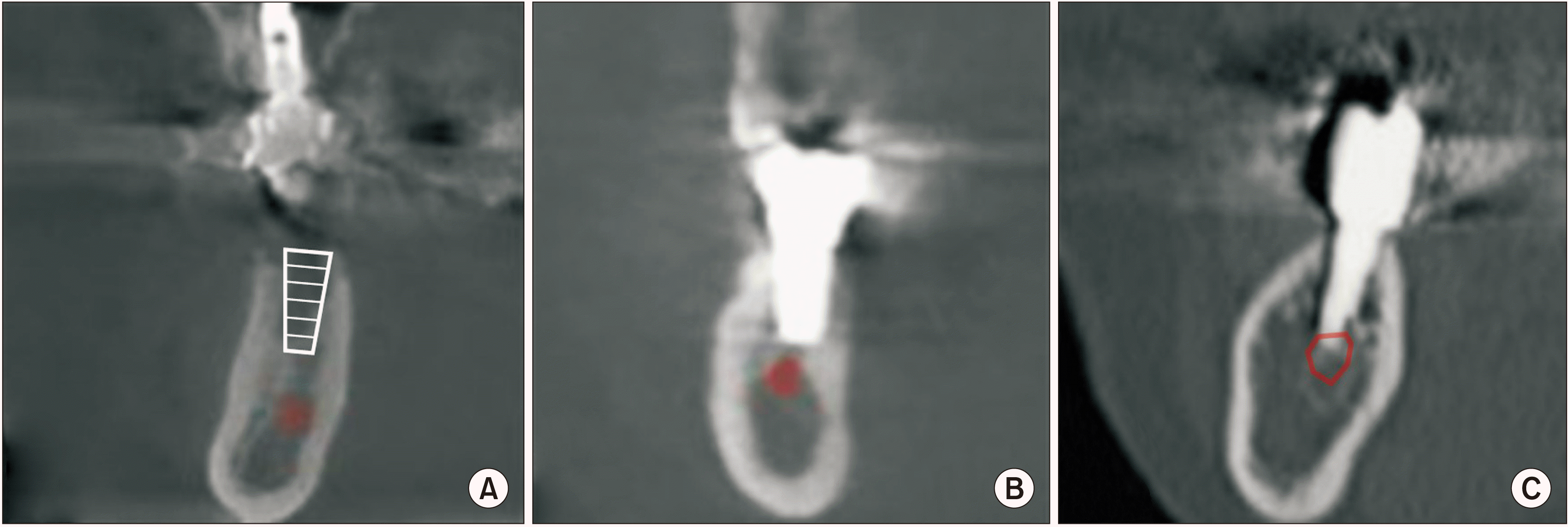
The data of 64 patients, including sex, age, implant surgery site, medication status, and whether the implant fixtures were removed or not, were examined through the electronic medical record. Based on the shortest distance (D) between the implant fixture or the drilling location and the IAN canal on CT images, the patients were classified into Group I (D>2 mm), Group II (2 mm≥D>0 mm), and Group III (D≤0 mm).(Fig. 1)
Among the 64 patients, 36 visited our clinic more than two times for symptoms of sensory change. For those 36 patients, the degree of sensory change was subjectively evaluated using the visual analog scale (VAS). Improvement in sensation was defined by increased feeling during probing using an explorer in the lower lip and chin. Based on the evaluation criteria, patients who expressed improvement in sensation at least once were classified as ‘Improved,’ those who did not exhibit improvement were classified as ‘Stationary,’ and those who complained of worsened sensation were classified as ‘Aggravated.’
Statistical and analytical processing was performed using IBM SPSS Statistics (ver. 26; IBM, Armonk, NY, USA). Subjective recovery, difference in recovery period between Group II and Group III, and status of implant fixture removal and recovery in Group III were identified using the chi-square and Fisher’s exact tests. Statistical significance was defined as P-value of 0.05 or less.
III. Results
The patients included 44 females and 20 males. The age distribution ranged from 32-82 years, and the mean age was 57.3±7.3 years. Dental implant surgery associated with sensory changes was more frequent in the molar area (72 fixtures) than in the premolar area (25 fixtures), with the second molar being the most frequent site (27 fixtures).(Table 1)
A total of 31 patients (86.1%) received oral medication treatment with a steroid (Solondo; Yuhan, Seoul, Korea) and vitamin B (Beecom [Yuhan] and Vitamedin [CJ HealthCare, Seoul, Korea]). Implant fixtures were removed in 20 patients (55.6%).
Among the 36 patients who visited our clinic more than two times, 21 patients (58.3%) reported improvement in sensation, 13 patients (36.1%) had no change in sensation, and 2 patients (5.6%) reported worsening sensation. The mean time to subjective improvement in sensation was 172.9±76.7 days. All cases of improvement involved medication. Of the 15 patients whose sensation did not improve or worsened, 10 were treated with medication. Among them, 9 patients (60.0%) visited our clinic 6 months after the occurrence of sensory changes.
Radiographic images were available for 3, 12, and 21 patients in Group I, Group II, and Group III, respectively. In Group II, removal of the implant fixture was performed in 4 patients, while improvement in sensation was achieved in all patients regardless of such removal. In addition, improvement was observed within 6 months. Among the Group III patients, 8 patients (40.0%) reported improvement in sensation with removal of the implant fixture, 2 patients (33.3%) exhibited improvement without removal of the implant fixture, and most of them showed improvement within 6 months. However, 1 patient demonstrated improvement between 6 and 12 months.
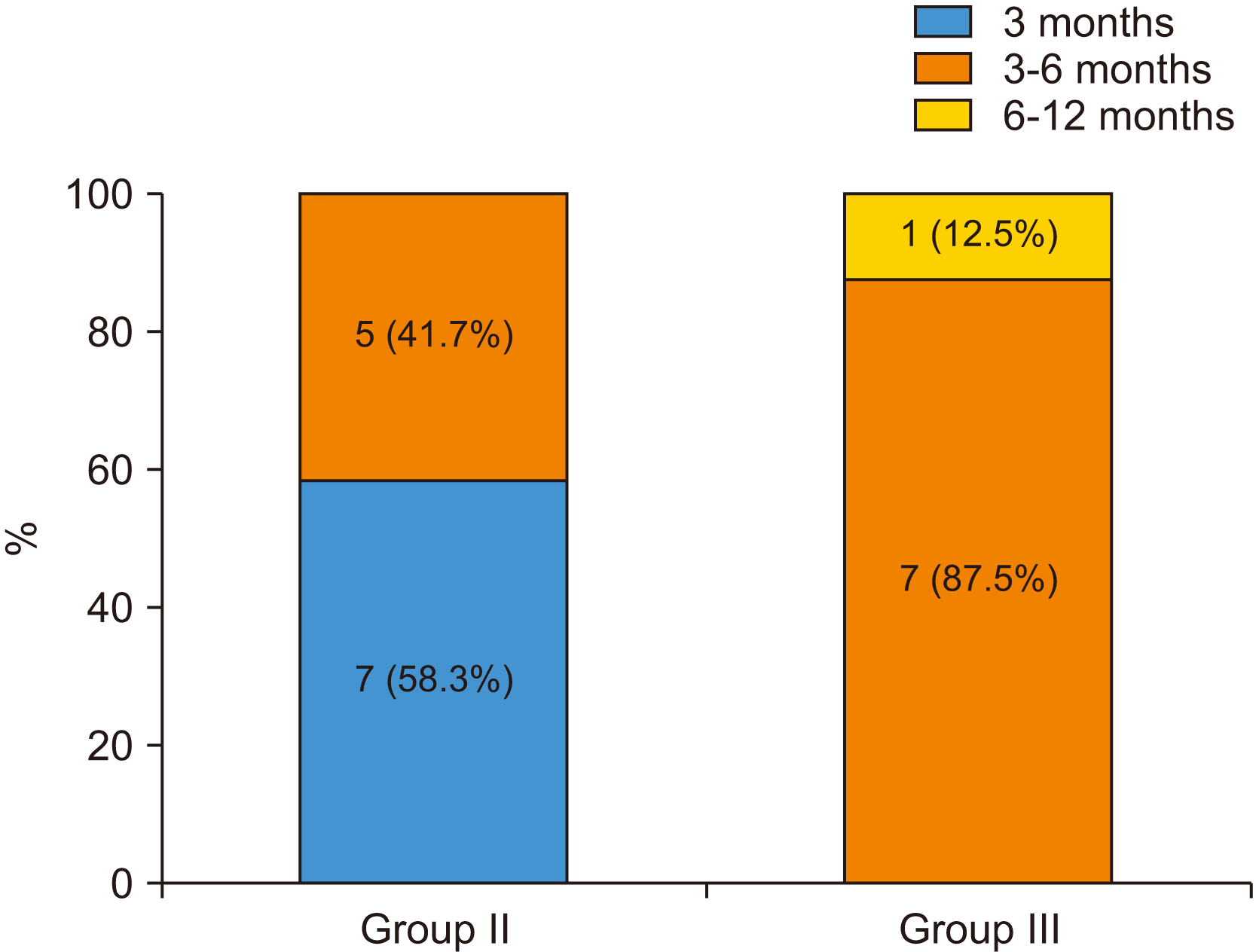
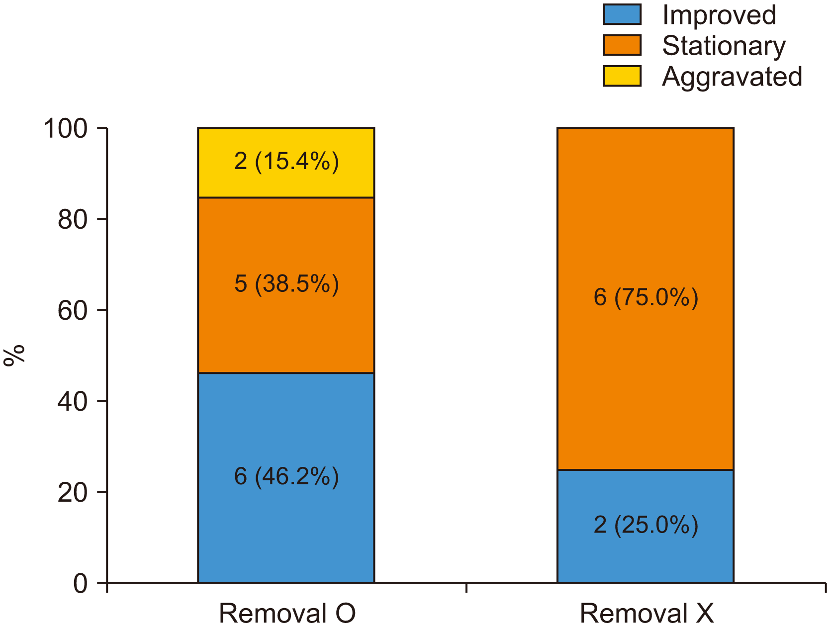
In Group II and Group III, there were no significant differences based on the results of the chi-square test for site of implant surgery regarding cause, sex, age, and sensory change.(Table 2) There was a significant difference in the rate of sensory recovery between Group II and Group III (P=0.001). All patients in Group II exhibited improvement in sensation, but 13 of the 21 patients (61.9%) in Group III did not.(Table 3) In addition, the difference in recovery period between Group II and Group III was significant (P=0.005). In Group II, more than half (58.3%) of the improved patients demonstrated improvement within 3 months. However, in patients who showed improvement in Group III, almost all (87.5%) exhibited improvement between 3 and 6 months.(Fig. 2) In Group III, removing the implant fixture did not result in any significant difference in recovery of sensation (P=0.337), although it did increase the rate of improvement in sensation.(Fig. 3)
IV. Discussion
Based on the results, sensory changes after dental implant surgery were twice as common in females than in males, and middle-aged (50-69 years) individuals accounted for more than half (59.4%) of the sensory changes. Consistent with several studies, this result may be attributed to proximity of implant placement to the IAN canal because the mandible is smaller in women and the elderly18-20. The most frequent site of sensory change after dental implant surgery is the mandibular second molar area, where the distance between the alveolar crest and the IAN canal is the shortest18. Therefore, professional diagnostic equipment such as CT must be used to measure the distance between the path of the IAN canal and the alveolar bone crest accurately in order to perform ideal dental implant surgery in the mandibular second molar area.
In this study, it was assumed that the prognosis of sensory changes after dental implant surgery would be affected by proximity to the IAN canal. The proximity of the implant fixture or drilling location to the IAN canal significantly contributed to sensory recovery. In Group II without direct injury to the IAN, all patients exhibited improvement in sensation regardless of removal of the implant fixture. However, in Group III with direct injury to the IAN, there was a much lower possibility of improvement in sensation compared to Group II, even in cases of aggravation. In addition, for recovery period, Group II demonstrated an improvement in sensation within a much shorter period of time than Group III. This indicates that direct injury to the IAN canal increases not only the recovery period, but also the risk of permanent sensory changes.
Khawaja and Renton21 reported that early removal of an implant fixture invading the IAN canal could reduce the possibility of neuropathy. In addition, Juodzbalys et al.17 suggested that removal of an implant fixture invading the IAN canal be performed within 36 hours after dental implant surgery to prevent infection of the damaged nerve and to reduce the degree of nerve compression due to edema. In addition, direct application of steroids (dexamethasone) to the nerve damage site or oral administration of steroids in the early stages of healing is recommended. This study also showed that removal of an implant fixture invading the IAN canal increased the possibility of improvement in sensation.
Several studies have mentioned that a safe distance of at least 2 mm should be maintained between the implant fixture or drilling location and the IAN canal during dental implant surgery in the mandible. They demonstrated that pressure on the IAN canal due to bone compression could occur at an implant fixture placed adjacent to the IAN canal14,18,22. The results showed a relatively higher possibility of sensory change even without direct invasion of the IAN canal in Group II. Therefore, surgeons must consider a safe distance between the implant fixture and the IAN canal.
In addition, removal of the implant fixture should be carefully considered since a 100% improvement rate was observed in Group II in this study, even without fixture removal. As shown in the results, sensory changes occurred in Group I with a distance greater than 2 mm between the implant fixture or drilling location and the IAN canal, and there were several cases with no improvement. This indicates that sensory changes could occur after dental implant surgery through indirect factors such as nerve injury from block anesthesia (chemical injury, mechanical injury from the needle, etc.), damage to the mental nerve through flap elevation, or damage from releasing the incision of the mandibular gingiva. Therefore, clinicians must consider several factors that could cause sensory changes after dental implant surgery to establish a better treatment plan for nerve complications.
The limitations of this study were the small number of study participants and the difficulty in determining the extent of nerve complications and duration of damage. In addition, the absence of objective indicators for sensory recovery and medication treatment without discriminating between the groups are limitations. Further studies should be conducted with more patients and include objective indicators for measuring sensory changes.
V. Conclusion
When the implant fixture or drilling location directly invaded the IAN canal, there was a higher possibility of improvement in sensation when the implant fixture was removed. In cases where the drilling location or the implant fixture did not invade the IAN canal, the sensory change may not be permanent. In such cases, other indirect factors, such as flap elevation and damage by anesthesia, should be considered as the cause of sensory changes after dental implant surgery. Removal of the implant fixture should be considered carefully in these instances.
In cases of sensory changes after dental implant surgery, clinicians must consider several factors that could cause sensory changes before removing an implant fixture such as distance between the implant fixture and the IAN canal on CT images.
 XML Download