

 PDF
PDF Citation
 Print
Print


I. Introduction
Mandibular fractures are the second-most common type of facial bone fracture after nasal bone fractures1,2. Mandibular condylar fractures account for 19%-52.5% of mandibular fractures depending on geographic region3. However, the surgical access to the mandibular condyle is relatively difficult due to the complexity of the surrounding anatomy. The lateral pterygoid muscle is attached to the medial surface of the mandibular condyle; therefore, in the case of a condylar fracture, the fragments are often displaced anteromedially, complicating proper reduction and fixation and allowing poor visualization of the surgical site4,5. Many extraoral approaches, including the most commonly used preauricular or endaural approach, are not conducive to an adequate visual field and carry a high incidence of postoperative facial palsy due to damage to the seventh cranial nerve6,7. In addition to nerve injury, there is much to consider when deciding to perform surgical intervention on and to choose the surgical technique for condylar fracture, such as age, medical status of the patient, injured region and its anatomy, and accompanying injuries. For these reasons, there is no common consensus about the treatment of mandibular condylar fractures, although some studies have suggested algorithms for decision-making, and the choice of treatment can be largely influenced by the individual clinician.
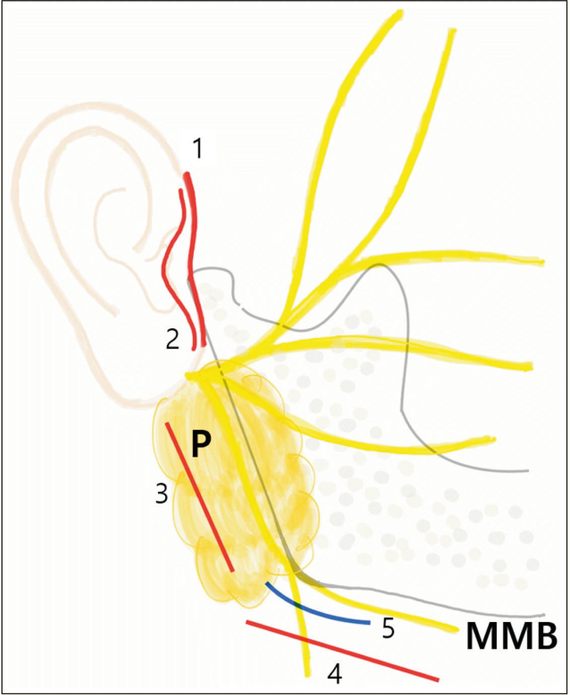
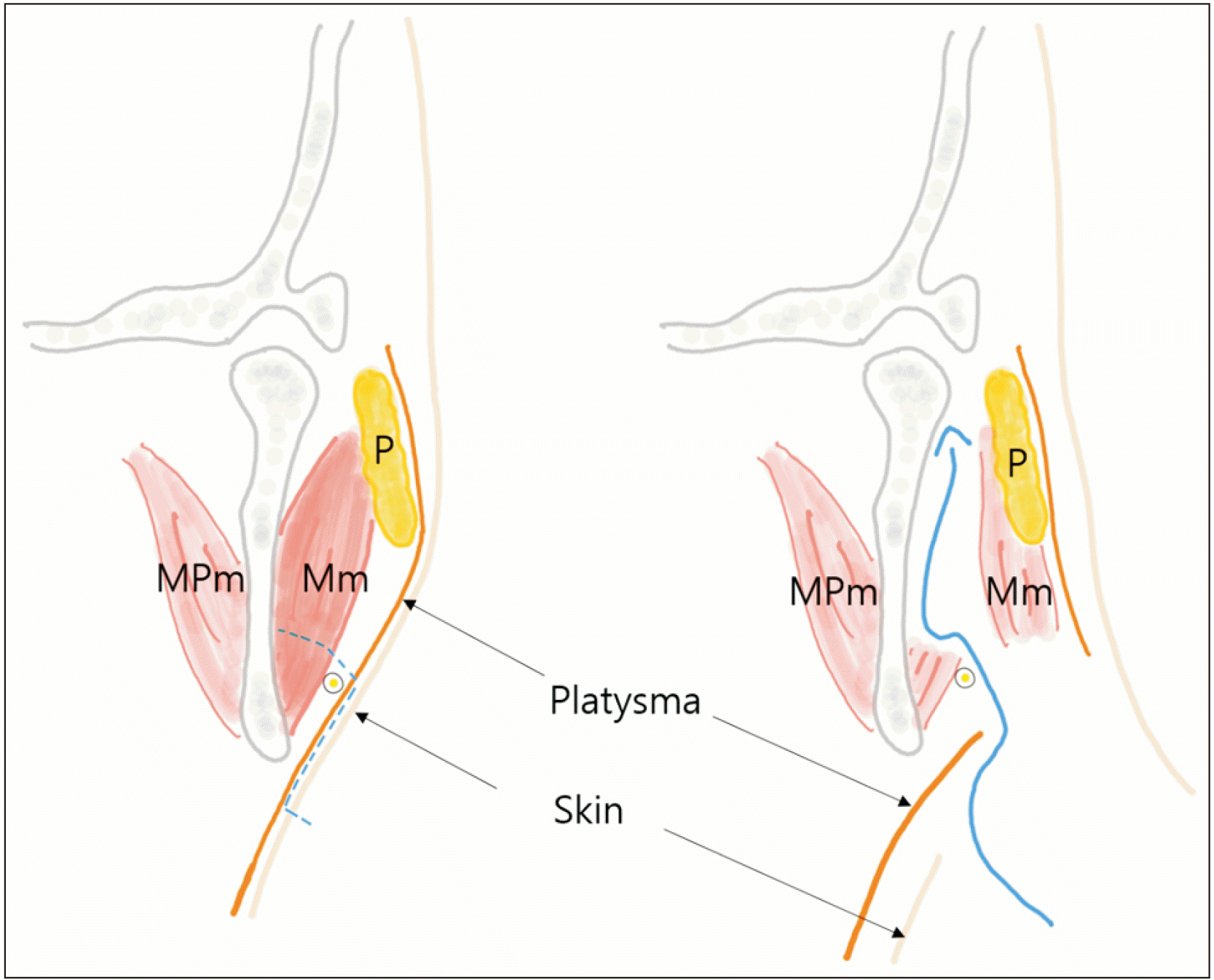
Nevertheless, there are cases in which open surgery must be given priority; therefore, numerous approaches to facilitate surgical access have been introduced and modified4,8-10. Among existing techniques, a high-submandibular transmasseteric approach was introduced recently but remains a more unpopular choice compared to preauricular, endaural, and retromandibular approaches. This method was originally developed for open reduction and internal fixation (ORIF) by Eckelt et al.11 based on the submandibular approach devised by Risdon12 in the 1930s to treat temporomandibular joint (TMJ) ankylosis, which was then modified by Meyer et al.13 in 2006. This modified high-submandibular approach (HSMA) features a higher incision line than that with previous submandibular or retromandibular methods, with an incision line closer to the fracture line.(Fig. 1) Another significant feature is that it can minimize possible damage to the marginal mandible branch of the facial nerve by visualizing the nerve while dissecting at the supraplatysmal level. There are some disadvantages; however, including that the scar is often visible, and there may be temporary trismus or impairment in jaw movement due to muscular scarring. Therefore, in the modified HSMA technique described by Wilk14 in 2009, to hide the incision behind the natural curvature of the mandibular angle, the incision is shorter and closer to the angle; more specifically, it is a 2- to 3-cm incision located right below the lower border of the mandible, dissecting through the supraplatysmal level and, if possible, detecting the marginal manbible branch of the facial nerve, with transection of the masster muscle14.
The surgical procedure of the modified HSMA technique we used is as follows. After placing the patient in a supine position, we tilted their head toward the opposite side of the fracture site to extend the neck at the surgical site. The inferior border of the mandibular angle was marked, and a 2-3 cm incision line (it can be extended to 5 cm if necessary) was made 1 cm below the drawn mandibular angle. The skin incision must remain in the subcutaneous plane so that the platysma muscle can be preserved. During this step, the facial nerve (usually a marginal mandibular branch) was deeper than the platysma muscle. After subcutaneous dissection toward 2-3 cm above the lower edge of the mandible, the platysma was incised to expose the masseter muscle, preserving the surrounding superficial fascia to prevent damage to the buccal branch of the facial nerve, which runs horizontally in the layer below the fascia. Use of a neurostimulator was considered to address possible damage to the nerve. The masseter muscle was dissected in the horizontal direction with sharp dissectors, at the level above the marginal mandibular branch if detected while undermining superficial fascia.(Fig. 2) To assess the fracture line and the anatomical structure of the ramus, dissection was extended to the posterior border of the ramus, and care was taken not to damage the parotid tail. The fracture site was exposed by dissecting the subperiosteal plane toward the condylar part. After osteosynthesis, repair work was performed layer by layer for closure. The masseter and platysma muscles were sutured with absorbable sutures, such as 3-0 polyglactin, and the skin wound was closed using synthetic monofilament sutures, such as nylon. A drainage tube typically was needed to minimize deadspace and prevent hematoma formation.
In this case report, we aimed to evaluate the clinical outcome of the modified HSMA technique for ORIF of mandibular condylar fractures.
II. Materials and Methods
From June 2021 to June 2022, medical records and images for patients who were diagnosed with mandibular condyle fracture at Korea University Anam Hospital and underwent ORIF through modified-HSMA were reviewed. All the surgeries were performed by a single surgeon (I.S.S.). Preoperative and postoperative radiographs and tomograph images were collected. Following information were reviewed from their medical records—sex, age, fractured site, degree of trauma at the time of injury, time elapsed from trauma to surgery, occlusion before and after surgery, whether facial nerve damage was present after surgery, TMJ symptom, postoperative scar assessment, and whether other complications were present.
III. Results
Total 6 cases were reviewed, all the demographic and clinical information are shown in Table 1.
1. Unilateral subcondylar fracture
1) Case 1
A 27-year-old male patient without systemic disease was referred to the department of oral and maxillofacial surgery via the emergency department for facial trauma due to a traffic accident. An initial clinical examination revealed multiple dislocations of the maxillary anterior teeth, and there was slight swelling and tenderness on the right side of the face. Mouth opening was restricted, with premature occlusion of the right posterior molars and an anterior open bite. From a subsequent radiographic examination, right subcondylar and parasymphysis fractures of the mandible were confirmed. There was no other accompanying trauma other than that to the oral and maxillofacial areas.(Fig. 3. A, 3. B)
As the trauma was accompanied by alveolar bone fracture and multiple tooth dislocations, intermaxillary fixation using both an arch bar and gingival screws was initially completed after admission. Open surgery was planned and performed on the fifth day after the injury. Reduction of the fractured alveolar bone and displaced teeth was performed, and bone fixation was completed with miniature titanium plates. Intermaxillary fixation was performed again during surgery, followed by parasymphysis fracture reduction and fixation with two miniature titanium plates. Access to the right subcondylar fracture was gained via the modified HSMA technique, and osteosynthesis was completed with two miniature titanium plates positioned in a lambdoidal shape. Insertion of a drainage tube and layered repair were performed, intraoral sutures were completed, and the surgery was finished.(Fig. 3. C)
Occlusion immediately after the surgery was stable; the patient’s movement was restricted using rubber bands. Root canal treatments and prosthetic restorations were performed during follow up after discharge. Mouth opening was 25 mm at two weeks postoperatively and 43 mm at one year after surgery. Neither TMJ noise nor discomfort was observed.
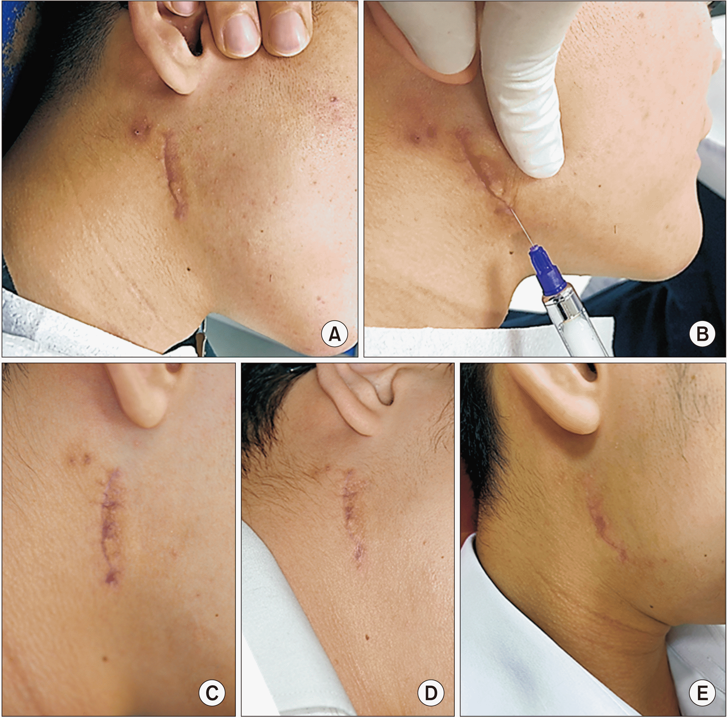
As a postoperative complication, a hypertrophic scar was observed at the previous skin incision area. Triamcinolone (40 mg) was injected subcutaneously three times at intervals of two weeks from the sixth week after surgery. A significant improvement was observed, and the patient was satisfied with the outcome.(Fig. 4) A mild degree of hypoesthesia (visual analog scale score, 2 points) thought to be related to traction damage to the mental nerve during osteosynthesis of the parasymphysis fracture was observed in the right mental region, but it had almost resolved by one year after the surgery.
2) Case 2
A 61-year-old female patient with a medical history of hypertension was referred to the department of oral and maxillofacial surgery via the emergency department for facial trauma from a fall. Clinical examination revealed swelling and tenderness at the right TMJ area and a left posterior open bite. Radiographic images revealed a right mandibular subcondylar fracture. No accompanying injury was found other than that in the oral and maxillofacial area. Early intermaxillary fixation was performed with gingival screws and boxing wires, and traction vector to original occlusion was applied using rubber bands. Open surgery was planned due to insufficient recovery of occlusion after traction. The ORIF procedure was performed on the fifth day after the injury. The modified HSMA technique was used to approach the condylar area; after intermaxillary fixation, reduction was performed, and osteosynthesis was achieved with two miniature titanium plates positioned nearly parallel to each other. Drainage tube insertion and repair of the masseter muscle, superficial muscular–aponeurotic system layer, and subcutaneous and skin layers completed the surgery.
Immediately after the surgery, occlusion was well maintained and stable; however, due to the mechanical disadvantage of the angle between the fixation plates, mouth opening was restricted using rubber bands for up to six weeks. No facial palsy or neurological symptom was observed postoperatively. Her TMJ discomfort was minimal after the surgery and subsided after one month. The scar was not seen in the front view, being well hidden behind the natural prominence of the mandibular angle.
3) Case 3
A 35-year-old male patient without systemic disease was referred to the department of oral and maxillofacial surgery via the emergency department for facial trauma. Clinical examination and radiographic images revealed multiple crown fractures of the anterior incisors and mandibular symphysis and left subcondylar fractures. The patient had mouth opening limitation <10 mm and a posterior open bite. No accompanying trauma other than that to the oral and maxillofacial area was found. ORIF was planned and performed on the third day after the injury. We first managed the patient’s mandibular symphysis; then, after intermaxillary fixation was completed with gingival screws and boxing wires, osteosynthesis was performed using two miniature titanium plates. The modified HSMA technique was used to approach the subcondylar fracture, and reduction and fixation were completed almost the same way as in Case 2.
Immediately after surgery, occlusion was well maintained. No facial palsy was observed, although there was mild hypoesthesia at the left lower lip, which resolved one month after the surgery. The patient reported mild TMJ discomfort during movement until six weeks from the surgery; his mouth opening range was 35 mm at that visit. The scar was not noticeable from the front of the patient, and the patient had no complaints regarding aesthetics.
2. Bilateral subcondylar fracture
1) Case 4
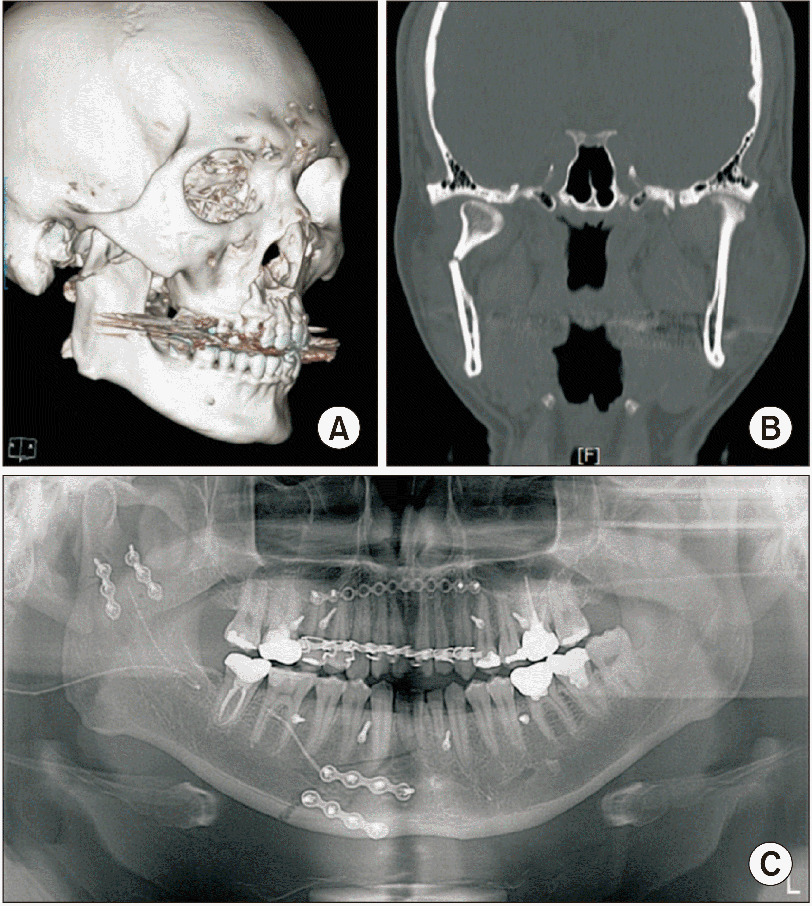
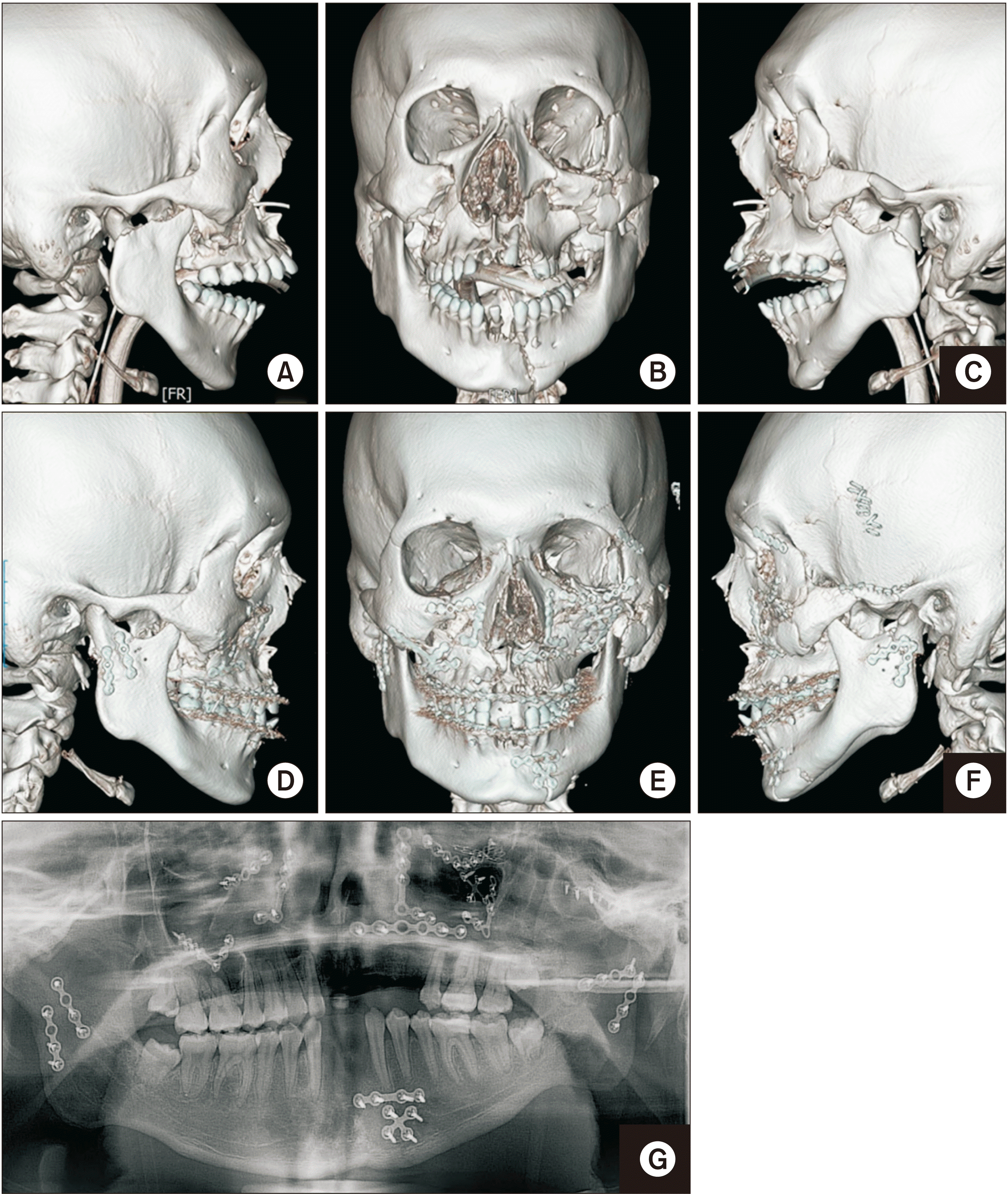
A 16-year-old male patient without systemic disease fell from a height of three-stories and was admitted to the intensive care unit via the emergency department. Clinical and radiographic examinations showed panfacial bone fractures with bilateral subcondylar fractures accompanied by a LeFort Ⅲ fracture, blowout fracture, and zygomatic arch and mandibular symphysis fractures in the oral and maxillofacial area.(Fig. 5. A-C) Tracheostomy was performed while the patient was in the intensive unit care, and he was referred to the department of oral and maxillofacial surgery for surgery. Panfacial comminuted fractures were seen, hindering proper occlusion and positioning of each segment, so reduction was performed in order from the orbit, zygomatic arch, maxilla, and symphysis of the mandible. Thereafter, intermaxillary fixation was performed, and the posterior molars were occluded as closely as possible. Approaches to the subcondyle at both sides via the modified HSMA technique were performed, and osteosynthesis was achieved with two miniature titanium plates at each side, similar to in the previous case in the form of a lambdoid. After drainage tube insertion, layered repair, and intra-oral sutures, the surgery was complete.(Fig. 5. D-F)
Intermaxillary fixation was maintained during the immediate postoperative period, and the patient was equipped with rubber bands at six weeks after the surgery. At this time, the mouth opening range was 25 mm, and posterior occlusion was stable in an acceptable range for functioning. The patient was educated on gradual mouth opening exercises to prevent TMJ ankylosis, and the opening range was 37 mm at 4 months after the surgery; however, he still complained of mild discomfort and a clicking sound from the joint. The patient was referred to the department of orthodontics and prosthetics for rehabilitation of optimized occlusion.(Fig. 5. G)
Except for malocclusion and mild noise during TMJ functioning, no major complications such as facial palsy or severe opening impairment were observed.
3. Unilateral condylar neck fracture
1) Case 5
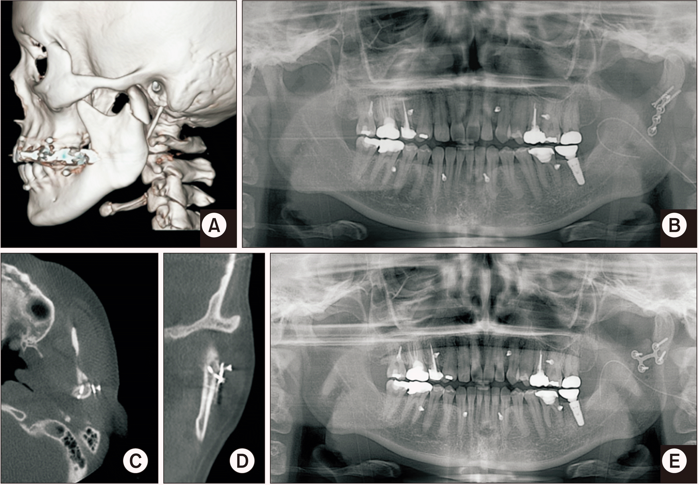
A 39-year-old female patient without systemic disease fell and bumped her lower jaw two weeks before her first visit to the hospital. Until two days before the first visit, no significant symptoms existed, the patient was not receiving any treatment, and she only took over-the-counter medications for a few days. From the day before her first visit, however, she felt pain in the joint area and a change in occlusion. The initial clinical examination showed tenderness at the left TMJ and a left posterior premature occlusion and anterior open bite. A left condylar neck fracture was confirmed by radiographic examination.(Fig. 6. A) After admission to the department of oral and maxillofacial surgery, intermaxillary fixation using gingival screws was performed. Traction was engaged using rubber bands before ORIF; the surgery was performed 18 days after the injury. The fractured condylar neck was approached via the modified HSMA technique, and osteosynthesis was performed using a miniature titanium plate and two lag screws. The surgery was completed after drain tube insertion and layered sutures.(Fig. 6. B)
Occlusion was stable during the immediate postoperative period; however, one month after the surgery, occlusion change and anterior open bite were observed.(Fig. 6. C) To correct the relapse, a reoperation was performed. Using the modified HSMA technique again, the previous hardware was removed, and osteosynthesis was completed using a cross-shaped miniature titanium plate.(Fig. 6. D) After the reoperation, occlusion was stable, and intermaxillary fixation using rubber bands was maintained until eight weeks postoperatively. Two months after the reoperation, her mouth opening range was 26 mm, and no TMJ noise, discomfort, or neurological complications were observed; however, due to the reoperation, scar tissue was relatively prominent compared to the other cases described. Subcutaneous steroid injection of 40 mg of triamcinolone was performed four times at two-week, but the scar did not show significant improvement. At four months after the reoperation, her mouth opening range was 35 mm, and occlusion was stable, but the scar remained without any improvement.
4. Unilateral simultaneous condylar neck and subcondylar fracture
1) Case 6
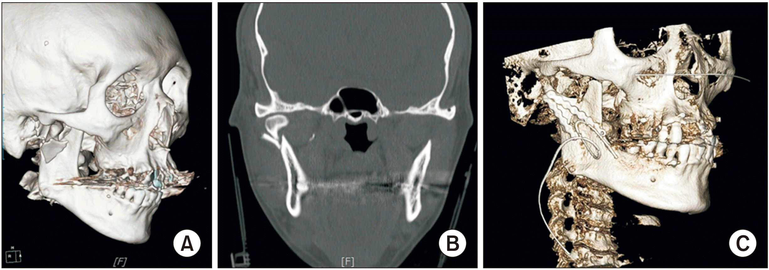
A 33-year-old male patient without systemic disease was referred from the emergency department to the department of oral and maxillofacial surgery after being injured in a traffic accident. The man showed tenderness at the right TMJ area and severe mouth opening limitation during the initial clinical examination. Multiple maxillary incisors were lost at the time of injury. Mandibular neck and subcondylar fractures were confirmed simultaneously by radiographic examination, with two free segments adjacent to each fracture, respectively.(Fig. 7. A, 7. B)
After admission, intermaxillary fixation was achieved using gingival screws. ORIF was performed on the third day after the injury. An approach to the fractured fragments via the modified HSMA technique was made. First, the two free segments were aligned and connected by a miniature metal plate; after this connection, the procedure was similar to a simple subcondylar fracture surgery. Intermaxillary fixation was performed using boxing wires, and osteosynthesis using two miniature titanium plates positioned in a lambdoid arrangement was completed. The surgery was finished after drainage tube insertion and layered sutures.(Fig. 7. C)
Postoperatively, occlusion was stable; however, severe swelling of the surgical site was observed beginning 6 hours after the surgery. This finding was considered indicative of hematoma formation, and evacuation under general anesthesia was performed within 12 hours of the first surgery. Although there was no vascular damage, it seemed that the hematoma had formed by muscular bleeding from the dissected masseter muscle. After thorough hemostasis control, a drainage tube was again inserted, and suturing was performed.
Occlusion remained stable, there was no facial palsy, and the scar was minimal. No other notable complications were observed after discharge.
IV. Discussion
HSMA was developed from the submandibular approach suggested by Risdon12, which was originally designed for treatment of TMJ ankylosis. Many surgeons, such as Meyer et al.13 and Wilk14, have adjusted the technique, and there are numerous modifications of HSMA. HSMA can minimize the risk of damage to the facial nerve and subsequent facial palsy, which is one of the most common and major complications of other surgical techniques to approach the mandibular condyle. Furthermore, via some modifications to transect the masseter muscle from the supraplatysmal layer, the risk of damage to the parotid gland and potential subsequent development of Frey syndrome can also be minimized. The technique also provides good visualization of the mandibular ramus, subcondyle, and condylar neck, although it seems there is no significant advantage in visualization compared to other approaches such as the preauricular or endaural approach.
Some disadvantages also exist, such as that the scar may not be well hidden by the angle of the mandible, which can impair aesthetics, and although it is temporary, trismus after masseter transection and postoperative scarring can be expected. However, the amount of scar tissue can be minimized by proper layer-by-layer dissection and gentle retraction of soft tissue; further, if optimized cases are chosen preoperatively while considering the patient’s sex, age, skin color or texture, mandibular morphology, masseteric bulging, etc., the postoperative aesthetics will be able to be well preserved. Temporary trismus can occur, but mostly only in the immediate postoperative period, not longer than 1-2 weeks after surgery; therefore, it rarely remains at the time when mouth opening is needed and does not significantly affect the treatment outcome.
In this case series, the modified HSMA technique, which transects the masseter muscle, is used for all the included cases. Among the cases, unilateral condylar neck, unilateral subcondyle, unilateral subcondylar simultaneous with condylar neck, and bilateral subcondylar fractures were present. The position of fractured segments and occlusion were all satisfactory postoperatively, and there were no complications involving the facial nerve or TMJ functioning. Notable skin scarring was observed in two patients, one of whom was significantly improved by serial subcutaneous triamcinolone injections; the others had minimum scars well hidden behind the prominence of the mandibular angle. In a patient with a remaining prominent scar, the occurrence was thought to have a significant relationship with healing failure due to a reoperation in the same area one month after the first surgery. Other complications included postoperative bleeding and hematoma formation after transection via the masseter muscle during surgery. Although this complication is not common, the risk should be recognized when transmasseteric approaches are used in patients with hypertrophic masseter muscles and those who have a higher bleeding tendency due to systemic issues such as anticoagulant medication requirements or some coagulopathies. Thorough hemostasis control during surgery and careful postoperative bleeding management are necessary for these patients.
There is no consensus on the evaluation method for occlusion after ORIF of mandible fractures. In most cases, the evaluation is performed by adopting the method in a general situation unrelated to trauma or by reflecting the subjective states displayed by the patients15. The most ideal treatment outcome would be to restore the occlusion to that seen before the fracture. The closer to the original occlusion the surgeon reaches, the better the treatment is in terms of occlusion; conversely, the greater is the difference from the previous state; the greater is the difficulty with normal functioning, the worse is the treatment outcome. In the cases included in this case series, occlusion was evaluated in three stages. First, a “stable” state of normal function is possible, or the patient does not experience discomfort or a significant difference compared to their previous occlusion. An “acceptable” state is when stable function is possible but mainly with a soft diet, and where the patient is able to recognize a change in occlusion. If there is a significant difference from the previous occlusion to the extent that mastication is difficult except with a liquid diet, the state was evaluated as “unstable.” If acceptable, the patient was referred for prosthetic or orthodontic treatment, and additional surgical treatment is a possible consideration for unstable occlusion after ORIF. Most of the cases in this study treated with the modified HSMA technique showed stable occlusion after surgery. In one case with a panfacial bone fracture, postoperative occlusion was acceptable, and the outcome was satisfactory because it is significantly difficult to achieve complete rehabilitation of proper occlusion when a panfacial bone fracture is present.
Because of the morphology of a condylar fracture, to gain stability of fixation and for stable load-sharing during functioning, it is recommended to position plates both at the buccal and retromandibular surfaces of the condyle. It is often hard, however, to approach the retromandibular surface and adequately visualize its surface to appropriately position the fixation plates. Two lambdoid forms or divergent positioning of the plates only at the buccal surface of the condyle can be options and were proved by this case series as reliable methods. Furthermore, a low angle between plates, or positioning them nearly parallel to each other, can reduce the dynamic stability of the fixation; however, this can be managed through a longer period of mouth opening restriction or intermaxillary fixation until enough osteosynthesis is completed, although considerations to prevent ankylosis of the TMJ remain necessary.
Intralesional injections are widely used for treatment of keloids or hypertrophic scars. Among medications that can be used for the intervention, corticosteroids—especially triamcinolone—are a safe and reliable option for treatment. They are thought to improve the aesthetics by inhibiting fibroblast growth and promoting collagen degradation16. One case in this study experienced an improvement of aesthetics after hypertrophic scar formation triggered by an incision for modified HSMA.
In one case, osteosynthesis failed one month after surgery, displacement of fracture fragments and changes in occlusion were observed, and reoperation was required. However, this seems to have occurred because the patient did not receive appropriate treatment or at least restricted their jaw movement using rubber elastics until >2 weeks after the injury, and eventually appropriate reduction and fixation were delayed. According to the literature, it is most desirable to undergo surgery within 72 hours of a fracture injury17. In this case, however, there was a delay of >2 weeks, and it is difficult to say that the delay did not affect the treatment outcome.
There is no consensus on the treatment of mandibular condylar fractures, and various options can be considered depending on the fractured site, shape, adjacent anatomy, systemic factors of patients, and the previous or remaining dentition constituting individual occlusion. Through this case series and its review, we found that a modified HSMA technique can be applied to most subcondylar or condylar neck fractures. We believe that the modified HSMA technique, which transects the masseter muscle, is a clinically reliable treatment option for successful ORIF of mandibular condylar fractures, especially in subcondylar and condylar neck fractures.
 XML Download