

 PDF
PDF Citation
 Print
Print


Introduction
The inferior vena cava is formed by the union of right and left common iliac veins. The common iliac veins are formed by the union of external iliac and internal iliac veins of the corresponding side. Some of the variation that have been reported about the iliac vein include absence of common iliac vein or its doubling, absence of external iliac vein, opening of renal veins into the iliac veins, and formation of the common iliac vein by the confluence of four veins [1]. Rarely a venous ladder is formed between the external iliac and internal iliac veins [2]. In case of a pelvic kidney, the renal vein might drain into internal iliac vein or to the junction of two common iliac veins forming the inferior vena cava. We report a rare combination of venous variations at the abdominopelvic junction and discuss the developmental, functional and clinical importance of these concurrent variations.
Case Report
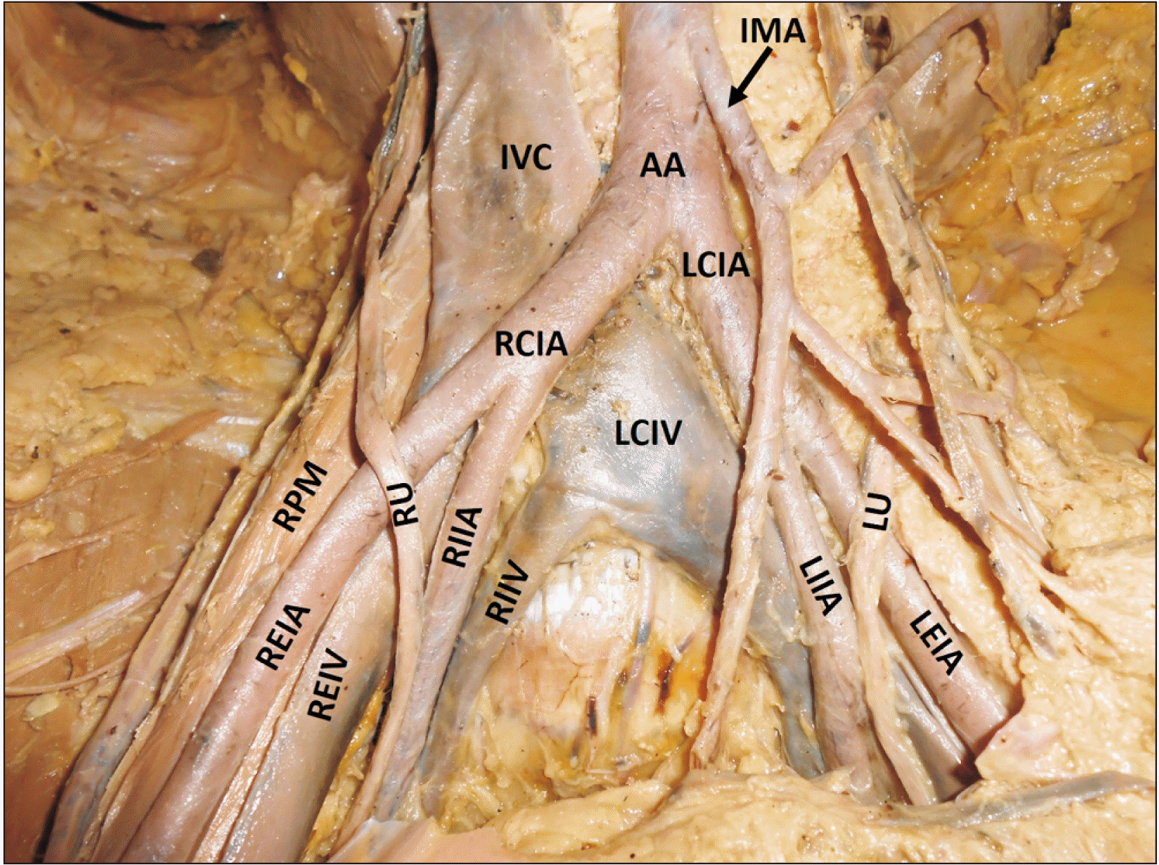
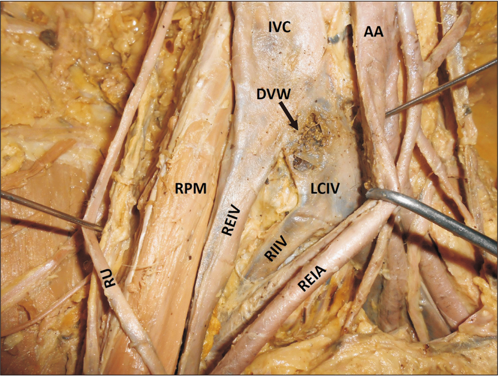
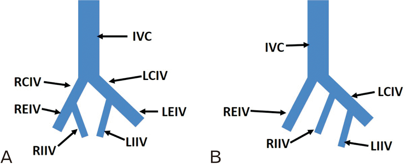
During routine dissection classes, we observed a rare combination of venous variations at the abdominopelvic junction of an adult male cadaver aged about 75 years. The right common iliac vein was absent. The inferior vena cava was formed by the union of the right external iliac vein and the left common iliac vein at the upper border of the fifth lumbar vertebra. The left common iliac vein was formed by the union of left external and internal iliac veins. The right internal iliac vein had a variant course and termination. It ran upwards and to the left, and joined the left common iliac vein 25 cm below the level of formation of the inferior vena cava (lower border of the fifth lumbar vertebra). The left common iliac vein was larger than usual in size and had its wall adhered to the right common iliac artery. The anterior wall of the left common iliac vein was very thin and it opened up easily while separating the right common iliac artery from it. The variations have been shown in the Figs. 1, 2. A simplified, schematic diagram of the variant veins has also been given (Fig. 3).
Discussion
Though morbidity and mortality rates in pelvic malignancies has reduced drastically due to the advances in imaging technologies, the surgeons often encounter significant bleeding during lateral pelvic surgeries. This bleeding is largely due to the presence of variations in the pelvic vasculature. Variations of the common iliac vein are less common compared to the variations of the external and internal iliac veins. In the current case, the inferior vena cava was formed by the union of right external iliac vein and the left common iliac vein. We can also interpret the formation of inferior vena cava in another way, i.e., the inferior vena cava was formed by the union of left common iliac vein and the right internal iliac vein at the level of lower border of the fifth lumbar vertebra and the right external iliac vein joined the inferior vena cava at the upper border of the fifth lumbar vertebra. Other congenital anomalies of the inferior vena cava include left inferior vena cava, double inferior vena cava and the azygos continuation of the inferior vena cava [3]. These anomalies were not found in the current case.
The inferior vena cava has a complex development by the contribution of three pairs of veins called posterior cardinal veins, subcardinal veins and supracardinal veins. The infra-renal segment of the inferior vena cava and the right common iliac vein develops from the right posterior cardinal vein. The left common iliac vein develops from the oblique anastomosis between the two posterior cardinal veins and the part of the left posterior cardinal vein lying below the level of the oblique anastomosis. The internal and external iliac veins are the veins that connect the lower limb veins with the abdominal veins. They develop from the plexus of the veins in front of the sacral region of the pelvic cavity. The veins of the lower limb develop from three sets of veins. The first set is called fibular vein which continues above as the sciatic vein, second one is the tibial vein and the third one is the femoral vein. In a later stage, a connecting vein develops between the sciatic vein and the femoral vein, thus the blood is diverted towards the femoral vein and the sciatic vein becomes smaller and it becomes a part of the inferior gluteal vein. The femoral vein establishes connections with the developing external iliac vein. The internal iliac vein and external iliac veins establish connections with the caudal part of the posterior cardinal veins. If there is any error in this process, the anomalous veins are seen in the pelvis as seen in the current case. In the current case, the vein forming the right internal iliac vein had failed to communicate with the external iliac vein of the right side. Instead of that, it had joined the left common iliac vein in the embryonic period.
On a functional viewpoint, the union of the right internal iliac vein with the left common iliac vein results in increased venous return through the left common iliac vein. All the blood from the pelvic organs and the left lower limb passes through the left common iliac vein in this case. This could result in enlargement of the vein. May-Thurner syndrome or Cockett syndrome is a syndrome in which the left common iliac vein is compressed between the right common iliac artery and the lower part of the vertebral column. In this syndrome, the veins of the pelvis and lower limbs enlarge and in some cases, it leads to deep vein thrombosis of lower limb. In the current case, there were no enlargement of the pelvic veins or the veins of lower limb. However, there was a fusion between the walls of right common iliac artery and the left common iliac vein. The wall of the vein adjacent to the artery was extremely thin. This was possibly due to the friction between the vein and the artery due to the large size of the vein. This type of fusion can lead to spontaneous rupture of the vein at any time of life.
Clinically, the knowledge of iliac vein variations is important in phlebography and retroperitoneal surgery as retroperitoneal pelvic surgeries are on a rise lately [4, 5]. Iliac veins and their tributaries must be paid attention during superior hypogastric neurectomy and hysterectomy. Orthopaedic procedures involving screw placement and obstetric surgeries can also endanger the anomalous course of the right internal iliac vein observed in the current case [6-8]. The anomalous veins in the pelvis could get injured also in aortic and iliac artery reconstruction procedures. Preoperative magnetic resonance imaging examination could minimise the iatrogenic injuries to some extent.
In conclusion, absence of right common iliac vein and opening of right internal iliac vein into the left common iliac vein is one of the rare venous variations in the abdominopelvic area [9, 10]. It was accompanied by the adhesion of the left common iliac vein with the right common iliac artery. This combination of variations may remain asymptomatic or could lead to May-Thurner syndrome. Knowledge of this variation could minimise bleeding in the retroperitoneal surgeries of abdominopelvic junction.
 XML Download