

 PDF
PDF Citation
 Print
Print


Introduction
The endothelial-lined trabeculated channels of the dural venous sinuses drain the superficial, deep, and posterior fossae and can be classified into two groups: (1) the posterosuperior group, which comprises the transverse, sigmoid, sagittal, and occipital sinuses, and (2) the anterior-inferior group, which comprises the cavernous and petrosal venous sinuses and basilar venous [1-3]. A long, slender dural venous sinus, the superior petrosal originates in the cavernous sinus, exits its posterosuperior aspect, and then runs caudally beneath the trochlear and oculomotor nerves to join posteriorly with the transverse sinus [4]. This long channel runs within the attached part of the tentorium cerebelli to the petrous part of the temporal bone from the cavernous sinus posteriorly to the transverse-sigmoid junction [5]. Crossing the superior portion of the trigeminal ganglion in the roof of Meckel’s cave, the superior petrosal sinus originates from a small cerebral vein and empties into the pro-otic vein [5, 6]. It continues in the posterosuperior edge of the sphenoid bone and connects the cavernous and transverse sinuses [7, 8]. The anterior component of the superior petrosal sinus attaches to the cavernous sinus near the trigeminal nerve proximally at the anterior part of the posterior clinoid process [7]. Veins of the pons, inner ear, cerebellum, and medulla oblongata constitute drain into the superior petrosal sinus [9]. The anterior part of the cerebellum is drained by the vein of the lateral recess of the cerebellomedullary fissure and superior petrosal sinus by the superior petrosal vein (SPV) [10]. The veins receive the contents of the anterior occipital, cavernous and circular sinuses and also act jointly to drain the bones of the skull with the inferior petrosal sinus [7]. The posterior cranial fossa is drained by the superior petrosal venous complex (SPVC) and is a landmark within the cerebellopontine angle [11].
The brainstem and cerebellum are drained by veins that form the following groups: anterior (petrosal), posterior (tentorial), and superior (galenic) [12]. Veins that drain into the superior petrosal sinus form the SPVC, each of them most commonly formed by the following tributaries: transverse pontine veins, veins of the cerebellopontine fissure, pontotrigeminal vein, and the middle cerebellar peduncle [5]. These veins drain the lateral pons and medulla, fourth ventricle and petrosal fissure [11]. The superior petrosal sinus drains the cranial cavity of cerebrospinal fluid and venous blood and empties it into the internal jugular vein, which then flows into the cardiovascular circulation via the superior vena cava [13]. Here, we describe a unique variation of the superior petrosal sinus identified at the time of routine dissection. As to our knowledge, such a case has not previously been reported, the current report would be of archival value.
Go to : 
Case Report
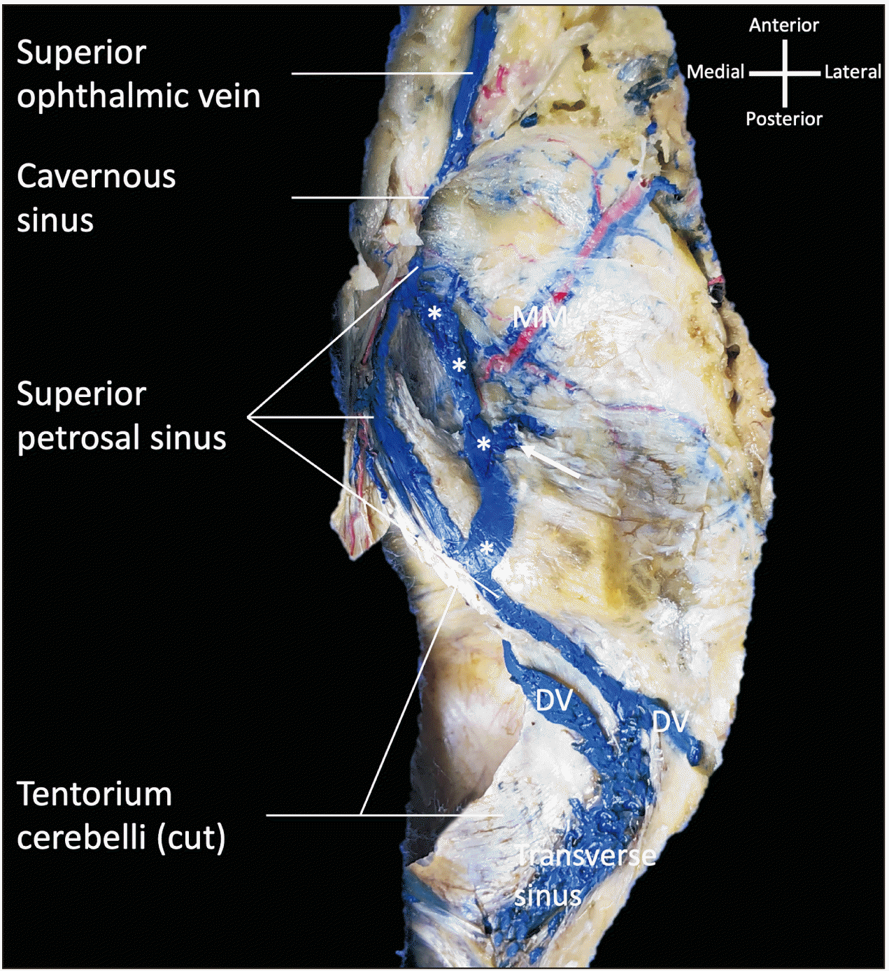
During the routine dissection of a latex injected cadaveric specimen (65-year-old at death male), on the right side of the skull base (middle cranial fossa), two superior petrosal sinuses were observed (Fig. 1). The more medial sinus traveled as does the typical superior petrosal sinus by traveling at the attachment of the tentorium cerebelli onto the petrous part of the temporal bone. In normal fashion, this sinus drains the transverse sinus posteriorly. Lateral to this sinus, a separate and more curvilinear superior petrosal sinus left the normally positioned superior petrosal sinus and traveled posteriorly near the foramen spinosum and then turned medially to drain into the normally positioned superior petrosal sinus. At about the middle part of the laterally positioned sinus, a small tributary of a diploic vein from the middle cranial fossa floor and middle meningeal vein both drained into it (Fig. 1). The normally positioned and laterally positioned superior petrosal sinuses traveled more or less in an oval shape and were approximately the same diameter (4 mm). Anteriorly, the two sinuses joined together and drained into the cavernous sinus. Posteriorly, the laterally positioned sinus drained into the normally positioned sinus which then traveled in normal fashion along the petrous ridge to end in the transverse sinus (Fig. 1). No intracranial pathology or contralateral anatomical variants were noted in this specimen.
Go to :
Discussion
Embryology
The head of the primary venous plexus splits into three layers: deep, middle, and outer. The basis of the vascular channels of the dura mater is formed by the outer plexus, while the deeper plexus connects with the superficial plexus via intermediate channels. As the meninges develop and grow from the skull base and gradually extend around the neural tube dorsally to the vertex of the head, the intermediate channels are eventually obliterated [4, 6, 10, 14]. The dural venous sinuses develop from the junction of the lateralis and venae capitis medialis and the middle cerebral vein to become the superior petrosal sinus [4]. During human development, it is the last major dural venous sinus to become defined [4] and is evolved from the pro-otic and mesencephalic vein during cerebellar development [13]. The superior petrosal sinus (Fig. 2) emerges at Carnegie stage 2 of development and thus, the variant identified in the present case would occur during this time. The formation of a large suprotic anastomosis appears at Carnegie stage 3, connecting the pro-otic and posterior rhombencephalic veins [15]. This newly formed channel and the terminal segment of the posterior rhombencephalic veins join to form the sigmoid sinus [16]. By Carnegie stage 6, the superior petrosal sinus runs along the otic capsule and inside Meckel’s cave over the trigeminal ganglion [16].
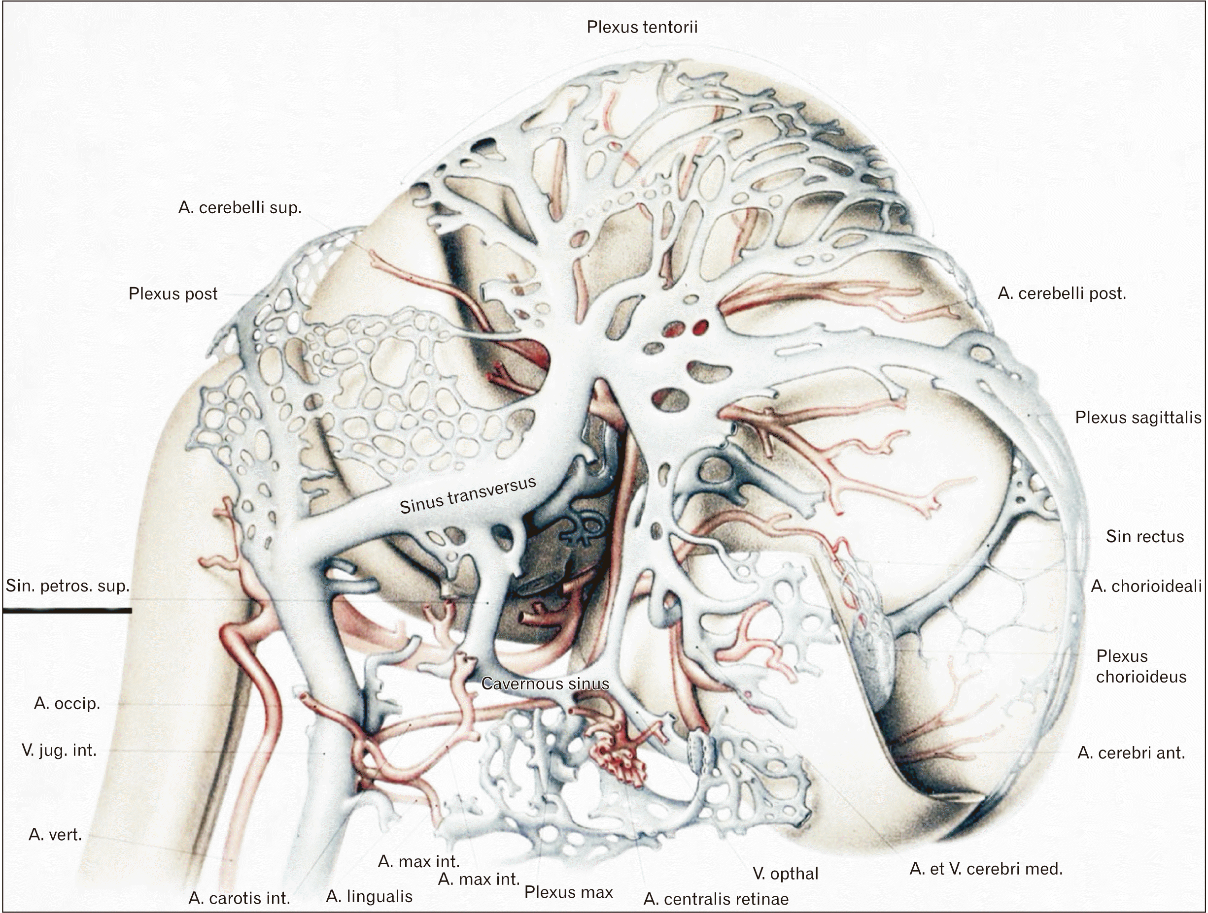 | Fig. 2Embryological development of the superior petrosal sinus (label underlined). Cited from Streeter (Am J Anat 1915;18:145-78) [6].
|
To summarize the developmental process: the trochlear nerve, oculomotor nerve, and superior petrosal sinus travel near the otic capsule and anterior dural fold during early development (Carnegie stage 2), with the trigeminal ganglion separating both [16]. As the temporal lobe enlarges, it pushes the anterior tentorial layer backwards, pushing the oculomotor, trigeminal ganglion, superior petrosal sinus and trochlear nerves toward the otic capsule [15]. The superior petrosal sinus and cranial nerves expand along the border of the otic capsule in order to attain their adult positions [13]. Later in development, the superior petrosal sinus travels over the trigeminal ganglion and beneath the trochlear and oculomotor nerves, the now-enlarged cavernous sinus projecting anterosuperiorly into the trigeminal ganglion and draining the superior petrosal sinus. The relationship between the superior petrosal sinus and the cavernous sinus occurs later in development and does not always form [15].
In a study of the hemodynamic features of the superior petrosal sinus and drainage variants from cavernous sinus dural arteriovenous fistulas (CSDAVFs), additional findings demonstrated duplication of the superior petrosal sinus in consequence of the plexiform feature of the remnant of the primitive tentorial sinus [17].
Anatomical variations
There are many anatomical variations and drainage patterns of the superior petrosal sinus. In some instances, the sinus can be enlarged or completely absent [18-20]. Matsushima et al. [5] observed that in the absence of the superior petrosal sinus and SPV, venous drainage was taken over by a bridging vein, the vein of Galen, into the transverse-sigmoid sinus. The pontotrigeminal and lateral mesencephalic veins drained into the vein of Galen tributaries without joining the SPV [5]. These different anatomical variations need to be considered, especially in transverse sinus dural arteriovenous fistula treatment and approaches, since obstruction of veins can cause venous complications [21]. Rarely, the basal vein of Rosenthal (BVR) drains directly into the superior petrosal sinus. The BVR emerges from intersection of the inferior striate veins, deep middle cerebral, and anterior cerebral and typically drains into the vein of Galen [22]. The BVR and superior petrosal sinus can be connected by the peduncular and lateral mesencephalic veins, although in this particular example there is a direct connection between the superior petrosal sinus and BVR in the absence of a tentorial sinus. Furthermore, Padget [23] noted that the anterior cerebellar vein drains considerable parts of the cerebellar hemisphere, including tributaries from the pons and medulla, and empties into the superior petrosal sinus. In cases where the great cerebral vein is occluded, this anastomotic vein (lateral mesencephalic vein) can drain cerebral blood into the sigmoid sinus via the superior petrosal sinus and the basal and mesencephalic veins. Additionally, the inferior anastomotic vein (vein of Labbé) can feed into the superior petrosal sinus [24]. This can make combined base approaches very difficult and sometimes impossible. Observations also suggest that the superior petrosal sinus occasionally communicates with an aberrant vein (ophthalmo-petrosal sinus) through the superior ophthalmic veins [25]. Another study demonstrated variable superior petrosal sinus drainage from CSDAVFs among patients: either CSDAVFs drained from the cavernous sinus through the entire superior petrosal sinus or dural arteriovenous fistulas (DAVFs) drained down the anterior segment of the superior petrosal sinus into petrosal vein tributaries, bypassing drainage through the posterior segment of the superior petrosal sinus [17]. The anterior and posterior segments of the superior petrosal sinus were disconnected in a few cases, indicating malfusion or hypoplasia of the posterior and anterior segments of the superior petrosal sinus during development. The plexiform feature of the primitive tentorial sinus remnant caused this duplication [17]. Additionally, Tanoue et al. [26] sorted the proximal communications of the superior petrosal sinus into three types.
Various arrangements of the superior petrosal sinus affect its relationship with the porous trigeminus. Tubbs et al. [16] observed superior petrosal sinus variations with reference to Meckel’s cave in cadaver heads: superior (68%), inferior (18%), and surrounding the opening of the cave (16%). Coates [27] examined the relationship of the trigeminal nerve to the superior petrosal sinus in cadavers and found the following variations: both right and left sinuses passed below the trigeminal nerve root and above the cranial surface of the nerve [27]. Furthermore, the sinus was divided in three specimens on both sides, with some parts passing over the trigeminal nerve root and some passing under.
Clinical significance
The clinical relevance of the superior petrosal sinus to abnormal connections such as carotid-cavernous fistulas and DAVFs have been noted [13]. DAVFs have been classified into three categories depending on their drainage pattern [13, 15]. Type I drains the dural venous sinuses or meningeal veins in an anterograde fashion. Type II drains into the subarachnoid veins in a retrograde or anterograde fashion. Type III does not have dural venous sinus or meningeal drainage and drains exclusively into the subarachnoid granulations. These drainage types are specific to DAVFs and are not associated with the drainage patterns of the SPVC into the superior petrosal sinus. DAVFs can be treated through either endovascular therapy or surgery; the therapy of choice for all arteriovenous fistula malformation (AVFM) types is transarterial embolization, a type of nonsurgical endovascular therapy. It is rarely a cure, but can make surgery less challenging by decreasing flow through the fistula [15]. Gamma knife radiosurgery is another type of surgery for superior petrosal sinus AVFM, though three years are needed for closure of the fistula and the treatment is not always viable for high-grade AVFMs [13, 28].
The superior petrosal sinus is in close proximity to critical intracranial structures, making it a critical landmark in surgical procedures involving the skull. Posterior fossa approaches often encounter the SPVC, one of the largest infratentorial venous channels [29]. The superior petrosal sinus can also be a helpful landmark for the tentorium cerebelli, which attaches to the edges of the superior petrosal groove [15]. Depending on the pathology and anatomical variations of the superior petrosal sinus, surgical approaches in accessing it can be direct or indirect. The different drainage pattern variants are distinguished by their entry site into the SPVC, internal acoustic meatus and Meckel’s cave and drainage into the superior petrosal sinus [13, 29, 30]. Type I enters the superior petrosal sinus superolateral to the boundaries of the internal acoustic meatus. The most common drainage pattern, Type II, empties between the medial edge of the facial nerve at the internal acoustic meatus and the lateral edge of the trigeminal nerve in Meckel’s cave. Type III enters above or medial to Meckel’s cave. Type I requires a subtemporal transtentorial approach, but this approach has limited use in types II and III [15]. In type III drainage pattern variants, a neurosurgical retrosigmoid suprameatal approach can be taken. Anterior transpetrosal, middle cranial fossa, retrosigmoid suboccipital, and petrosal-tentorial triangular approaches can also provide access to the superior petrosal sinus [1]. Additionally, for conditions such as classical trigeminal neuralgia, microvascular decompression of the trigeminal nerve has been shown to be a safe and efficacious treatment, the SPV being an important landmark for the retrosigmoid surgical approach [31].
However, there are risks with attempting to ligate the superior petrosal sinus. A major risk is blocking flow in the vein of Labbé, which drains the inferior parietal and posterior temporal lobes; this can lead to blood coagulation along the superior petrosal sinus. Moreover, temporal lobe infarction can result, potentially leading to brain herniation and death [32].
A venous phase computerized tomography angiogram or preoperative magnetic resonance (MR) venogram can determine the drainage pattern and location of SPVC entry into the superior petrosal sinus and further indicate which surgical approach can be used to decrease the probability of venous complications [15].
Understanding the variations of the intracranial venous sinuses is important for anatomists and clinicians [33]. Of these sinuses, the superior petrosal sinus is an important landmark requiring consideration in many pathologies and surgical approaches. Clinically, such a variant could result in misinterpretation on imaging studies such as MR venography, potentially be injured during skull base surgery such as tumor removal, or result in a complication during intravenous interventional procedures for treatment of, for example, arteriovenous malformations. Therefore, knowledge of its anatomical variations such as described in the case reported herein can avoid misdiagnosis on imaging and iatrogenic surgical injury.
Go to :
 XML Download