

 PDF
PDF Citation
 Print
Print


Introduction
The foramen of Vesalius (sphenoidal emissary foramen) is a variable and small bony passage located between the foramen ovale and foramen rotundum in the greater wing of the sphenoid bone [1-4]. It lies anteromedial to the foramen ovale and inferolateral to the foramen rotundum unilaterally or bilaterally. Andreas Vesalius was presumably the first anatomist to describe this structure foramen which gives transmits emissary veins from the pterygoid venous plexus to the cavernous sinus. This venous connection represents an important extra to intracranial anastomosis connecting the venous systems of the face and brain. Reviews of studies of dry specimens show between 5% and 80% of skulls contain at least one foramen of Vesalius [1, 2].
Endochondral ossification of the skull base takes place through a series of “primary” ossification centers which arise in a general caudal to rostral pattern beginning with ossification centers in the occipital bone and at lastly developing ossification centers in the alisphenoid giving rise to the greater wing of the sphenoid bone. These centers of ossification arise between 12 and 17 weeks of development [5]. The positions of pre-existing vessels are progressively fixed by the ossification of the skull base, and reciprocally, the foramina of the skull base are positioned in accordance with their contained vessels. Emissary sphenoidal veins occupy this role relative to when present, the foramen of Vesalius [1, 2]. Some have also considered that when present, the foramen of Vesalius transmits the venous component normally found traveling through the foramen ovale [6].
Here, we describe, to our knowledge, the largest foramen of Vesalius reported in the English literature and describe the potential clinical consequences of such a finding.
Go to : 
Case Report
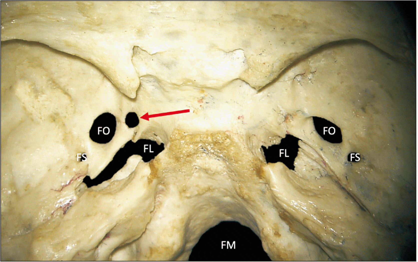
During the routine evaluation of an adult female skull housed in the osteological collection of our medical school, a large foramen of Vesalius was identified (Fig. 1). The specimen was approximately 50-year-old at the time of death and was derived from a collection of roughly 100 human skulls from primarily a North American background. This roughly circular shaped foramen of Vesalius was 4.34 mm in diameter and was located 7.5 mm posteromedial to the foramen rotundum and 1.49 mm anteromedial to the foramen of ovale. This foramen was 2.6 mm anterior to the foramen lacerum. The contralateral foramen of Vesalius measured 0.92 mm in diameter. No other gross anatomical variations were noted at the skull base in this specimen. All measurements were made using microcalipers (Mitutoyo, Kawasaki, Japan).
Go to :
Discussion
Morphometric studies of foramen of Vesalus have documented mean widths of 0.67–2.22 mm when measured across their maximum dimension [1]. The specimen presented here had a diameter (4.34 mm) that was approximately twice the maximally reported mean diameter found in the literature. Most studies do not show a significant difference in size between left and right foramina Vesalli. A large foramen of Vesalius, such as the variant described in this study, has the potential to disorient a clinician passing a needle through the nearby foramen ovale into the middle cranial fossa as the foramina are seen in the operating room using fluoroscopy [6, 7]. Therefore, the quality of visualization of the skull base foramina is generally moderate at best. Such procedures are used for rhizotomy of the trigeminal nerve, temporal lobe electrode placement, balloon deployment to treat trigeminal neuralgia i.e., compression of the trigeminal ganglion, or cavernous sinus tumor biopsy [8-11]. In attempt to avoid surrounding foraminal during transcutaneous needle approaches to the foramen ovale, Tubbs et al. [9], described a “safe zone” of the foramen ovale extending 6 mm around the circumference and excluding the entire territory around the foramen spinosum posterolaterally. If intraoperative imaging fails to resolve a small bony bridge separating a large and nearby foramen of Vesalius and the foramen ovale, a clinician will be at greater risk of passing their needle too medially, potentially injurying the cavernous sinus or at least, the emissary vein traveling through the foramen of Vesalius. This would be compounded by an enlarged foramen of Vesalius as reported here. The distance between the foramen ovale and the enlarged foramen of Vesalius in our specimen was only 1.49 mm. Although there will be a protective effect from cartilage filling the foramen lacerum, the ICA is also at risk of damage if the needle passes more than 6 mm medial to the foramen ovale [9, 11].
Our case helps illustrate the wide variation in morphology of an inconsistent foramen found near significant clinical anatomy of the skull base. Clinicians and surgeons should be mindful of variations such as the enlarged foramen of Vesalius reported here when attempting transcutaneous puncture of the foramen ovale.
Go to :
 XML Download